

Myosin cross-bridge kinetics slow at longer muscle lengths during isometric contractions in intact soleus from mice
- PMID: 33975478
- PMCID: PMC8190544
- DOI: 10.1098/rspb.2020.2895
Myosin cross-bridge kinetics slow at longer muscle lengths during isometric contractions in intact soleus from mice
Abstract
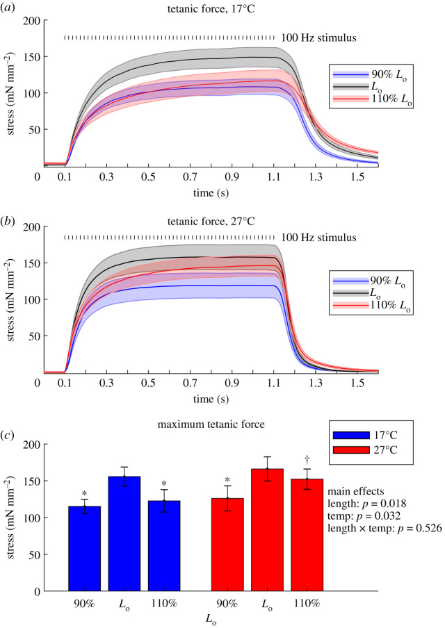
Muscle contraction results from force-generating cross-bridge interactions between myosin and actin. Cross-bridge cycling kinetics underlie fundamental contractile properties, such as active force production and energy utilization. Factors that influence cross-bridge kinetics at the molecular level propagate through the sarcomeres, cells and tissue to modulate whole-muscle function. Conversely, movement and changes in the muscle length can influence cross-bridge kinetics on the molecular level. Reduced, single-molecule and single-fibre experiments have shown that increasing the strain on cross-bridges may slow their cycling rate and prolong their attachment duration. However, whether these strain-dependent cycling mechanisms persist in the intact muscle tissue, which encompasses more complex organization and passive elements, remains unclear. To investigate this multi-scale relationship, we adapted traditional step-stretch protocols for use with mouse soleus muscle during isometric tetanic contractions, enabling novel estimates of length-dependent cross-bridge kinetics in the intact skeletal muscle. Compared to rates at the optimal muscle length (Lo), we found that cross-bridge detachment rates increased by approximately 20% at 90% of Lo (shorter) and decreased by approximately 20% at 110% of Lo (longer). These data indicate that cross-bridge kinetics vary with whole-muscle length during intact, isometric contraction, which could intrinsically modulate force generation and energetics, and suggests a multi-scale feedback pathway between whole-muscle function and cross-bridge activity.
Keywords: myosin cross-bridges intact muscle.
Figures


References
Publication types
MeSH terms
Substances
Associated data
Grants and funding
LinkOut - more resources
Full Text Sources
Other Literature Sources