. 2021 May 11;12(3):e03443-20.
doi: 10.1128/mBio.03443-20.

The DnaK/DnaJ Chaperone System Enables RNA Polymerase-DksA Complex Formation in Salmonella Experiencing Oxidative Stress

Affiliations

The DnaK/DnaJ Chaperone System Enables RNA Polymerase-DksA Complex Formation in Salmonella Experiencing Oxidative Stress

Ju-Sim Kim et al. mBio. .

Abstract

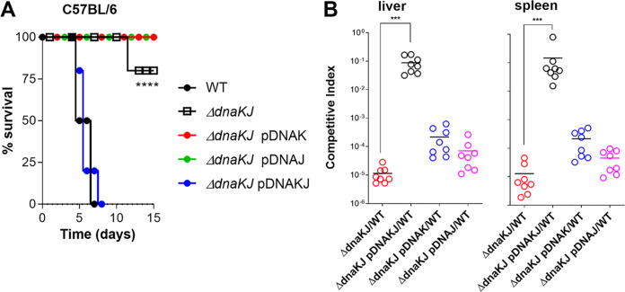
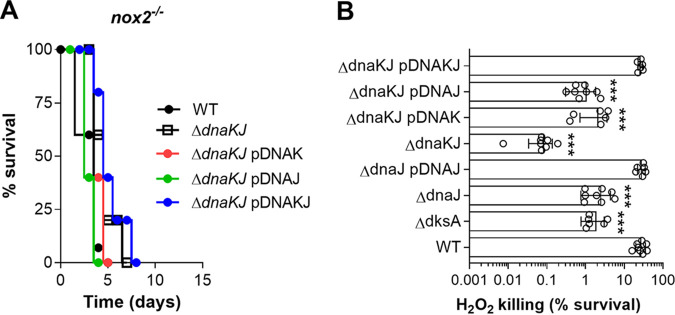
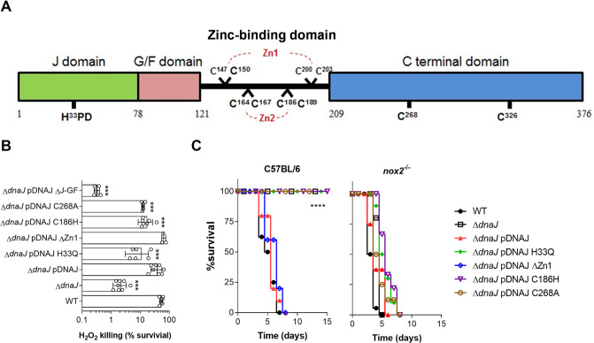
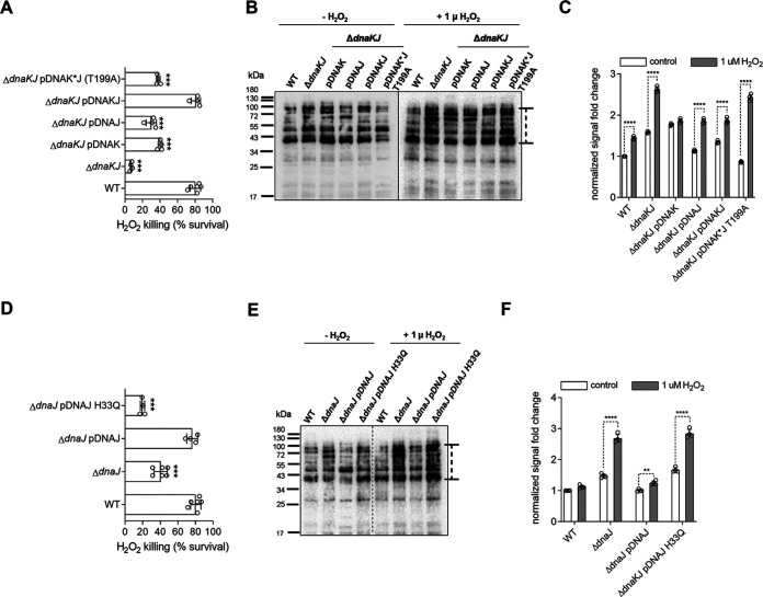
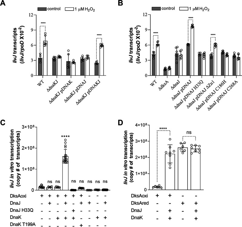
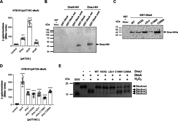
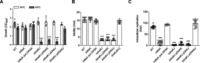
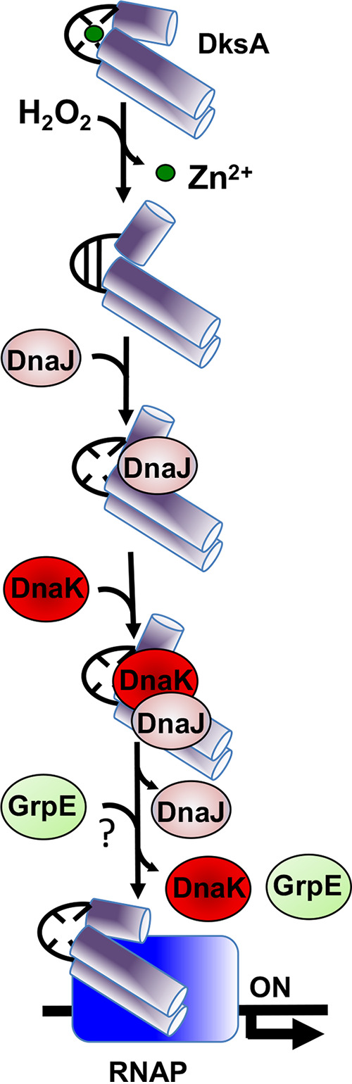
Our previous biochemical approaches showed that the oxidoreductase activity of the DnaJ protein facilitates the interaction of oxidized DksA with RNA polymerase. Investigations herein demonstrate that under biologically relevant conditions the DnaJ- and DksA-codependent activation of the stringent response in Salmonella undergoing oxidative stress involves the DnaK chaperone. Oxidation of DksA cysteine residues stimulates redox-based and holdase interactions with zinc-binding and C-terminal domains of DnaJ. Genetic and biochemical evidence indicates that His33 in the HPD motif in the J domain of DnaJ facilitates interactions of unfolded DksA with DnaK. A mutation in His33 in the J domain prevents the presentation of unfolded DksA to DnaK without limiting the oxidoreductase activity mapped to DnaJ's zinc-2 site. Thr199 in the ATPase catalytic site of DnaK is required for the formation of the DksA/RNA polymerase complex. The DnaK/DnaJ/DksA complex enables the formation of an enzymatically active RNA polymerase holoenzyme that stimulates transcription of branched-chain amino acid and histidine metabolic genes in Salmonella exposed to reactive oxygen species. The DnaK/DnaJ chaperone protects Salmonella against the cytotoxicity associated with reactive oxygen species generated by the phagocyte NADPH oxidase in the innate host response. The antioxidant defenses associated with DnaK/DnaJ can in part be ascribed to the elicitation of the DksA-dependent stringent response and the protection this chaperone system provides against protein carbonylation in Salmonella undergoing oxidative stress.IMPORTANCE DksA was discovered 30 years ago in a screen for suppressors that reversed the thermosensitivity of Escherichia coli mutant strains deficient in DnaK/DnaJ, raising the possibility that this chaperone system may control DksA function. Since its serendipitous discovery, DksA has emerged as a key activator of the transcriptional program called the stringent response in Gram-negative bacteria experiencing diverse adverse conditions, including nutritional starvation or oxidative stress. DksA activates the stringent response through the allosteric control this regulatory protein exerts on the kinetics of RNA polymerase promoter open complexes. Recent investigations have shown that DksA overexpression protects dnaKJ mutant bacteria against heat shock indirectly via the ancestral chaperone polyphosphate, casting doubt on a possible complexation of DnaK, DnaJ, and DksA. Nonetheless, research presented herein demonstrates that the cochaperones DnaK and DnaJ enable DksA/RNA polymerase complex formation in response to oxidative stress.

Keywords: DksA; DnaJ; DnaK; Salmonella Typhimurium; chaperone; hydrogen peroxide; oxidative stress; redox; stringent response.


Figures

FIG 1
FIG 1
DnaK and DnaJ contribute to Salmonella pathogenesis. (A) Survival of C57BL/6 mice after intraperitoneal (i.p.) inoculation of ∼150 CFU of the indicated Salmonella strains. Mouse survival was monitored over 2 weeks. The data are from 8 to 10 mice from 2 independent experiments. P values were determined by log-rank analysis (****, P < 0.0001) compared to controls infected with wild-type Salmonella. (B) The competitive index was measured in livers and spleens of C57BL/6 mice 96 h after i.p. inoculation of ∼1,000 CFU of a mixture containing equal numbers of the indicated strains. Horizontal bars represent the medians from 8 mice collected in 2 independent experiments. ***, P < 0.001, as determined by one-way ANOVA.
FIG 2
FIG 2
DnaK and DnaJ protect Salmonella from oxidative stress. (A) Survival of phagocyte NADPH oxidase-deficient nox2−/− mice after i.p. challenge with ∼150 CFU of the indicated Salmonella strains. The data are from 8 to 10 mice in 2 independent experiments. (B) Survival of Salmonella grown overnight in LB broth after 2 h of treatment with 200 μM H2O2 in PBS. The data are the means ± standard deviations (SD) (n = 6) from 6 to 10 independent experiments. ***, P < 0.001, as determined by one-way ANOVA compared to wild-type Salmonella.
FIG 3
FIG 3
Impact of dnaJ variants in the antioxidant defenses of Salmonella. (A) Schematic representation of DnaJ regions consisting of the J, G/F (Gly/Phe-rich region), zinc-binding, and C-terminal domains. The J domain, via the evolutionarily conserved HPD motif, stimulates DnaK ATPase activity, and the G/F domain assists binding of DnaJ to DnaK. Four CXXCXGXG consensus sequences (where with C represents Cys, X is any other amino acid, and G is glycine) assemble into two zinc-binding domains, Zn1 and Zn2. The C-terminal domain contains 2 additional cysteine residues. (B) Susceptibility of wild-type and the indicated Salmonella mutants grown overnight in LB broth after 2 h of treatment with 200 μM H2O2 in PBS. The data are the means ± SD (n = 6 to 8) from 3 or 4 independent experiments. ***, P < 0.001, as determined by one-way ANOVA compared to wild-type Salmonella. (C) C57BL/6 and nox2/− mice were i.p. inoculated with ∼150 CFU of the indicated Salmonella strains. Mouse survival was monitored over 2 weeks. The data are from 8 to 18 mice collected in 2 or 3 independent experiments. ****, P < 0.0001, as determined by log-rank analysis compared to mice infected with wild-type Salmonella.
FIG 4
FIG 4
DnaK and DnaJ lessen protein carbonylation in anaerobic Salmonella exposed to H2O2. (A and D) Survival of anaerobic Salmonella exposed to 1 μM H2O2 for 2 h in an anaerobic chamber. ***, P < 0.001, as determined by one-way ANOVA compared to wild-type Salmonella. The data are the means ± SD from 4 to 6 biological replicates collected in 2 or 3 days. (B and E) Protein carbonylation in anaerobic Salmonella treated with or without 1 μM H2O2 for 2 h was assessed by immunoblotting of DNP-derivatized proteins. The data are representative of 2 or 3 independent experiments. (C and F) Density of protein carbonylation was measured with ImageJ, and the density comparison region is indicated by a dotted line with a cap next to the blots. Fold change in density was calculated as density of mutant/density of wild-type controls. The data are the means ± SD (n = 4) from 4 independent measurements. ****, P < 0.0001, as determined by one-way ANOVA.
FIG 5
FIG 5
DnaK and DnaJ activate DksA-redox dependent gene expression. (A and B) The indicated Salmonella strains were grown in EGCA medium in an anaerobic chamber. Abundance of livJ transcripts in RNA isolated from anaerobic Salmonella treated with or without 1 μM H2O2 for 30 min was measured by qRT-PCR. The expression of the housekeeping gene rpoD was used as an internal control. The data are the means ± SD from 4 to 6 biological replicates collected in 2 or 3 independent days. ****, P < 0.0001, as determined by one-way ANOVA. (C and D) Activation of livJ transcripts in in vitro transcription reaction mixtures containing 5 μM oxidized DksA, 50 nM DnaJ, and 500 nM DnaK proteins in the presence of RNA polymerase. Interactions of DnaJ and DnaK proteins with oxidized or reduced DksA are shown in panel D. Reduced DksA (5 μM) showed sufficiently activated in vitro livJ transcripts in the absence of DnaJ (50 nM) and DnaK (500 nM) proteins. The abundance of livJ transcripts was analyzed by qRT-PCR. The data are the means ± SD (n = 6 to 20) from at least 3 independent experiments. ****, P < 0.0001, as determined by one-way ANOVA. ns, nonsignificant compared to reaction mixtures containing 5 μM oxidized DksA (C) or reduced DksA (D).
FIG 6
FIG 6
Interactions of DnaK and DnaJ proteins with DksA. Interactions of DnaK and DnaJ proteins with DksA were evaluated by a bacterial two-hybrid system (A and D) and biochemical pulldown assays (B and C). The data in panel A are the means ± SD from 12 to 16 biological replicates from 3 or 4 independent experiments ***, P < 0.001, and ****, P < 0.0001, as determined by one-way ANOVA. ns, nonsignificant compared to the negative vector control. Blots in panels B and C are representative of 3 or 4 independent experiments. (E) Reduced DksA (5 μM) residues were alkylated with AMS, and the proteins were visualized on nonreducing SDS-PAGE gels stained with Coomassie brilliant blue. Samples were treated with or without 500 μM H2O2 at 37°C for 1 h in the presence and absence of 5 μM DnaJ variants. The data are from 2 or 3 independent experiments.
FIG 7
FIG 7
Contribution of dnaK, dnaJ, and dksA to thermotolerance, motility, and pathogenesis of Salmonella. (A) Growth of the indicated Salmonella strains in LB broth for 6 h at 30°C or 45°C in a shaker incubator. The data are the means ± SD (n = 4 to 10) from 4 to 8 independent experiments. (B) Motility was assessed by spotting 107 CFU of the indicated Salmonella strains in 0.3% LB agar plates for 3 h at 37°C. The swimming zone was measured in millimeters. The data are the means ± SD (n =14) from at least 4 independent experiments. (C) Intracellular replication of Salmonella in J774 cells 18 h postinfection was determined by CFU measurement. The data are the means ± SD (n = 8 to 20) from at least 3 independent experiments. ***, P < 0.001, and ****, P < 0.0001, as determined by one-way ANOVA.
FIG 8
FIG 8
Model for the DnaK/DnaJ-mediated assembly of a DksA-RNA polymerase (RNAP) complex in response to reactive oxygen species. Hydrogen peroxide (H2O2) induces disulfide formation and zinc release in the transcriptional regulator DksA. DnaJ recognizes misfolded, disulfide-bonded DksA as a client protein. The oxidoreductase activity of DnaJ reduces the disulfide bonds in DksA, transferring the reduced, unfolded DksA protein to ATP-bound DnaK. The J domain of DnaJ stimulates ATPase activity of DnaK, which firmly binds to unfolded DksA. Although not proven in our investigations, it is possible that the nucleotide exchange factor GrpE facilitates refolding of DksA by DnaK. Together, the DnaK/DnaJ/GrpE chaperone system enables the formation of RNA polymerase-DksA complexes in response to oxidative stress.

References

    1. Rosenzweig R, Nillegoda NB, Mayer MP, Bukau B. 2019. The Hsp70 chaperone network. Nat Rev Mol Cell Biol 20:665–680. doi:10.1038/s41580-019-0133-3. - DOI - PubMed
    1. Szabo A, Langer T, Schroder H, Flanagan J, Bukau B, Hartl FU. 1994. The ATP hydrolysis-dependent reaction cycle of the Escherichia coli Hsp70 system DnaK, DnaJ, and GrpE. Proc Natl Acad Sci U S A 91:10345–10349. doi:10.1073/pnas.91.22.10345. - DOI - PMC - PubMed
    1. Mayer MP, Kityk R. 2015. Insights into the molecular mechanism of allostery in Hsp70s. Front Mol Biosci 2:58. doi:10.3389/fmolb.2015.00058. - DOI - PMC - PubMed
    1. Mayer PM. 2013. Hsp70 chaperone dynamics and molecular mechanism. Trends Biochem Sci 38:507–514. doi:10.1016/j.tibs.2013.08.001. - DOI - PubMed
    1. Szabo A, Korszun R, Hartl FU, Flanagan J. 1996. A zinc finger-like domain of the molecular chaperone DnaJ is involved in binding to denatured protein substrates. EMBO J 15:408–417. doi:10.1002/j.1460-2075.1996.tb00371.x. - DOI - PMC - PubMed

Publication types

MeSH terms

Substances

LinkOut - more resources