. 2021 May 11;12(5):470.
doi: 10.1038/s41419-021-03750-4.

Knockout RAGE alleviates cardiac fibrosis through repressing endothelial-to-mesenchymal transition (EndMT) mediated by autophagy

Affiliations

Knockout RAGE alleviates cardiac fibrosis through repressing endothelial-to-mesenchymal transition (EndMT) mediated by autophagy

Lu Zhang et al. Cell Death Dis. .

Abstract

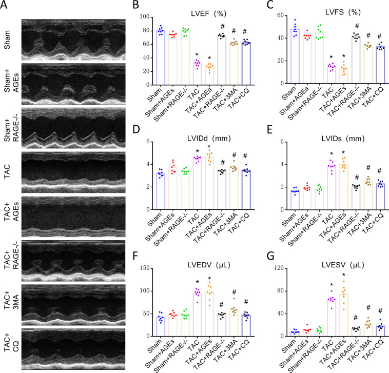
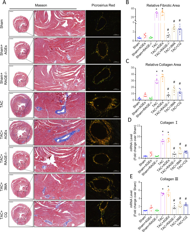
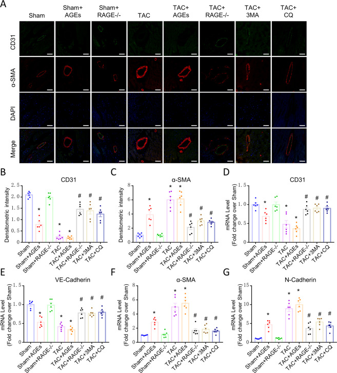
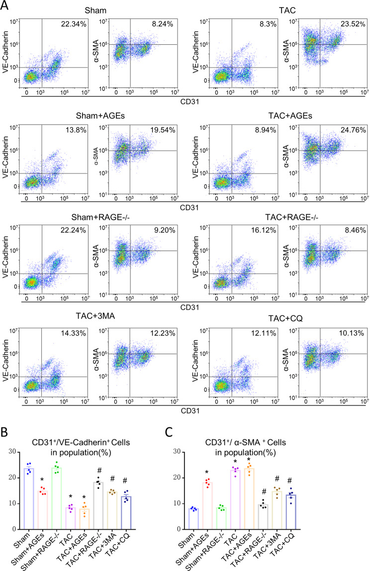
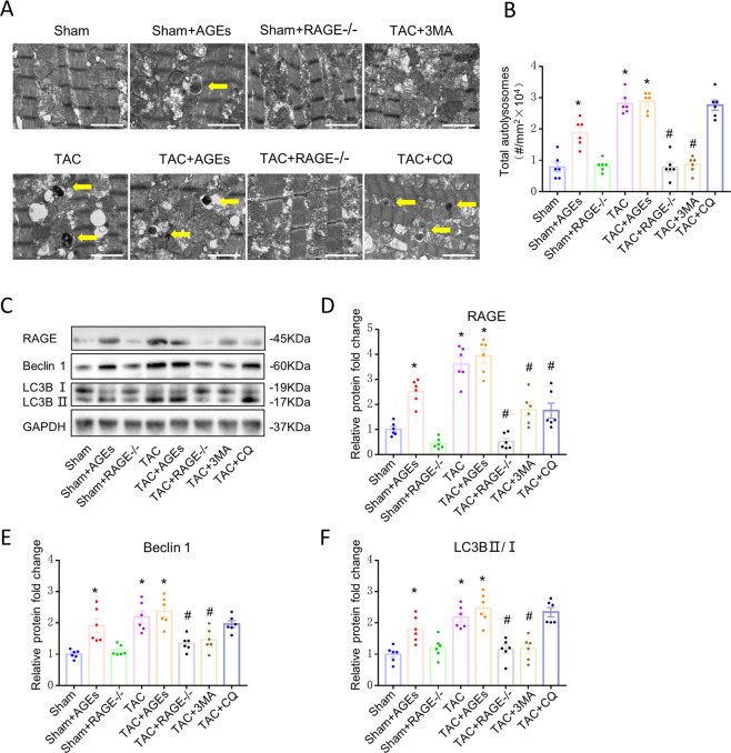
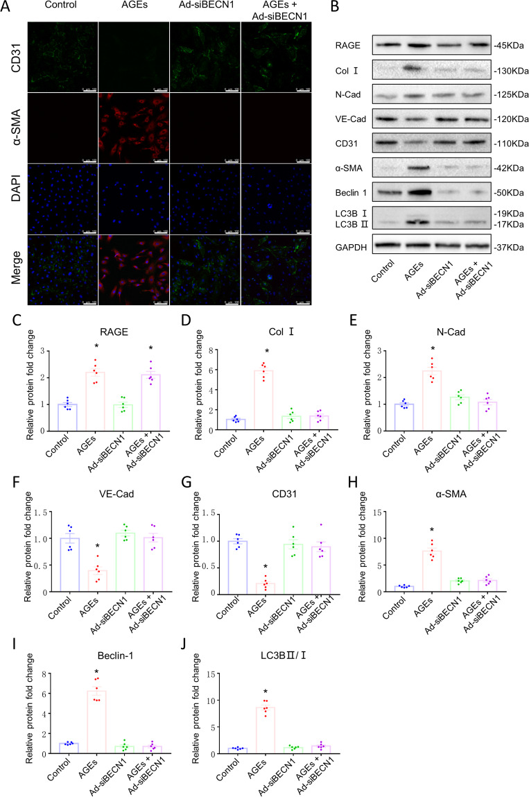
Endothelial-to-mesenchymal transition (EndMT) has been shown to contribute to cardiac fibrosis and heart failure (HF). Recent studies have demonstrated that EndMT is regulated by autophagy, and we previously showed suppression of excessive autophagy and alleviation of cardiac fibrosis in HF mice with inactivated receptor for advanced glycation end products (RAGE). Thus, we investigated whether reduced cardiac fibrosis due to RAGE knockout occurred by inhibiting EndMT mediated by excessive autophagy. We found a decrease in endothelial cells (CD31+/VE-Cadherin+) and an increase in cells co-expressing CD31 and α-smooth muscle actin (α-SMA, myofibroblast marker) at 8 weeks in heart tissue of mice subjected to transverse aortic constriction (TAC), which implied EndMT. Knockout RAGE decreased EndMT accompanied by decreased expression of autophagy-related proteins (LC3BII/I and Beclin 1), and alleviated cardiac fibrosis and improved cardiac function in TAC mice. Moreover, 3-methyladenine (3-MA) and chloroquine (CQ), inhibitors of autophagy, attenuated EndMT, and cardiac fibrosis in TAC mice. Importantly, EndMT induced by AGEs could be blocked by autophagy inhibitor in vivo and in vitro. These results suggested that AGEs/RAGE-autophagy-EndMT axis involved in the development of cardiac fibrosis and knockout RAGE ameliorated cardiac fibrosis through decreasing EndMT regulated by autophagy, which could be a promising therapeutic strategy for HF.


Conflict of interest statement

The authors declare no competing interests.

Figures

Fig. 1
Fig. 1. Improvement of cardiac function at 8 weeks after TAC by genetic deletion of RAGE or an autophagy inhibitor (3-MA or CQ).
A Representative M-mode images. Echocardiography was performed to measure LVEF (B), LVFS (C), LVIDd (D), LVIDs (E), LVEDV (F), and LVESV (G). n = 8. Data are presented as mean ± SEM. *p < 0.05 vs. sham group, #p < 0.05 vs. TAC group.
Fig. 2
Fig. 2. Effect of AGEs/RAGE or autophagy on cardiac fibrosis after TAC.
A Myocardial fibrosis was detected by Masson’s trichrome staining (scale bar: transverse sections = 1000 µm, perivascular sections = 200 µm), and perivascular collagen synthesis was detected by a Sirius red-polarized method (scale bar = 50 µm). Blue areas indicate fibrosis. Yellow areas indicate collagen I, and green areas indicate collagen III. The fibrosis area was measured with a quantitative digital image analysis system: fibrotic area (B) and collagen deposition area (C). mRNAs for fibrosis associated genes collagen I (D) and collagen III (E) were measured by qPCR. n = 6. Data are presented as mean ± SEM. *p < 0.05 vs. sham group, #p < 0.05 vs. TAC group.
Fig. 3
Fig. 3. Endothelial cells may transition to mesenchymal cells in HF.
A Confocal microscopic image of double immunofluorescence staining with CD31 (green) and α-SMA (red); nuclei were counter stained with DAPI (blue); Scale bars = 100 µm. B, C Quantification of immunofluorescence analysis. mRNAs for endothelial cells-associated genes [CD31 (D), VE-Cadherin (E)] and mesenchymal cells-associated gene [α-SMA (F) and N-Cadherin (G)] were measured by qPCR. n = 6. Data are presented as mean ± SEM. *p < 0.05 vs. sham group, #p < 0.05 vs. TAC group.
Fig. 4
Fig. 4. EndMT is one origin of myofibroblasts in HF and could be regulated by AGEs/RAGE and autophagy.
A Flow cytometry analysis of primary isolates of mouse left ventricular cells. After mechanical disaggregation and enzymatic digestion, single-cell suspensions were incubated with combination of fluorescent antibodies to CD31, VE-Cadherin, and α-SMA. B, C Quantification of co-expressed cell analysis. n = 5. Data are presented as mean ± SEM. *p < 0.05 vs. sham group, #p < 0.05 vs. TAC group.
Fig. 5
Fig. 5. Reduction of autophagy at 8 weeks after TAC by genetic deletion of RAGE or an autophagy inhibitor (3-MA or CQ).
A Representative autophagic ultrastructure of the heart tissue under transmission electron microscopy (Scale bars = 2 µm; arrow: autolysosome). B Quantification of autophagy lysosomes analysis. CF Left ventricular levels of RAGE and autophagy-related proteins (LC3BII/I and Beclin 1) were assessed by western blot. Quantification of RAGE (D), Beclin 1 (E), and LC3BII/I (F). GAPDH was used as the internal control. n = 6. Data are presented as mean ± SEM. *p < 0.05 vs. sham group, #p < 0.05 vs. TAC group.
Fig. 6
Fig. 6. AGEs-RAGE induced EndMT by mediating endothelial autophagy in HUVECs.
A Confocal microscopic image of double immunofluorescence staining for CD31 (green) and α-SMA (red); nuclei were counter stained with DAPI (blue); (Scale bars = 100 µm). B Protein expression of Beclin 1, LC3BII/I, RAGE, Collagen I, N-Cadherin, VE-Cadherin, CD31, and α-SMA assessed by western blotting. Quantification of RAGE (C), COL I (D), N-Cadherin (E), VE-Cadherin (F), CD31 (G), α-SMA (H), Beclin 1 (I), and LC3BII/I (J). GAPDH was used as the internal control. n = 6. Data are presented as mean ± SEM. *p < 0.05 vs. control group.

Similar articles

Cited by

References

    1. Wu QQ, et al. Mechanisms contributing to cardiac remodelling. Clin. Sci. 2017;131:2319–2345. doi: 10.1042/CS20171167. - DOI - PubMed
    1. van Putten S, Shafieyan Y, Hinz B. Mechanical control of cardiac myofibroblasts. J. Mol. Cell. Cardiol. 2016;93:133–142. doi: 10.1016/j.yjmcc.2015.11.025. - DOI - PubMed
    1. Travers JG, Kamal FA, Robbins J, Yutzey KE, Blaxall BC. Cardiac fibrosis: the fibroblast awakens. Circ. Res. 2016;118:1021–1040. doi: 10.1161/CIRCRESAHA.115.306565. - DOI - PMC - PubMed
    1. Delaunay M., Osman H., Kaiser S. & Diviani D. The role of cyclic AMP signaling in cardiac fibrosis. Cells9, 69 (2020). - PMC - PubMed
    1. MacLean J, Pasumarthi KB. Signaling mechanisms regulating fibroblast activation, phenoconversion and fibrosis in the heart. Indian J. Biochem. Biophys. 2014;51:476–482. - PubMed

Publication types

Substances