. 2021 May 11;7(1):33.
doi: 10.1038/s41421-021-00270-5.

Phase separation of EML4-ALK in firing downstream signaling and promoting lung tumorigenesis

Affiliations

Phase separation of EML4-ALK in firing downstream signaling and promoting lung tumorigenesis

Zhen Qin et al. Cell Discov. .

Abstract

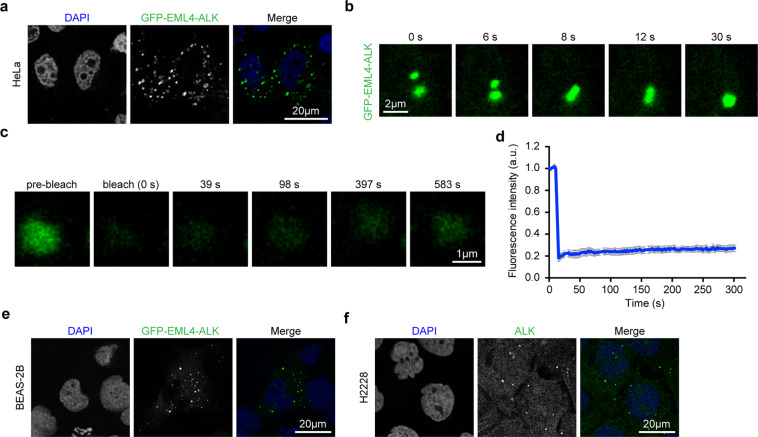
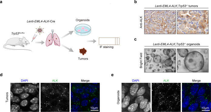
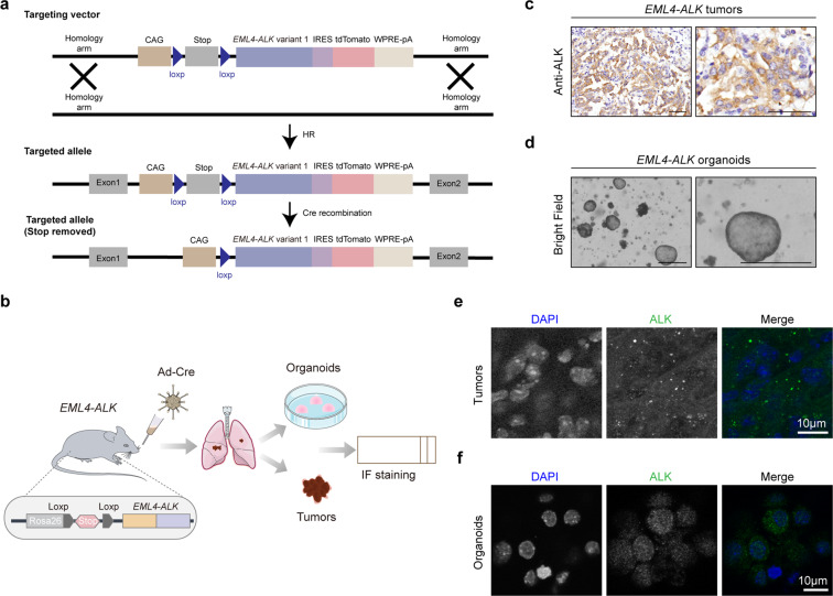
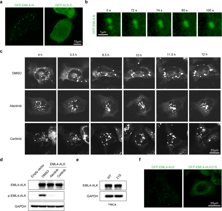
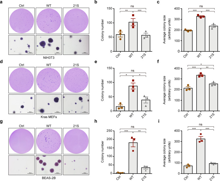
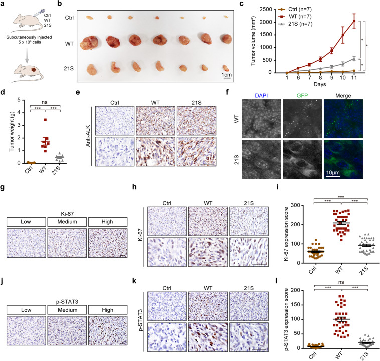
EML4-ALK fusion, observed in about 3%-7% of human lung adenocarcinoma, is one of the most important oncogenic drivers in initiating lung tumorigenesis. However, it still remains largely unknown about how EML4-ALK fusion exactly fires downstream signaling and drives lung cancer formation. We here find that EML4-ALK variant 1 (exon 1-13 of EML4 fused to exon 20-29 of ALK) forms condensates via phase separation in the cytoplasm of various human cancer cell lines. Using two genetically engineered mouse models (GEMMs), we find that EML4-ALK variant 1 can drive lung tumorigenesis and these murine tumors, as well as primary tumor-derived organoids, clearly show the condensates of EML4-ALK protein, further supporting the findings from in vitro study. Mutation of multiple aromatic residues in EML4 region significantly impairs the phase separation of EML4-ALK and dampens the activation of the downstream signaling pathways, especially the STAT3 phosphorylation. Importantly, it also significantly decreases cancer malignant transformation and tumor formation. These data together highlight an important role of phase separation in orchestrating EML4-ALK signaling and promoting tumorigenesis, which might provide new clues for the development of clinical therapeutic strategies in treating lung cancer patients with the EML4-ALK fusion.


Conflict of interest statement

The authors declare no competing interests.

Figures

Fig. 1
Fig. 1. Phase separation of EML4–ALK variant 1 in human cancer cell lines.
a HeLa cells were transfected with GFP–EML4–ALK for 24 h and the GFP–EML4–ALK was visualized by confocal microscopy. Nucleus was stained with DAPI (blue). Scale bar, 20 μm. b HeLa cells were transfected with GFP–EML4–ALK for 12 h and GFP fluorescence was monitored through live imaging. Snapshots at indicated time points showed the fusion event. Scale bar, 2 μm. c Representative FRAP images of GFP–EML4–ALK condensates in HeLa cells. The images were taken before and after photobleaching. Scale bar, 1 μm. d FRAP recovery curve of GFP–EML4–ALK condensates in HeLa cells. n = 12. Data were shown as mean ± SEM. e BEAS-2B cells were transfected with GFP–EML4–ALK for 24 h and the GFP–EML4–ALK was visualized by confocal microscopy. Nucleus was stained with DAPI (blue). Scale bar, 20 μm. f Immunofluorescence staining analysis of endogenous EML4–ALK in H2228 cells. ALK was indicated in green. Nucleus was stained with DAPI (blue). Scale bar, 20 μm.
Fig. 2
Fig. 2. EML4–ALK forms condensates in lung tumors and tumor-derived organoids in Lenti-EML4-ALK;Trp53/ mouse model.
a Schematic illustration of Lenti-EML4-ALK;Trp53−/ mouse model. Trp53flox/flox mice at 6–8 weeks were treated with 1 × 106 PFU of Lenti-EML4-ALK-Cre lentivirus via nasal inhalation and analyzed 28 weeks afterward for immunofluorescence staining of lung tumors and tumor-derived organoids. b Representative photos for ALK immunostaining in Lenti-EML4-ALK;Trp53/− lung tumors. Scale bar, 50 μm. c Representative photos for Lenti-EML4-ALK;Trp53−/− organoids derived from lung tumors. Scale bar, 500 μm. d Immunofluorescence staining analysis of EML4–ALK in Lenti-EML4-ALK;Trp53/− tumors. ALK was indicated in green. Nucleus was stained with DAPI (blue). Scale bar, 10 μm. e Immunofluorescence staining analysis of EML4–ALK in Lenti-EML4-ALK;Trp53−/− organoids. ALK was indicated in green. Nucleus was stained with DAPI (blue). Scale bar, 10 μm.
Fig. 3
Fig. 3. EML4–ALK forms condensates in EML4–ALK tumors and organoids.
a Schematic illustration of the Rosa26-Loxp-Stop-Loxp-EML4–ALK mice. See “Materials and methods” for details. b Schematic illustration of EML4–ALK mouse model. EML4–ALK mice at 6–8 weeks were treated with 2 × 106 PFU of Ad-Cre via nasal inhalation and analyzed 5 weeks afterward for immunofluorescence staining of tumors and tumor-derived organoids. c Representative photos for ALK immunostaining in EML4–ALK tumors. Scale bar, 50 μm. d Representative photos for EML4–ALK organoids derived from lung tumors. Scale bar, 500 μm. e Immunofluorescence staining analysis of EML4–ALK in EML4–ALK tumors. ALK was indicated in green. Nucleus was stained with DAPI (blue). Scale bar, 10 μm. f Immunofluorescence staining analysis of EML4–ALK in EML4–ALK organoids. ALK was indicated in green. Nucleus was stained with DAPI (blue). Scale bar, 10 μm.
Fig. 4
Fig. 4. The EML4–ALK21S mutant fails to phase separate.
a Representative fluorescent images of HeLa cells expressing GFP–EML4-N or GFP–ALK-C. Either GFP–EML4-N or GFP–ALK-C was visualized by confocal microscopy. Scale bar, 10 μm. b HeLa cells were transfected with GFP–EML4-N for 12 h and GFP fluorescence was monitored through live imaging. Snapshots at indicated time points showed the fusion event. Scale bar, 1 μm. c BEAS-2B cells were transfected with GFP–EML4–ALK for 24 h. Cells were treated with DMSO or ALK inhibitors, alectinib (500 nM), ceritinib (500 nM), and GFP fluorescence was monitored through live imaging for up to 12 h. Scale bar, 20 μm. d Western blot analysis of EML4–ALK phosphorylation after ALK inhibitor treatment for 12 h. e HeLa cells were transfected with GFP–EML4–ALK or GFP–EML4–ALK21S and analyzed by western blot. WT, GFP–EML4–ALK; 21S, GFP–EML4–ALK21S. f HeLa cells were transfected with GFP–EML4–ALK or GFP–EML4–ALK21S and analyzed by confocal microscopy. Scale bar, 20 μm.
Fig. 5
Fig. 5. The 21S mutations markedly attenuate the EML4–ALK-induced hyperactivation of downstream signaling pathways.
a Schematic illustration of EML4–ALK downstream signaling pathways. bd Western blot analysis of AKT, ERK1/2, STAT3 phosphorylation. NIH3T3 (b), Kras MEFs (c) and BEAS-2B (d) cells, stably expressing EML4–ALK or EML4–ALK21S, were deprived of serum and glucose for 2 h and then subjected to western blot analysis. Ctrl, control; WT, GFP–EML4–ALK; 21 S, GFP–EML4–ALK21S. e, f H2228 cells were transfected with the GFP–EML4–ALK (e) or GFP–EML4–ALK21S (f) and analyzed by confocal microscopy. P-STAT3 was indicated in red. Scale bar, 10 μm.
Fig. 6
Fig. 6. The 21S mutations impair the tumorigenic capability of EML4–ALK in soft-agar colony formation.
ac NIH3T3 cells were stably transfected with empty vector, EML4–ALK or EML4–ALK21S and analyzed by soft-agar colony formation assay. Representative images for soft-agar colonies (a). Scale bar, 500 μm. Statistical analysis of colony numbers (b). Statistical analysis of colony sizes (c). df Kras MEFs cells were stably transfected with empty vector, EML4–ALK or EML4–ALK21S and analyzed by soft-agar colony formation assay. Representative images for soft-agar colonies (d). Scale bar, 500 μm. Statistical analysis of colony numbers (e). Statistical analysis of colony sizes (f). gi BEAS-2B cells were stably transfected with empty vector, EML4–ALK or EML4–ALK21S and analyzed by soft-agar colony formation assay. Representative images for soft-agar colonies (g). Scale bar, 500 μm. Statistical analysis of colony numbers (h). Statistical analysis of colony sizes (i). All data were shown as mean ± SEM. *P < 0.05; **P < 0.01; ***P < 0.001; ns: not significant. Ctrl, control; WT, GFP–EML4–ALK; 21S, GFP–EML4–ALK21S.
Fig. 7
Fig. 7. The EML4–ALK21S mutant exhibits impaired capability in tumor formation.
a Schematic illustration of tumor formation assay in nude mice. See “Materials and methods” for details. b Photos of subcutaneous tumors derived from control, GFP–EML4–ALK and GFP–EML4–ALK21S groups. Scale bar, 1 cm. n = 7 for each group. c Growth curves of the subcutaneous tumors. d Statistical analysis of tumor weights. e Representative photos for ALK immunostaining in subcutaneous tumors derived from control, GFP–EML4–ALK and GFP–EML4–ALK21S groups. Scale bar, 50 μm. f Representative fluorescence photos for subcutaneous tumors derived from GFP–EML4–ALK and GFP–EML4–ALK21S groups. The GFP–EML4–ALK or GFP–EML4–ALK21S was visualized by confocal microscopy. Nucleus was stained with DAPI (blue). Scale bar, 10 μm. g Representative images of low, medium, high expression of Ki-67. Scale bar, 50 μm. h Representative photos for Ki-67 immunostaining in subcutaneous tumors derived from control, EML4–ALK and EML4–ALK21S groups. Scale bar, 50 μm. i Statistical analysis of Ki-67 staining. j Representative images of low, medium, high expression of p-STAT3. Scale bar, 50 μm. k Representative photos for p-STAT3 immunostaining in subcutaneous tumors derived from control, GFP–EML4–ALK and GFP–EML4–ALK21S groups. Scale bar, 50 μm. l Statistical analysis of p-STAT3 immunostaining. All data were shown as mean ± SEM. *P < 0.05; **P < 0.01; ***P < 0.001. Ctrl, control; WT, GFP–EML4–ALK; 21S, GFP–EML4–ALK21S.

Similar articles

Cited by

References

    1. Siegel RL, Miller KD, Jemal A. Cancer statistics, 2020. CA. Cancer J. Clin. 2020;70:7–30. doi: 10.3322/caac.21590. - DOI - PubMed
    1. Chen Y, Tang WY, Tong X, Ji H. Pathological transition as the arising mechanism for drug resistance in lung cancer. Cancer Commun. 2019;39:1–13. doi: 10.1186/s40880-019-0402-8. - DOI - PMC - PubMed
    1. Ding L, et al. Somatic mutations affect key pathways in lung adenocarcinoma. Nature. 2008;455:1069–1075. doi: 10.1038/nature07423. - DOI - PMC - PubMed
    1. Soda M, et al. Identification of the transforming EML4-ALK fusion gene in non-small-cell lung cancer. Nature. 2007;448:561–566. doi: 10.1038/nature05945. - DOI - PubMed
    1. Horn L, Pao W. EML4-ALK: Honing in on a new target in non-small-cell lung cancer. J. Clin. Oncol. 2009;27:4232–4236. doi: 10.1200/JCO.2009.23.6661. - DOI - PMC - PubMed