. 2021 May 11;11(1):9964.
doi: 10.1038/s41598-021-87924-2.

Multimodal integrated approaches in low grade glioma surgery

Affiliations

Multimodal integrated approaches in low grade glioma surgery

Tamara Ius et al. Sci Rep. .

Abstract

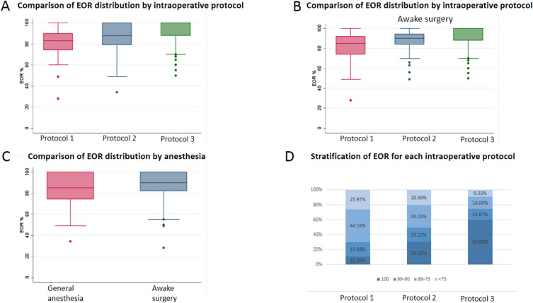
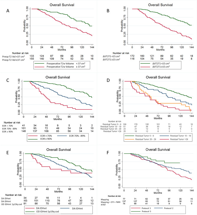
Surgical management of Diffuse Low-Grade Gliomas (DLGGs) has radically changed in the last 20 years. Awake surgery (AS) in combination with Direct Electrical Stimulation (DES) and real-time neuropsychological testing (RTNT) permits continuous intraoperative feedback, thus allowing to increase the extent of resection (EOR). The aim of this study was to evaluate the impact of the technological advancements and integration of multidisciplinary techniques on EOR. Two hundred and eighty-eight patients affected by DLGG were enrolled. Cases were stratified according to the surgical protocol that changed over time: 1. DES; 2. DES plus functional MRI/DTI images fused on a NeuroNavigation system; 3. Protocol 2 plus RTNT. Patients belonging to Protocol 1 had a median EOR of 83% (28-100), while those belonging to Protocol 2 and 3 had a median EOR of 88% (34-100) and 98% (50-100) respectively (p = 0.0001). New transient deficits with Protocol 1, 2 and 3 were noted in 38.96%, 34.31% and 31,08% of cases, and permanent deficits in 6.49%, 3.65% and 2.7% respectively. The average follow-up period was 6.8 years. OS was influenced by molecular class (p = 0.028), EOR (p = 0.018) and preoperative tumor growing pattern (p = 0.004). Multimodal surgical approach can provide a safer and wider removal of DLGG with potential subsequent benefits on OS. Further studies are necessary to corroborate our findings.


Conflict of interest statement

The authors declare no competing interests.

Figures

Figure 1
Figure 1
Difference achieved in tumor resection according to the intraoperative protocol used. (A) shows the median EOR achieved in patients operated with general anesthesia and awake craniotomy. In (B) the EOR data are stratified according to the intraoperative surgical protocol. Protocol 1 (Mapping) had a median EOR of 83%, while Protocol 2 (Mapping + DTI + fMRI) had a median EOR of 88% and Protocol 3 had a median EOR of 100%. (C) displays the median EOR achieved in the subgroup of patients who underwent awake craniotomy: Protocol 1 (Mapping) 85%, Protocol 2 (Mapping + DTI + fMRI) 90% and Protocol 3 100%. The circles represent the outlier values. (D) is a bar chart representing the distribution of EOR as a categorical variable in the three surgical protocols. EOR Extent Of Resection.
Figure 2
Figure 2
Kaplan–Meier curves displaying overall survival (OS) according to the preoperative tumor volume (A), preoperative tumor growing pattern, as expressed by ΔT2T1 (B), extent of resection (C), intraoperative protocol (D), residual tumor (E) and molecular class (F). Preop T2 Vol  preoperative tumor volume computed on T2-weighted images, ΔT2T1 preoperative tumor volume segmented on T2-weighted MRI images – preoperative tumor volume segmented on T1-weighted images, EOR extent of resectionm DA IDHwt Diffuse Astrocytoma Isocitrate Dehydrogenase wild type, DA IDHmt Diffuse Astrocytoma Isocitrate Dehydrogenase mutated, OD IDHmt 1p19q cod Oligodendroglioma Isocitrate Dehydrogenase mutated, 1p 19q codeleted.

References

    1. Capelle L, et al. Spontaneous and therapeutic prognostic factors in adult hemispheric World Health Organization Grade II gliomas: a series of 1097 cases: clinical article. J. Neurosurg. 2013;118:1157–1168. doi: 10.3171/2013.1.JNS121. - DOI - PubMed
    1. Clark VE, Cahill DP. Extent of resection versus molecular classification: What matters when? Neurosurg. Clin. N. Am. 2019;30:95–101. doi: 10.1016/j.nec.2018.08.006. - DOI - PubMed
    1. Claus EB, et al. Survival rates in patients with low-grade glioma after intraoperative magnetic resonance image guidance. Cancer. 2005;103:1227–1233. doi: 10.1002/cncr.20867. - DOI - PubMed
    1. Hervey-Jumper SL, Berger MS. Maximizing safe resection of low- and high-grade glioma. J. Neurooncol. 2016;130:269–282. doi: 10.1007/s11060-016-2110-4. - DOI - PubMed
    1. Ius T, et al. An NF-κB signature predicts low-grade glioma prognosis: A precision medicine approach based on patient-derived stem cells. Neuro Oncol. 2017;20:776–787. doi: 10.1093/neuonc/nox234. - DOI - PMC - PubMed