

Sustained release of usnic acid from graphene coatings ensures long term antibiofilm protection
- PMID: 33976310
- PMCID: PMC8113508
- DOI: 10.1038/s41598-021-89452-5
Sustained release of usnic acid from graphene coatings ensures long term antibiofilm protection
Abstract
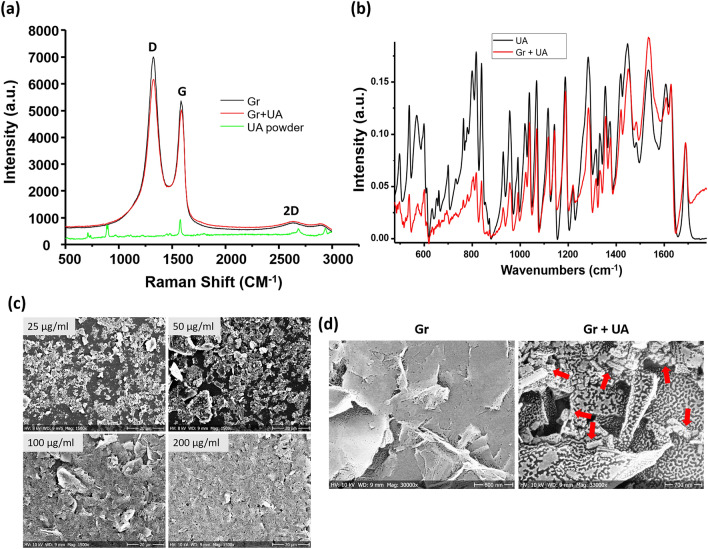
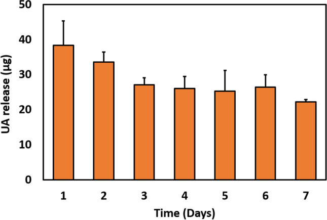
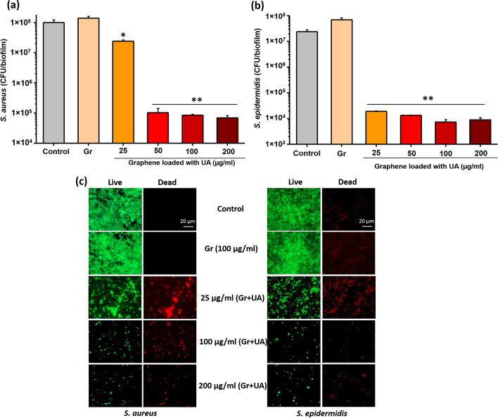
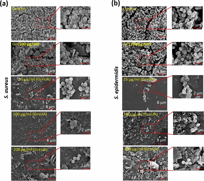
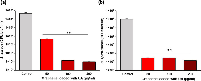
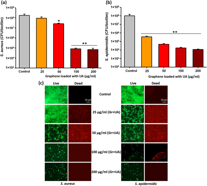
Protecting surfaces from bacterial colonization and biofilm development is an important challenge for the medical sector, particularly when it comes to biomedical devices and implants that spend longer periods in contact with the human body. A particularly difficult challenge is ensuring long-term protection, which is usually attempted by ensuring sustained release of antibacterial compounds loaded onto various coatings. Graphene have a considerable potential to reversibly interact water insoluble molecules, which makes them promising cargo systems for sustained release of such compounds. In this study, we developed graphene coatings that act as carriers capable of sustained release of usnic acid (UA), and hence enable long-term protection of surfaces against colonization by bacterial pathogens Staphylococcus aureus and Staphylococcus epidermidis. Our coatings exhibited several features that made them particularly effective for antibiofilm protection: (i) UA was successfully integrated with the graphene material, (ii) a steady release of UA was documented, (iii) steady UA release ensured strong inhibition of bacterial biofilm formation. Interestingly, even after the initial burst release of UA, the second phase of steady release was sufficient to block bacterial colonization. Based on these results, we propose that graphene coatings loaded with UA can serve as effective antibiofilm protection of biomedical surfaces.
Conflict of interest statement
The authors declare no competing interests.
Figures

Similar articles
-
Release behavior and antibiofilm activity of usnic acid-loaded carboxylated poly(L-lactide) microparticles.Eur J Pharm Biopharm. 2014 Oct;88(2):415-23. doi: 10.1016/j.ejpb.2014.06.002. Epub 2014 Jun 12. Eur J Pharm Biopharm. 2014. PMID: 24929210
-
Layer-by-layer self-assembly of minocycline-loaded chitosan/alginate multilayer on titanium substrates to inhibit biofilm formation.J Dent. 2014 Nov;42(11):1464-72. doi: 10.1016/j.jdent.2014.06.003. Epub 2014 Jun 12. J Dent. 2014. PMID: 24930872
-
Nanoporous Superhydrophobic Coatings that Promote the Extended Release of Water-Labile Quorum Sensing Inhibitors and Enable Long-Term Modulation of Quorum Sensing in Staphylococcus aureus.ACS Biomater Sci Eng. 2015 Oct 12;1(10):1039-1049. doi: 10.1021/acsbiomaterials.5b00313. Epub 2015 Aug 26. ACS Biomater Sci Eng. 2015. PMID: 26501126 Free PMC article.
-
Recent Advances in Surface Nanoengineering for Biofilm Prevention and Control. Part II: Active, Combined Active and Passive, and Smart Bacteria-Responsive Antibiofilm Nanocoatings.Nanomaterials (Basel). 2020 Aug 4;10(8):1527. doi: 10.3390/nano10081527. Nanomaterials (Basel). 2020. PMID: 32759748 Free PMC article. Review.
-
Antibiofouling Activity of Graphene Materials and Graphene-Based Antimicrobial Coatings.Microorganisms. 2021 Aug 30;9(9):1839. doi: 10.3390/microorganisms9091839. Microorganisms. 2021. PMID: 34576733 Free PMC article. Review.
Cited by
-
Graphene Properties, Synthesis and Applications: A Review.JOM (1989). 2023;75(3):614-630. doi: 10.1007/s11837-022-05505-8. Epub 2022 Oct 14. JOM (1989). 2023. PMID: 36267692 Free PMC article. Review.
-
Effectiveness of a Self-Decontaminating Coating Containing Usnic Acid in Reducing Environmental Microbial Load in Tertiary-Care Hospitals.Int J Environ Res Public Health. 2023 Apr 7;20(8):5434. doi: 10.3390/ijerph20085434. Int J Environ Res Public Health. 2023. PMID: 37107716 Free PMC article.
-
Phytochemical Analysis, Antioxidant, Antimicrobial, and Cytotoxic Activity of Different Extracts of Xanthoparmelia stenophylla Lichen from Stara Planina, Serbia.Plants (Basel). 2022 Jun 21;11(13):1624. doi: 10.3390/plants11131624. Plants (Basel). 2022. PMID: 35807576 Free PMC article.
-
Strategies and applications of antibacterial surface-modified biomaterials.Bioact Mater. 2025 Jul 9;53:114-140. doi: 10.1016/j.bioactmat.2025.07.009. eCollection 2025 Nov. Bioact Mater. 2025. PMID: 40688018 Free PMC article. Review.
-
Graphene-based nanomaterials for cancer therapy and anti-infections.Bioact Mater. 2022 Feb 5;14:335-349. doi: 10.1016/j.bioactmat.2022.01.045. eCollection 2022 Aug. Bioact Mater. 2022. PMID: 35386816 Free PMC article. Review.
References
-
- Vickery K, Hu H, Jacombs AS, Bradshaw DA, Deva AK. A review of bacterial biofilms and their role in device-associated infection. Healthc. Infect. 2013;18:61–66. doi: 10.1071/HI12059. - DOI
Publication types
LinkOut - more resources
Full Text Sources
Other Literature Sources
