. 2021 May 11;4(1):550.
doi: 10.1038/s42003-021-02086-1.

Parameter-free rendering of single-molecule localization microscopy data for parameter-free resolution estimation

Affiliations

Parameter-free rendering of single-molecule localization microscopy data for parameter-free resolution estimation

Adrien C Descloux et al. Commun Biol. .

Abstract

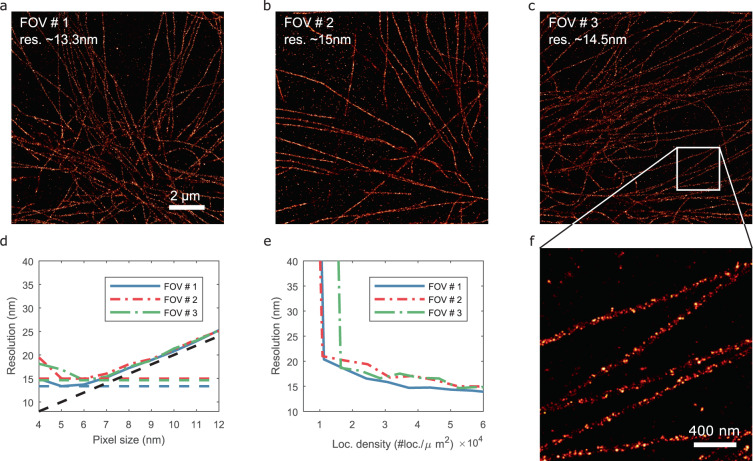
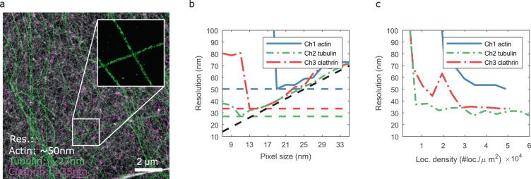
Localization microscopy is a super-resolution imaging technique that relies on the spatial and temporal separation of blinking fluorescent emitters. These blinking events can be individually localized with a precision significantly smaller than the classical diffraction limit. This sub-diffraction localization precision is theoretically bounded by the number of photons emitted per molecule and by the sensor noise. These parameters can be estimated from the raw images. Alternatively, the resolution can be estimated from a rendered image of the localizations. Here, we show how the rendering of localization datasets can influence the resolution estimation based on decorrelation analysis. We demonstrate that a modified histogram rendering, termed bilinear histogram, circumvents the biases introduced by Gaussian or standard histogram rendering. We propose a parameter-free processing pipeline and show that the resolution estimation becomes a function of the localization density and the localization precision, on both simulated and state-of-the-art experimental datasets.


Conflict of interest statement

The authors declare no competing interests.

Figures

Fig. 1
Fig. 1. Illustration of bilinear rendering of localization microscopy data.
a Schematic of the rendering of localizations with positions [2.3, 1.8], [5.2, 1.5], [4.2, 5.2] (black crosses) on the image grid, with the weights indicated. b Standard histogram rendering applied on the center region (pixel size of 15 nm; FOV of 3×3 µm) of the cSir Phalloidin dataset (see Fig. 2 for details). c Bilinear histogram rendering of the same region as in b. Scale bar 500 nm.
Fig. 2
Fig. 2. Experimental resolution estimate using bilinear histogram rendering.
ae Selection of Shareloc.xyz data rendered with a pixel size of 5 nm (field-of-view of 12 × 12 µm2), scale bar: 2 µm. The resolution indicated corresponds to the smallest resolution estimated from all the tested pixel sizes. fj Zoom-in of ae of 2.5 × 2.5 µm2 regions indicated by the white squares, scale bar: 500 nm. k Resolution vs pixel size. l Resolution as a function of number of localizations. m Resolution as a function of localization density.
Fig. 3
Fig. 3. Resolution estimation of DNA-PAINT data.
a–c Bilinear histogram of microtubule data at 5 nm pixel size, scale bar: 2 µm. d Resolution as a function of the pixel size. e Resolution as a function of the loc. density. f Zoom in of c, scale bar: 400 nm.
Fig. 4
Fig. 4. Resolution estimation of multi-color STORM data.
a Bilinear histogram rendering at 5 nm pixel size. Actin (phalloidin-AF647; STORM; gray colormap), microtubules (two anti-α-tubulin antibodies; STORM; green colormap) and clathrin-coated pits (anti-clathrin light chain; Atto655 imager; DNA-PAINT; red colormap). b Resolution vs pixel size. c Resolution as a function of the loc. density. Scale bar: 2 µm.

References

    1. Descloux AC, Grussmayer KS, Radenovic A. Parameter-free image resolution estimation based on decorrelation analysis. Nat. Methods. 2019;16:918–924. doi: 10.1038/s41592-019-0515-7. - DOI - PubMed
    1. Coelho S, et al. Ultraprecise single-molecule localization microscopy enables in situ distance measurements in intact cells. Sci. Adv. 2020;6:eaay8271. doi: 10.1126/sciadv.aay8271. - DOI - PMC - PubMed
    1. Cnossen J, et al. Localization microscopy at doubled precision with patterned illumination. Nat. Methods. 2020;17:59–63. doi: 10.1038/s41592-019-0657-7. - DOI - PMC - PubMed
    1. Gwosch KC, et al. MINFLUX nanoscopy delivers 3D multicolor nanometer resolution in cells. Nat. Methods. 2020;17:217–224. doi: 10.1038/s41592-019-0688-0. - DOI - PubMed
    1. Li Y, et al. Real-time 3D single-molecule localization using experimental point spread functions. Nat. Methods. 2018;15:367–369. doi: 10.1038/nmeth.4661. - DOI - PMC - PubMed

Publication types

MeSH terms

Substances

LinkOut - more resources