. 2021 Dec;35(12):3561-3567.
doi: 10.1038/s41375-021-01259-5.

Enhanced differentiation of functional human T cells in NSGW41 mice with tissue-specific expression of human interleukin-7

Affiliations

Enhanced differentiation of functional human T cells in NSGW41 mice with tissue-specific expression of human interleukin-7

Emilie Coppin et al. Leukemia. 2021 Dec.

Abstract

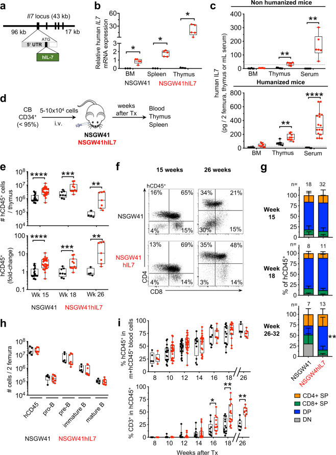
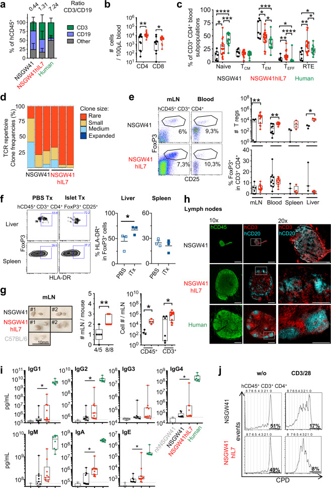
Humanized mouse models have become increasingly valuable tools to study human hematopoiesis and infectious diseases. However, human T-cell differentiation remains inefficient. We generated mice expressing human interleukin-7 (IL-7), a critical growth and survival factor for T cells, under the control of murine IL-7 regulatory elements. After transfer of human cord blood-derived hematopoietic stem and progenitor cells, transgenic mice on the NSGW41 background, termed NSGW41hIL7, showed elevated and prolonged human cellularity in the thymus while maintaining physiological ratios of thymocyte subsets. As a consequence, numbers of functional human T cells in the periphery were increased without evidence for pathological lymphoproliferation or aberrant expansion of effector or memory-like T cells. We conclude that the novel NSGW41hIL7 strain represents an optimized mouse model for humanization to better understand human T-cell differentiation in vivo and to generate a human immune system with a better approximation of human lymphocyte ratios.


Conflict of interest statement

The authors declare no competing interests.

Figures

Fig. 1
Fig. 1. Improved intrathymic human T cell differentiation in NSGW41hIL7 mice.
a Scheme of BAC constructs for the generation of NSGW41hIL7 mice. b Abundance of hIL-7 transcript in bone marrow (BM), spleen, and thymus from humanized NSGW41 or NSGW41hIL7 mice. c hIL-7 protein levels in bone marrow, thymus, and serum isolated from non-humanized NSGW41 (black) or NSGW41hIL7 mice (red, top) and from NSGW41 or NSGW41hIL7 mice that have received human CD34+ HSPCs 26-38 weeks before (bottom). Gray lines indicate the limit of assay sensitivity. d Scheme of transplantation experiments. e Numbers (top) and fold-change (bottom) of human (h)CD45+ cells in thymi of humanized NSGW41 or NSGW41hIL7 mice at the indicated time points after humanization. Fold-changes were calculated by dividing hCD45+ thymocyte numbers from humanized NSGW41hIL7 mice by the thymocyte numbers from humanized NSGW41 mice. This was conducted separately for each experiment and the results pooled. f Analysis of CD4 and CD8 expression on hCD45+ thymocytes from NSGW41 or NSGW41hIL7 mice that have received human HPSCs 15 (left) or 26 (right) weeks before. g Composition of thymocyte subsets in NSGW41 or NSGW41hIL7 mice at the indicated time points after humanization. Frequencies of DN populations were significantly decreased in NSGW41hIL7 compared to NSGW41 counterparts (week 15 and 18: P < 0.01 and week 32: P < 0.001). h Numbers of B cell subsets in the bone marrow of NSGW41 or NSGW41hIL7 mice 26–32 weeks after humanization. i Kinetics of the appearance of human CD45+ cells (hCD45+, top) and hCD3+ T cells within human leukocytes (bottom) in the blood after humanization. b, c, e, h, i Each dot represents an individual mouse. Boxes and whiskers indicate quartiles and median.
Fig. 2
Fig. 2. hIL-7-BAC transgene increases numbers of functional peripheral T cells in the absence of excessive lymphoproliferation.
a Frequencies of T cells, B cells, and non-defined other cells of human origin in the blood of NSGW41 (n = 13), NSGW41hIL7 (n = 16) mice 18 weeks after humanization and human controls. Numbers on top indicate T vs. B cell ratios. b Human CD4+ and CD8+ T cells in the blood of humanized NSGW41 or NSGW41hIL7 mice 26 weeks after humanization. c Composition of blood CD4+ T cell subpopulations 26 weeks after humanization: Naïve T cells, central memory (TCM), effector memory (TEM), T effector (TEFF), and recent thymic emigrants (RTE) in NSGW41, NSGW41hIL7 mice, or human blood. d T cell receptor (TCR) repertoire diversity in splenic αβ T cells in NSGW41 or NSGW41hIL7 mice. Clones were binned into rare (0 < X ≤ 0.001), small (0.001 < X ≤ 0.01), medium (0.01 < X ≤ 0.1), and expanded (0.1 < X ≤ 1) (n = 3 per group). e Human CD4+ FoxP3+ Treg cells in humanized mice. Representative dot plots analyzing mesenteric lymph nodes (mLN) and blood (left) and numbers (right, top) and frequencies (right, bottom) of Tregs in mLN, blood, spleen, and liver of humanized NSGW41 or NSGW41hIL7 mice. f Human activated Tregs after intraportal xenotransplantation of porcine pancreatic islets into NSGW41hIL7 mice 26 weeks after humanization. Representative dot plots of liver and spleen analysis (left). Frequencies of HLA-DR+ FoxP3+ T cells in spleen and liver of humanized NSGW41hIL7 mice (right) 18 h after transplantation of islets (iTx) or PBS. g Photographs of mLN from humanized NSGW41 or NSGW41hIL7 mice isolated 26 weeks after humanization, or C57BL/6 controls (left). Quantity of mLNs per mouse (middle). Human CD45+ and hCD3+ cell numbers in mLNs from NSGW41 or NSGW41hIL7 mice (right). h Immunofluorescent images from lymph nodes from humanized NSGW41 or NSGW41hIL7 mice that were humanized 30 weeks before and a human cervical lymph node stained for human CD45 (green, left), CD3 (red), and CD20 (turquoise, middle and right). Areas of blow-up pictures (20× magnification) correspond to the white squares indicated in the 10× magnification images. Scale-bar: 500 µm. Data are representative of the analysis of lymph nodes from three mice per group. i Concentration of serum immunoglobulins in humanized mice. Levels of IgG1, IgG2, IgG3, IgG4, IgM, IgA, and IgE were determined from non-humanized (nh)NSGW41, humanized NSGW41, or humanized NSGW41hIL7 mice at 24–32 weeks after humanization. Human serum was used as positive control. Gray lines indicate the limit of assay sensitivity. j Activation of human T cells from NSGW41 (top) or NSGW41hIL7 mice (bottom). Histograms depict division of CPD-labeled spleen hCD3+ T cells 6 days after stimulation with CD3/28 beads (right) or control (w/o, left). Frequencies of non-divided human T cells and T cells that have divided 4, 5, or 6 times 6 days after stimulation. b, c, e, f, g, i Each dot represents an individual mouse.

References

    1. Puel A, Ziegler SF, Buckley RH, Leonard WJ. Defective IL7R expression in T(−)B(+)NK(+) severe combined immunodeficiency. Nat Genet. 1998;20:394–7. doi: 10.1038/3877. - DOI - PubMed
    1. Freeden-Jeffry von U, Vieira P, Lucian LA, McNeil T, Burdach SE, Murray R. Lymphopenia in interleukin (IL)-7 gene-deleted mice identifies IL-7 as a nonredundant cytokine. J Exp Med. 1995;181:1519–26. doi: 10.1084/jem.181.4.1519. - DOI - PMC - PubMed
    1. Peschon JJ, Morrissey PJ, Grabstein KH, Ramsdell FJ, Maraskovsky E, Gliniak BC, et al. Early lymphocyte expansion is severely impaired in interleukin 7 receptor-deficient mice. J Exp Med. 1994;180:1955–60. doi: 10.1084/jem.180.5.1955. - DOI - PMC - PubMed
    1. van Lent AU, Dontje W, Nagasawa M, Siamari R, Bakker AQ, Pouw SM, et al. IL-7 enhances thymic human T cell development in “human immune system” Rag2-/-IL-2Rgammac-/- mice without affecting peripheral T cell homeostasis. J Immunol. 2009;183:7645–55. doi: 10.4049/jimmunol.0902019. - DOI - PubMed
    1. Rich BE, Campos-Torres J, Tepper RI, Moreadith RW, Leder P. Cutaneous lymphoproliferation and lymphomas in interleukin 7 transgenic mice. J Exp Med. 1993;177:305–16. doi: 10.1084/jem.177.2.305. - DOI - PMC - PubMed

Publication types