. 2021 Sep;14(5):1183-1202.
doi: 10.1038/s41385-021-00407-5. Epub 2021 May 11.

IL-17 mediates protective immunity against nasal infection with Bordetella pertussis by mobilizing neutrophils, especially Siglec-F+ neutrophils

Affiliations

IL-17 mediates protective immunity against nasal infection with Bordetella pertussis by mobilizing neutrophils, especially Siglec-F+ neutrophils

Lisa Borkner et al. Mucosal Immunol. 2021 Sep.

Erratum in

Abstract

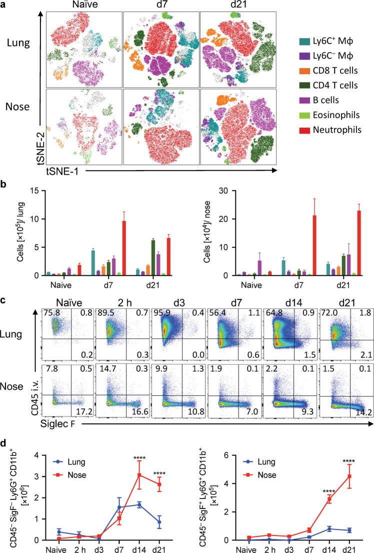
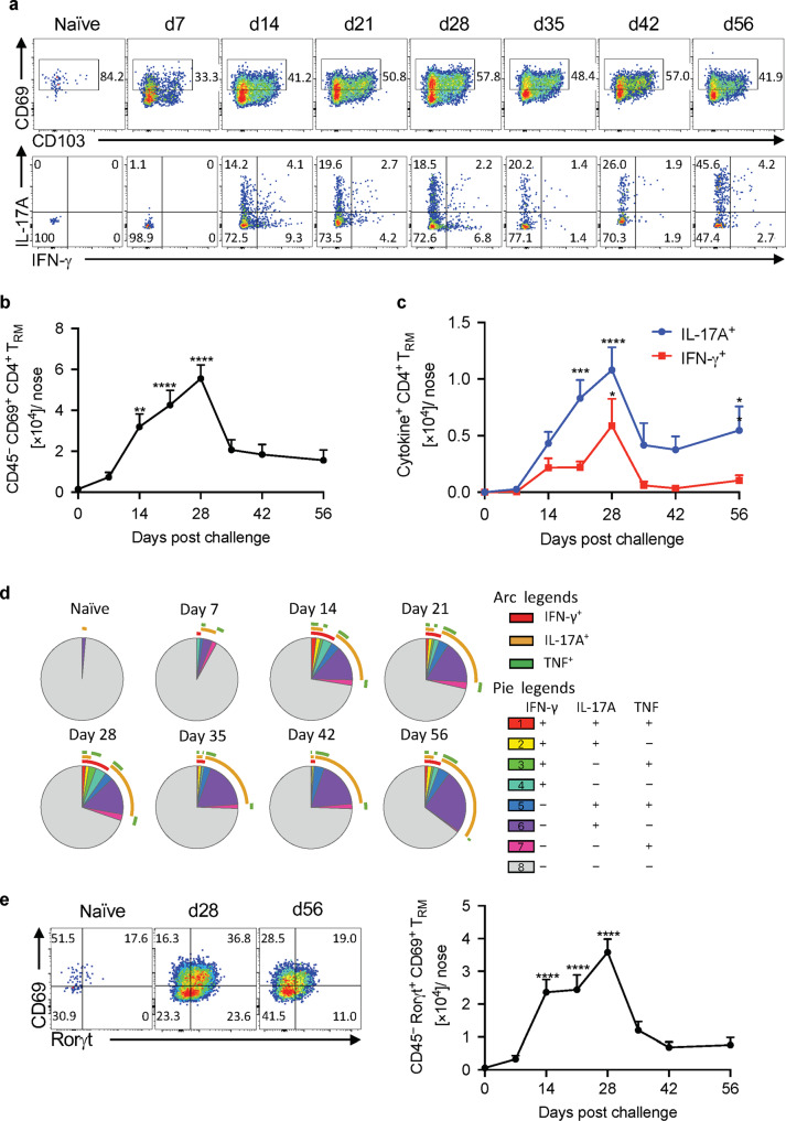
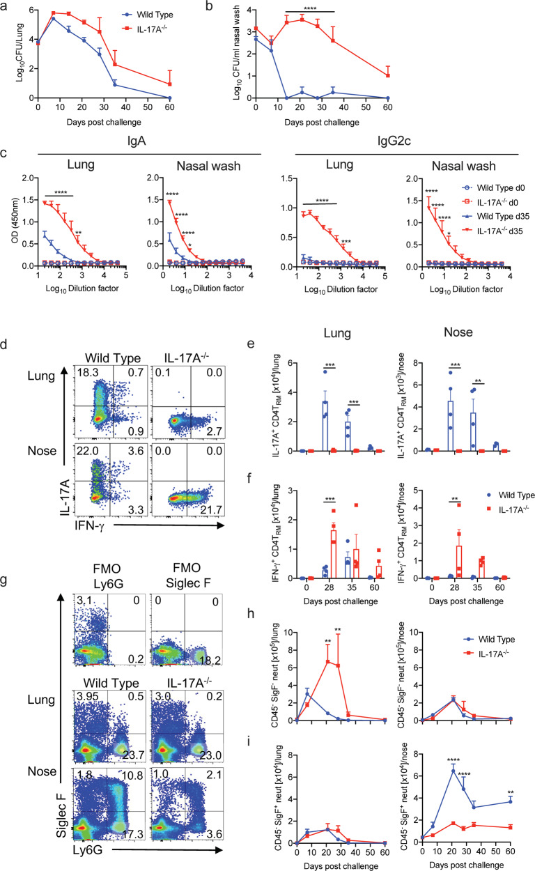
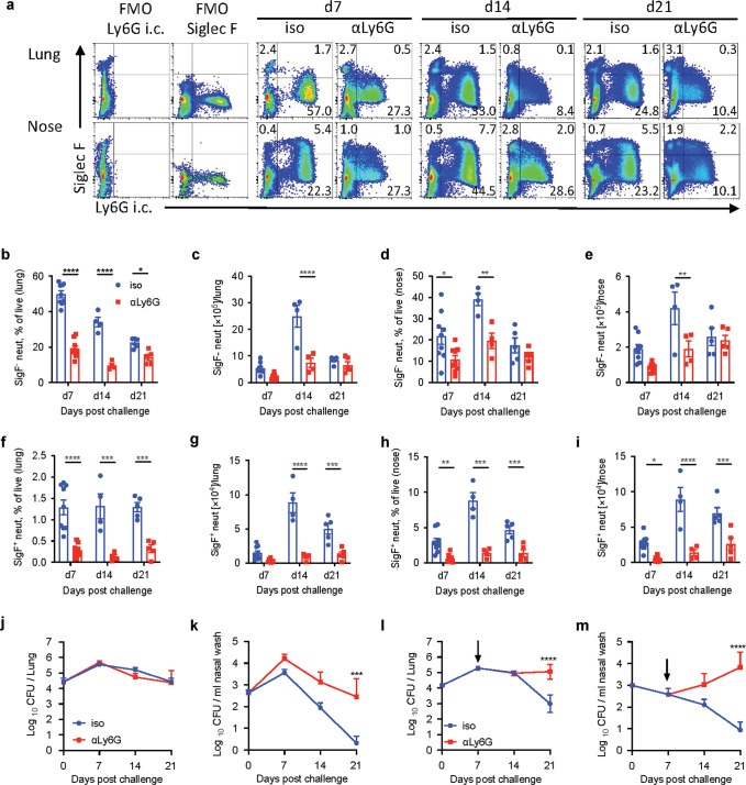
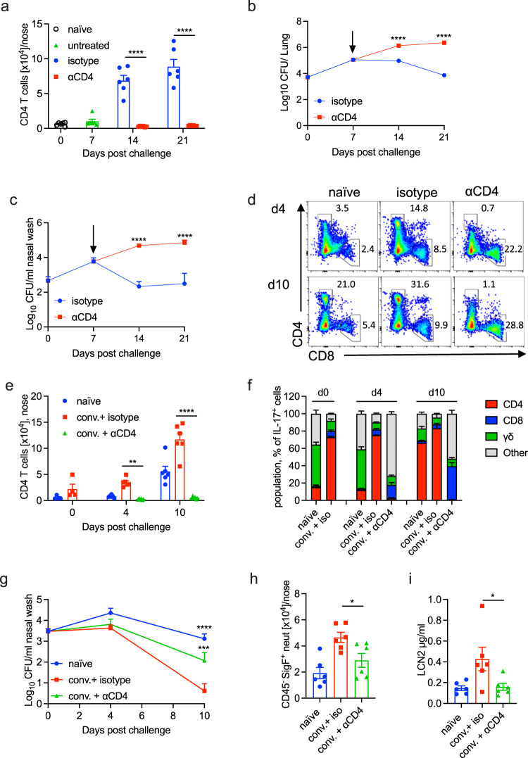
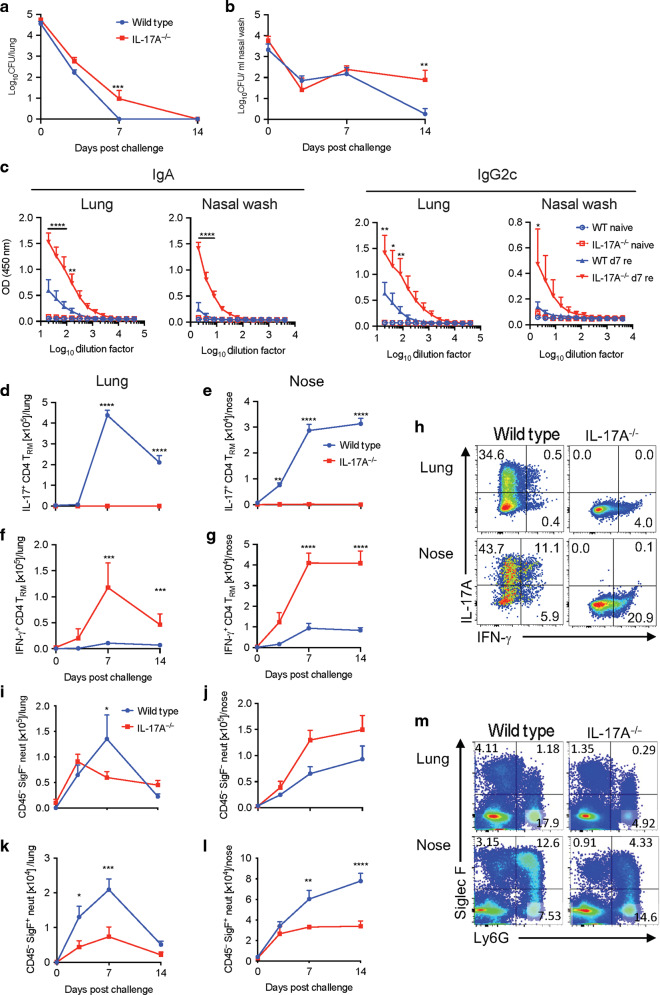
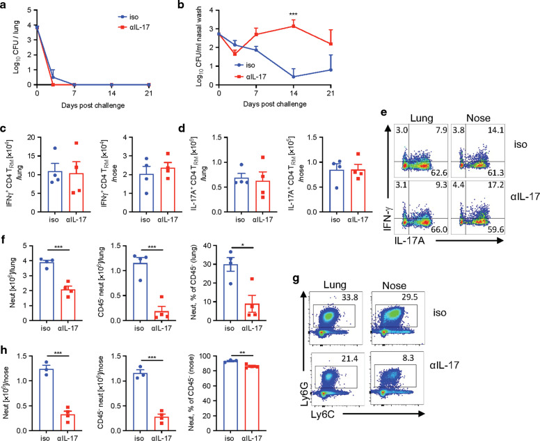
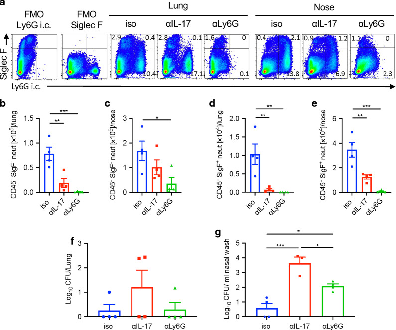
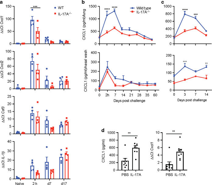
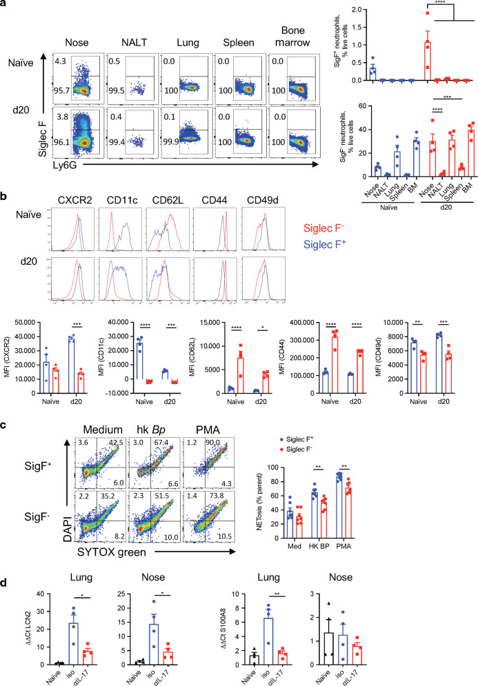
Understanding the mechanism of protective immunity in the nasal mucosae is central to the design of more effective vaccines that prevent nasal infection and transmission of Bordetella pertussis. We found significant infiltration of IL-17-secreting CD4+ tissue-resident memory T (TRM) cells and Siglec-F+ neutrophils into the nasal tissue during primary infection with B. pertussis. Il17A-/- mice had significantly higher bacterial load in the nasal mucosae, associated with significantly reduced infiltration of Siglec-F+ neutrophils. Re-infected convalescent mice rapidly cleared B. pertussis from the nasal cavity and this was associated with local expansion of IL-17-producing CD4+ TRM cells. Depletion of CD4 T cells from the nasal tissue during primary infection or after re-challenge of convalescent mice significantly delayed clearance of bacteria from the nasal mucosae. Protection was lost in Il17A-/- mice and this was associated with significantly less infiltration of Siglec-F+ neutrophils and antimicrobial peptide (AMP) production. Finally, depletion of neutrophils reduced the clearance of B. pertussis following re-challenge of convalescent mice. Our findings demonstrate that IL-17 plays a critical role in natural and acquired immunity to B. pertussis in the nasal mucosae and this effect is mediated by mobilizing neutrophils, especially Siglec-F+ neutrophils, which have high neutrophil extracellular trap (NET) activity.


Conflict of interest statement

Kingston Mills is an inventor on a patent application around a novel vaccine adjuvant and has collaborative research funding from and acts as consultant to Vaccine manufacturers. The funders had no role in the writing of the manuscript or in the decision to publish.

Figures

Fig. 1
Fig. 1. Siglec-F+ neutrophils are recruited to the nasal tissue during B. pertussis infection.
C57BL/6 mice were aerosol infected with B. pertussis. At different time points, mice were injected i.v. with fluorochrome-labeled CD45 antibody and euthanized 10 min later. Cell suspensions were prepared from lung and nasal tissue and immune cells were analyzed by flow cytometry. a Representative tSNE plots for cells from lung and nasal tissue in naive mice and 7 or 21 days post infection. Neutrophils (Ly6G+Ly6C+), eosinophils (Siglec-F+ CD11c), B cells (B220+MHCII+), CD4 T cells (CD3+CD4+), CD8 T cells (CD3+CD8+), Ly6C macrophages (F4/80+CD11b+Ly6C), Ly6C+ macrophages (F4/80+CD11b+Ly6C+). b Absolute cell counts for immune cell populations in lung and nose. c Cells were pre-gated on total neutrophils (Ly6G+CD11b+), dot plots show the intravital CD45 stain on the y-axis and Siglec-F on the x-axis. d Absolute cell numbers of tissue-resident Siglec-F neutrophils (CD45 i.v. Siglec-FLy6G+CD11b+) and Siglec-F+ neutrophils in lung and nose. n = 4/group, mean ± SEM, statistical analysis: Two-way ANOVA followed by Sidak’s post-test, significances are indicated in comparison to naive mice of the same population, ****p < 0.0001.
Fig. 2
Fig. 2. IL-17A-secreting CD4 TRM cells accumulate in the nasal tissue during B. pertussis infection.
C57BL/6 mice were aerosol infected with B. pertussis. At different time points, mice were injected i.v. with fluorochrome-labeled CD45 antibody and sacrificed 10 min later. Cell suspensions were prepared from nasal tissue and cells were stimulated with sBP and anti-CD49d/CD28 and analyzed by flow cytometry. a CD69 and CD103 expression on tissue-resident CD44+ CD4+ T cells and IL-17A and IFN-γ-production in the CD69+ sub-population. b Total number of CD4 TRM (CD45 i.v. CD4+ CD44+ CD69+) in nasal tissue during B. pertussis infection. c Total number of B. pertussis-specific IL-17A- or IFN-γ-producing CD4 TRM cells. d SPICE analysis of cytokine production in sBP-stimulated CD4 TRM. e Analysis of RORγT expression in CD4 TRM cells in naive mice and 28 and 56 days post infection. Absolute numbers of tissue-resident RORγt+ CD69+ CD4 T cells. Statistical analysis: b One-way ANOVA followed by Dunnett’s post-test, significances are indicated in comparison to naive mice; c Two-way ANOVA followed by Sidak’s post-test, significances are indicated in comparison to naive mice of the same population; *p < 0.05, **p < 0.01, ***p < 0.001, ****p < 0.0001, n = 4/group, mean ± SEM.
Fig. 3
Fig. 3. Il17A−/− mice fail to clear B. pertussis infection in the nasal cavity.
WT and Il17A−/− mice were aerosol infected with B. pertussis. At different time points, mice were injected i.v. with fluorochrome-labeled CD45 antibody and euthanized 10 min later. Cell suspensions were prepared from lung and nasal tissue and cells were stimulated with sBP and CD49d/CD28 and analyzed by flow cytometry. CFU counts in lungs (a) and nasal washes (b) of WT and Il17A−/− mice. c B. pertussis-specific IgA and IgG2c in nasal washes and lung homogenates. d Representative dot plots of IL-17A- and IFN-γ-production in CD4 TRM cells in lung and nose of WT and Il17A−/− on d 28 post infection. Total number of IL-17A (e) and IFN-γ (f) producing CD4 TRM in lung and nose. g Representative dot plots of Ly6G and Siglec-F expression in lung and nose cell suspensions of WT and Il17A−/− mice on d 28 post infection. Cell counts of tissue-resident Siglec-F (h) and Siglec-F+ (i) neutrophils in lungs and nasal tissue. Statistical analysis: a, b, e, f, h, i Two-way ANOVA followed by Sidak’s post-test, c Two-way ANOVA comparing WT and IL-17A−/− d35 followed by Sidak’s post-test, *p < 0.05, **p < 0.01, ***p < 0.001, ****p < 0.0001, n = 4/group, mean ± SEM.
Fig. 4
Fig. 4. Depletion of neutrophils during primary infection with B. pertussis impairs bacterial clearance from nasal mucosae.
C57BL/6 mice were treated with anti-Ly6G or isotype control antibody (iso) in combination with secondary antibody MAR 18.5 starting from d −1, and aerosol infected with B. pertussis. At 7, 14 and 21 days post infection, mice were injected i.v. with fluorochrome-labeled CD45 antibody and euthanized 10 min later. Cell suspensions were prepared from lung and nasal tissue and immune cells were analyzed by flow cytometry. a Representative dot plots showing the reduction of Siglec-F+ and Siglec-F neutrophils (intracellular Ly6G+) in anti-Ly6G treated mice compared with isotype control antibody treated mice 7, 14 and 21 days post infection. Percentage of Siglec-F neutrophils in lung (b) and nasal tissue (c). Absolute numbers of Siglec-F neutrophils in lung (d) and nasal tissue (e). Percentage of Siglec-F+ neutrophils in lung (f) and nasal tissue (g). Absolute numbers of Siglec-F+ neutrophils in lung (h) and nasal tissue (i). CFU counts in lung (j) and nasal washes (k) of anti-Ly6G and isotype control treated mice. C57BL/6 mice were aerosol infected with B. pertussis and treated with anti-Ly6G or isotype control in combination with secondary antibody MAR 18.5 starting from d 7 post infection. CFU counts in lung (l) and nasal washes (m) of anti-Ly6G and isotype control antibody treated mice. Statistical analysis: Two-way ANOVA followed by Sidak’s post-test, *p < 0.05, **p < 0.01, ***p < 0.001, ****p < 0.0001. n = 4–8/group, pooled data from two independent experiments, mean ± SEM.
Fig. 5
Fig. 5. Depletion of CD4 T cells impairs clearance of B. pertussis from the nasal mucosa in primary infection and after challenge of convalescent mice.
C57BL/6 mice were aerosol infected with B. pertussis. Beginning on day 7 post infection, mice were treated by i.p. and i.n administration of a neutralizing anti-CD4 antibody or an isotype control antibody. At 7, 14 and 21 days post infection, mice were injected i.v. with fluorochrome-labeled CD45 antibody and euthanized 10 min later. Cell suspensions were prepared from lung and nasal tissue and immune cells were analyzed by flow cytometry. CFU counts in lung (a) and nasal washes (b) of anti-CD4 or isotype control antibody treated mice. c Absolute counts of CD4 T cells in nasal tissue of anti-CD4 or control antibody treated mice. C57BL/6 mice were aerosol infected with B. pertussis and allowed to clear the infection. Convalescent mice were treated by i.p. and i.n administration of a neutralizing anti-CD4 antibody or an isotype control antibody from day −1 of re-challenge with B. pertussis. Nasal tissue cells were prepared 4 and 10 days after re-challenge or from control mice given a primary challenge on the same day. All mice were injected i.v. with fluorochrome-labeled CD45 antibody 10 min before euthanasia. Cell suspensions were prepared from nasal tissue, stimulated with PMA and ionomycin and immune cells were analyzed by flow cytometry. d Representative dots plot of nasal CD4 versus CD8 T cells pre-gated on CD3. e Absolute counts of CD4 T cells in nasal tissue of anti-CD4 or isotype control antibody treated mice. f Relative proportion of IL-17-secreting CD4 T cells, CD8 T cells, γδ T cells or other cell types in the nasal cavity on days 0, 4 or 10 after B. pertussis challenge of naive or convalescent mice. g CFU counts in the nasal wash of naive mice or convalescent mice treated with anti-CD4 or isotope control antibody. h Tissue-resident Siglec-F+ neutrophils in nasal tissue and i concentration of LCN2 in nasal wash 4 days post challenge. Statistical analysis: a, b, c, e, g Two-way ANOVA followed by Sidak’s post-test; h, i One-way ANOVA followed by Tukey’s post-test, *p < 0.05, **p < 0.01, ***p < 0.001, ****p < 0.0001. di Pooled data from two independent experiments, n = 6–10/group, mean ± SEM.
Fig. 6
Fig. 6. The role of IL-17 in protective acquired immunity to B. pertussis.
WT and Il17A−/− mice were aerosol infected with B. pertussis and left for 6 months to clear the infection, and then re-infected. Prior to re-infection and 3, 7 and 14 days post challenge, mice were injected i.v. with fluorochrome-labeled CD45 antibody and euthanized 10 min later. Cell suspensions were prepared from lung and nasal tissue and immune cells were analyzed by flow cytometry. CFU counts in lungs (a) and nasal wash (b) of re-infected WT and Il17A−/− mice. c B. pertussis-specific IgA and IgG2c titers in nasal washes and lung homogenates 7 days post re-challenge. IL-17A-producing CD4 TRM cells in lungs (d) and nasal tissue (e), and IFN-γ-producing CD4 TRM in lungs (f) and nasal tissue (g). h Representative dot plots showing IL-17A and IFN-γ production in CD4 TRM cells on day 7 post re-challenge. Siglec-F neutrophils in lung (i) and nasal tissue (j), and Siglec-F+ neutrophils in lung (k) and nasal tissue (l) during re-infection. m Representative dot plots showing Ly6G and Siglec-F expression on live cells in lungs and nasal tissue on day 7 of re-challenge. Statistical analysis: Two-way ANOVA followed by Sidak’s post-test comparing WT and IL-17A−/− (a, b, dg, il) or WT d7 re and IL-17A−/− d7 re (c) *p < 0.05, **p < 0.01, ***p < 0.001, ****p < 0.0001. n = 4/group, mean ± SEM.
Fig. 7
Fig. 7. Depletion of IL-17 impairs clearance from the nasal cavity following re-challenge of convalescent mice.
C57BL/6 mice were aerosol infected with B. pertussis and allowed to clear the infection. 3 months later, convalescent mice were re-challenged with B. pertussis. Mice were treated with anti-IL-17A or isotype control from one day before infection until the end of the experiment. Prior to infection and 7 days post challenge, mice were injected i.v. with fluorochrome-labeled CD45 antibody and euthanized 10 min later. Cell suspensions were prepared from lung and nasal tissue and immune cells were analyzed by flow cytometry. CFU counts in lungs (a) and nasal washes (b) of anti-IL-17A treated re-infected convalescent mice. IFN-γ- (c) and IL-17A (d) -producing CD4 TRM cells in lungs and nasal tissue. e Representative dot plots of IL-17A- or IFN-γ-producing CD4 TRM cells on day 7 post re-challenge of anti-IL-17A or isotype treated convalescent mice. f Absolute numbers of neutrophils, CD45 i.v. tissue-resident neutrophils, and % of CD45 i.v. cells in total neutrophils in the lungs. g Representative dot plots of total neutrophils (Ly6C+ Ly6G+) in lungs and noses on day 7 post re-challenge of anti-IL-17A treated convalescent mice. h Absolute numbers of neutrophils, CD45 i.v. tissue-resident neutrophils, and % of CD45 i.v. cells in total neutrophils in the nasal tissue. Statistical analysis: a, b Two-way ANOVA followed by Sidak’s post-test, ***p < 0.001; c, d, f, h unpaired, two-tailed t-test, *p < 0.05, **p < 0.01, ***p < 0.001; n = 4/group, mean ± SEM.
Fig. 8
Fig. 8. Depletion of neutrophils during re-challenge impairs clearance in the nose of convalescent mice.
C57BL/6 mice were aerosol infected with B. pertussis and allowed to clear the infection. 4.5 months later, convalescent mice were re-challenged with B. pertussis. Mice were treated with anti-IL-17A or anti-Ly6G and 2nd antibody or isotype control from one day before infection until the end of the experiment. 7 days post challenge, mice were injected i.v. with fluorochrome-labeled CD45 antibody and euthanized 10 min later. Cell suspensions were prepared from lung and nasal tissue and immune cells were analyzed by flow cytometry. a Representative dot plots showing expression of Ly6G (intracellular) and Siglec-F on live cells in lungs and nasal tissue on day 7 post re-challenge. Absolute numbers of Siglec-F neutrophils in lung (b) or nasal tissue (c). Absolute numbers of Siglec-F+ neutrophils in lung (d) or nasal tissue (e). CFU counts in lungs (f) and nasal washes (g) on day 7 post re-challenge. Statistical analysis: One-way ANOVA followed by Tukey’s post-test, *p < 0.05, **p < 0.01, ***p < 0.001; n = 4/group, mean ± SEM.
Fig. 9
Fig. 9. IL-17 induces chemokines and cytokines necessary for neutrophil recruitment.
WT and Il17A−/− mice were aerosol infected with B. pertussis. Prior to and 2 h, 7 and 17 d post challenge mice were sacrificed, and mRNA was extracted from nasal tissue for RT-PCR analysis. a Gene expression of Cxcl1, Cxcl2, Cfs3 and IL-1β in nasal tissue of WT and Il17A−/− mice was determined by RT-PCR and normalized to 18S RNA. b WT and Il17A−/− mice were aerosol infected with B. pertussis. Up to day 60 post challenge nasal washes and lung homogenates were prepared and protein concentration of CXCL1 was determined by ELISA. c Convalescent WT and Il17A−/− were aerosol infected with B. pertussis. On day 3, 7 and 14 post re-challenge nasal washes and lung homogenates were prepared and protein concentration of CXCL1 was determined by ELISA. d C57BL/6 mice were treated intranasally with IL-17A and euthanized 2 h later. CXCL1 concentration in nasal washes was determined by ELISA and Cxcl1 gene expression was analyzed by RT-PCR and normalized to 18S RNA. Statistical analysis: Two-way ANOVA followed by Sidak’s post-test, d unpaired, two-tailed t-test, *p < 0.05, **p < 0.01, ***p < 0.001; ac n = 4/ group; d n = 8/group, pooled data from two independent experiments, mean ± SEM.
Fig. 10
Fig. 10. Siglec-F+ neutrophils have an activated phenotype and high NET activity.
C57BL/6 mice were aerosol infected with B. pertussis and euthanized on 20 d post challenge. Cell suspensions were prepared from several tissues and immune cells were analyzed by flow cytometry. a Representative dot plots showing Siglec-F+ expression on CD11b+Ly6G+ pre-gated cells and percentages of Siglec-F+ and Siglec-F neutrophils in naive mice and on day 20 post challenge in nasal tissue, NALT, lung, spleen, and bone marrow. b Histograms and MFIs showing the expression of the surface markers CXCR2, CD11c, CD62L, CD44, and CD49d on Siglec-F+ and Siglec-F neutrophils in nasal tissue of naive mice and mice 20 d post challenge with B. pertussis. c Single cell suspension was prepared from nasal tissue of B. pertussis infected mice up to 1-month post challenge, was stimulated with heat-killed B. pertussis or PMA and then stained with the intracellular DNA dye (DAPI) and the cell-impermeant DNA dye (SYTOXgreen). Percentage of cells undergoing NETosis (double positive for DAPI and SYTOXgreen). d mRNA was prepared on day 6 post re-challenge from lungs and nasal tissue of convalescent mice treated with anti-IL-17A or isotype. Gene expression of Lcn2 and S100a8 was determined by RT-PCR and normalized against 18S RNA. Statistical analysis: a Two-way ANOVA followed by Tukey’s post-test, b, c Two-way ANOVA followed by Sidak’s post-test, d One-way ANOVA followed by Tukey’s post-test, *p < 0.05, **p < 0.01, ***p < 0.001; a, b, d n = 4/ group, c n = 7/group, pooled from two independent experiments, mean ± SEM.

References

    1. Dorji D, et al. Bordetella Pertussis virulence factors in the continuing evolution of whooping cough vaccines for improved performance. Med. Microbiol. Immunol. 2018;207:3–26. doi: 10.1007/s00430-017-0524-z. - DOI - PubMed
    1. Gill C, Rohani P, Thea DM. The relationship between mucosal immunity, nasopharyngeal carriage, asymptomatic transmission and the resurgence of Bordetella pertussis. F1000Res. 2017;6:1568. doi: 10.12688/f1000research.11654.1. - DOI - PMC - PubMed
    1. Klein NP, et al. Waning protection following 5 doses of a 3-component diphtheria, tetanus, and acellular pertussis vaccine. Vaccine. 2017;35:3395–3400. doi: 10.1016/j.vaccine.2017.05.008. - DOI - PubMed
    1. Klein NP, Bartlett J, Rowhani-Rahbar A, Fireman B, Baxter R. Waning protection after fifth dose of acellular pertussis vaccine in children. N. Engl. J. Med. 2012;367:1012–1019. doi: 10.1056/NEJMoa1200850. - DOI - PubMed
    1. Zerbo O, et al. Acellular pertussis vaccine effectiveness over time. Pediatrics. 2019;144:e20183466. doi: 10.1542/peds.2018-3466. - DOI - PMC - PubMed

Publication types

MeSH terms