Review
. 2021 Dec;21(12):798-813.
doi: 10.1038/s41577-021-00543-w. Epub 2021 May 11.

Autoantibodies in neurological disease

Affiliations
Review

Autoantibodies in neurological disease

Harald Prüss. Nat Rev Immunol. 2021 Dec.

Abstract

The realization that autoantibodies can contribute to dysfunction of the brain has brought about a paradigm shift in neurological diseases over the past decade, offering up important novel diagnostic and therapeutic opportunities. Detection of specific autoantibodies to neuronal or glial targets has resulted in a better understanding of central nervous system autoimmunity and in the reclassification of some diseases previously thought to result from infectious, 'idiopathic' or psychogenic causes. The most prominent examples, such as aquaporin 4 autoantibodies in neuromyelitis optica or NMDAR autoantibodies in encephalitis, have stimulated an entire field of clinical and experimental studies on disease mechanisms and immunological abnormalities. Also, these findings inspired the search for additional autoantibodies, which has been very successful to date and has not yet reached its peak. This Review summarizes this rapid development at a point in time where preclinical studies have started delivering fundamental new data for mechanistic understanding, where new technologies are being introduced into this field, and - most importantly - where the first specifically tailored immunotherapeutic approaches are emerging.


Conflict of interest statement

The author declares no competing interests.

Figures

Fig. 1
Fig. 1. Diagnosing autoantibodies in neurological disease.
a,b | Cell-based assays show high sensitivity for defined autoantigens. Antigens of interest (here leucine-rich glioma-inactivated 1 (LGI1)) are expressed in HEK293 cells, which are fixed, incubated with a patient sample containing autoantibodies (orange) and visualized with fluorescently labelled secondary antibodies (green). c,d | Using the same labelling technique, live neurons can be incubated with a patient sample for detection of autoantibodies. Enlarged insert: synaptic clusters of bound N-methyl-d-aspartate receptor (NMDAR) autoantibodies (green). el | Tissue-based assays using rodent brain sections detect autoantibody binding to a large variety of target epitopes on neurons, glia cells and endothelium. Examples include autoantibodies to NMDAR (parts e,f), GAD (part g), Hu protein (part h), amphiphysin (part i), flotillin (part j), glial fibrillary acidic protein (GFAP) (part k) and an as yet undefined antigen on catecholaminergic fibres around brain arteries in a patient with immunotherapy-responsive dementia (part l).
Fig. 2
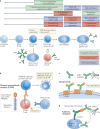
Fig. 2. Development of autoantibodies in neurological disease.
ac | Emerging data suggest defective B cell tolerance checkpoints in several antibody-mediated neurological diseases, increasing autoreactive immature B cells (part a) that are not removed (part b) but can be activated and enter germinal centres (part c). d,e | Several established or suspected triggers related to tissue injury or infection (part d) lead to transport of autoantigen to lymph nodes (part e). f | Germinal centres form after stimulation of autoreactive B cells with their antigen together with T cell help. T cells might be activated by the same autoantigen, a viral antigen or yet to be determined unrelated antigens. g | Enhanced T cell activation by immune checkpoint inhibitors is a recent trigger for antibody-mediated neurological disease. h | Affinity-matured and class-switched B cells become plasmablasts and secrete large amounts of autoantibodies. i,j | In a possible alternative route, autoreactive extrafollicular B cells recognize neuronal autoantigens via their B cell receptor (BCR) and are simultaneously stimulated via virus-induced B cell-intrinsic Toll-like receptor (TLR) activation, leading to maturation even without T cell help. k | In many autoantibody-mediated neurological diseases, plasma cells migrate into the brain and release large amounts of autoantibodies. CNS, central nervous system; TCR, T cell receptor.
Fig. 3
Fig. 3. Different disease mechanisms by pathogenic autoantibodies.
a | N-methyl-d-aspartate receptor (NMDAR) autoantibodies lead to receptor cross-linking, internalization and degradation, thus reducing the number of NMDARs on the neuronal surface. b | Aquaporin 4 (AQP4) autoantibodies bind to clustered AQP4 and induce complement-dependent cytotoxicity. c | Binding of GABAB receptor autoantibodies directly block receptor signalling. d | Leucine-rich glioma-inactivated 1 (LGI1) autoantibodies induce neuronal dysfunction by interrupting the trans-synaptic binding of LGI1 to its postsynaptic receptor ADAM22 (and likewise ADAM23 at the presynaptic site; not shown). e | Myelin oligodendrocyte glycoprotein (MOG) autoantibodies target the myelin sheath of axons and induce FcR-mediated antibody-dependent cellular cytotoxicity. f | Selected autoantibodies, such as to synapsin, are internalized via neuronal FcRs and lead to target inactivation intracellularly. AMPA, α-amino-3-hydroxy-5-methyl-4-isoxazolepropionic acid; GABA, γ-aminobutyric acid.
Fig. 4
Fig. 4. Innovative immunotherapies in neurological antibody-mediated diseases.
a | A large variety of treatment approaches are currently available (green, red) or in clinical development (blue), focusing on different targets along the B cell lineage. b | An exciting new route is the development of highly antibody-selective immunotherapies. Chimeric autoantibody receptor (CAAR) T cells are engineered to detect and deplete B cells monospecific for a given autoantibody, such as N-methyl-d-aspartate receptor (NMDAR) autoantibodies, via extracellular presentation of the target autoantigen. c | The non-pathogenic aquaporin 4 (AQP4)-selective antibody ‘aquaporumab’ lacks complement-dependent cytotoxicity (CDC)/antibody-dependent cellular cytotoxicity (ADCC) effector functions and can outcompete pathogenic AQP4 patient antibodies. d | In an experimental approach using anti-CD138 antibodies conjugated to an autoantigen, binding of pathogenic autoantibodies may induce CDC/ADCC-mediated depletion of long-lived plasma cells. CAR, chimeric antigen receptor; FcRn, neonatal Fc receptor; i.v., intravenous.

Similar articles

Cited by

References

    1. Lennon VA, Kryzer TJ, Pittock SJ, Verkman AS, Hinson SR. IgG marker of optic-spinal multiple sclerosis binds to the aquaporin-4 water channel. J. Exp. Med. 2005;202:473–477. - PMC - PubMed
    1. Dalmau J, et al. Paraneoplastic anti-N-methyl-d-aspartate receptor encephalitis associated with ovarian teratoma. Ann. Neurol. 2007;61:25–36. - PMC - PubMed
    1. Pollak TA, et al. Autoimmune psychosis: an international consensus on an approach to the diagnosis and management of psychosis of suspected autoimmune origin. Lancet Psychiatry. 2019;7:93–108. - PubMed
    1. Graus F, et al. A clinical approach to diagnosis of autoimmune encephalitis. Lancet Neurol. 2016;15:391–404. - PMC - PubMed
    1. Graus F, Dalmau J. Paraneoplastic neurological syndromes in the era of immune-checkpoint inhibitors. Nat. Rev. Clin. Oncol. 2019;16:535–548. - PubMed