

Collision Tumor of the Stomach
- PMID: 33976618
- PMCID: PMC8077467
- DOI: 10.1159/000514395
Collision Tumor of the Stomach
Abstract
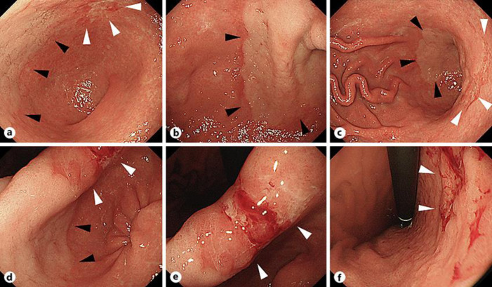
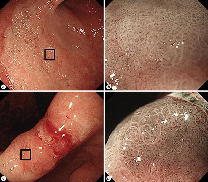
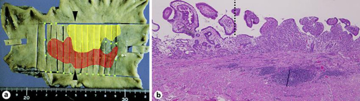
Collison tumor of the stomach is rare, and its endoscopic and pathological features remain poorly described. A 70-year-old woman was referred to our hospital for examination and treatment of undifferentiated gastric cancer. Esophagogastroduodenoscopy revealed a whitish, superficial elevated lesion in contact with a reddish, superficial depressed lesion from the anterior wall of the gastric angle and antrum to the lesser curvature. Laparoscopic distal gastrectomy was performed for preoperative diagnosis of suspected early gastric cancer presenting as a differentiated and undifferentiated collision tumor, which led to the lesion being diagnosed as collision tumor, tub1-tub2+por1-sig, pT1a (M), ly0, v0, N0, stage IA. To our knowledge, this report represents a valuable addition to the collision tumor literature describing a rare case of preoperatively diagnosed collision tumor of the stomach.
Keywords: Collision carcinoma; Collision tumor; Stomach.
Copyright © 2021 by S. Karger AG, Basel.
Conflict of interest statement
The authors have no conflicts of interest to disclose in association with this study.
Figures
Similar articles
-
Endoscopic and clinicopathological features of intramucosal, histologically mixed-type, low-grade, well-differentiated gastric tubular adenocarcinoma with the potential for late-onset lymph node metastasis.BMC Gastroenterol. 2018 Dec 27;18(1):189. doi: 10.1186/s12876-018-0919-3. BMC Gastroenterol. 2018. PMID: 30587141 Free PMC article.
-
Synchronous Early Gastric Cancer/Neuroendocrine Tumor Associated with Autoimmune Gastritis Completely Resected with Endoscopic Submucosal Dissection.Intern Med. 2019 Sep 15;58(18):2633-2637. doi: 10.2169/internalmedicine.2679-19. Epub 2019 Jun 7. Intern Med. 2019. PMID: 31178500 Free PMC article.
-
[A case of remnant gastric cancer seven years after initial diagnosis].Gan To Kagaku Ryoho. 2014 Nov;41(12):2414-6. Gan To Kagaku Ryoho. 2014. PMID: 25731541 Japanese.
-
[A case report of endosonography used for the diagnosis of early gastric cancer and gastrointestinal stromal tumor].Nihon Shokakibyo Gakkai Zasshi. 2014 Oct;111(10):1976-82. Nihon Shokakibyo Gakkai Zasshi. 2014. PMID: 25283226 Review. Japanese.
-
Mass-like Dieulafoy's lesion associated with advanced gastric cancer at the antrum of stomach: a case report and literature review.Diagn Pathol. 2017 Oct 10;12(1):73. doi: 10.1186/s13000-017-0663-y. Diagn Pathol. 2017. PMID: 29017601 Free PMC article. Review.
Cited by
-
Gastric collision tumor of adenocarcinoma and MALT lymphoma: A rare case report and literature review.Int J Surg Case Rep. 2024 Dec;125:110556. doi: 10.1016/j.ijscr.2024.110556. Epub 2024 Nov 3. Int J Surg Case Rep. 2024. PMID: 39500138 Free PMC article.
References
-
- Meyer R. Beitrag zur Verstandigung uber die Namengebung in der Geschwulstlehre. Zentralbl Allg Pathol. 1919;30:291–6.
-
- Spagnolo DV, Heenan PJ. Collision carcinoma at the esophagogastric junction: report of two cases. Cancer. 1980 Dec;46((12)):2702–8. - PubMed
-
- Schizas D, Katsaros I, Michalinos A, Damaskos C, Garmpis N, Ntomi V, et al. Collision tumors of the gastrointestinal tract: a systematic review of the literature. Anticancer Res. 2018 Nov;38((11)):6047–57. - PubMed
-
- Nambu S, Tanaka M, Shibuya T, Fujikura S, Sasaki H, Hirokawa S, et al. A case study of colliding gastric cancer. Gastroenterol Endosc. 1984;26:1118–25.
-
- Aoyagi K, Hashimoto K, Kohfuji K, Tanaka T, Kodama I, Yano S, et al. Two cases of colliding carcinoma of the stomach. Jpn J Gastroenterol Surg. 1992;25((8)):2152–6.
Publication types
LinkOut - more resources
Full Text Sources
Other Literature Sources
