

Rapidly Progressive Periorbital Oedema: A Case of Cutaneous Angiosarcoma
- PMID: 33976630
- PMCID: PMC8077485
- DOI: 10.1159/000514304
Rapidly Progressive Periorbital Oedema: A Case of Cutaneous Angiosarcoma
Abstract
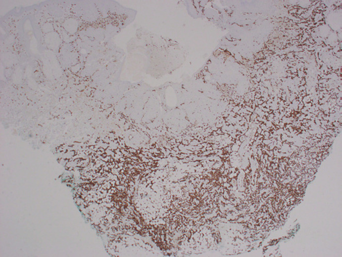
Angiosarcoma is a rare form of malignant endothelial cell tumour characterised by rapidly infiltrating anaplastic cells of vascular or lymphatic origin. We report an uncommon case of cutaneous angiosarcoma (cAS) manifesting as rapidly progressive unilateral periorbital oedema. Due to the acute onset of disease, the patient was initially treated with antibiotics for presumed periorbital cellulitis. The lack of response to conservative management raised the suspicion of a more serious condition, which eventually revealed the diagnosis of angiosarcoma through skin biopsy. As suggested by several previous case reports, the subtle manifestation of cAS made it a great mimicker of benign skin conditions. This case report serves as a reminder to the aggressive nature of angiosarcoma which can lead to marked facial swelling within several weeks. As the tumour was not resectable by the time of diagnosis, the patient was offered palliative radiotherapy.
Keywords: Angiosarcoma; Eyelid; Oedema; Periorbital.
Copyright © 2021 by S. Karger AG, Basel.
Conflict of interest statement
The authors have no conflicts of interest to declare.
Figures


Similar articles
-
Cutaneous Angiosarcoma in an Unusual Location and Without Predisposing Factors.Eur J Case Rep Intern Med. 2020 Sep 25;7(12):001939. doi: 10.12890/2020_001939. eCollection 2020. Eur J Case Rep Intern Med. 2020. PMID: 33313005 Free PMC article.
-
Angiosarcoma presenting with minor erythema and swelling.Case Rep Ophthalmol. 2013 Apr 5;4(1):59-63. doi: 10.1159/000346952. Print 2013 Jan. Case Rep Ophthalmol. 2013. PMID: 23616765 Free PMC article.
-
Cutaneous angiosarcoma presenting as bilateral periorbital edema.Orbit. 2023 Dec;42(6):621-623. doi: 10.1080/01676830.2022.2056901. Epub 2022 Apr 25. Orbit. 2023. PMID: 35467482
-
Atypical presentation of cutaneous angiosarcoma: review of the literature.Clin Exp Dermatol. 2022 Sep;47(9):1636-1641. doi: 10.1111/ced.15256. Epub 2022 Jun 22. Clin Exp Dermatol. 2022. PMID: 35548936 Review.
-
Low-grade dermal angiosarcoma of the breast following radiotherapy.Am Surg. 1996 Aug;62(8):668-72. Am Surg. 1996. PMID: 8712566 Review.
References
-
- Penel N, Marréaud S, Robin YM, Hohenberger P. Angiosarcoma: State of the art and perspectives. Crit Rev Oncol Hematol. 2011;80((2)):257–63. - PubMed
-
- Mackenzie IJ. Angiosarcoma of the face. Arch Dermatol. 1985;((121)):549–550. - PubMed
-
- Gunduz K, Shields JA, Shields CL, et al. Cutaneous angiosarcoma with eyelid involvement. Am J Ophthalmol. 1998;125:870–871. - PubMed
Publication types
LinkOut - more resources
Full Text Sources
Other Literature Sources
Research Materials
