Dextran Sulfate Effects EMT of Human Gastric Cancer Cells by Reducing HIF-1α/ TGF-β
- PMID: 33976746
- PMCID: PMC8100798
- DOI: 10.7150/jca.55550
Dextran Sulfate Effects EMT of Human Gastric Cancer Cells by Reducing HIF-1α/ TGF-β
Abstract
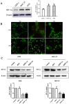
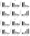
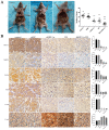
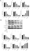
The peritoneal implant metastasis is one of the main pathway and main cause for high mortality for gastric cancer metastasis. Researchs show that epithelial-mesenchymal transition (EMT) playing essential role in modulating gastric cancer metastasis, and the expression of hypoxia inducible factor-1α (HIF-1α) can promote EMT in tumor cells. This research aims to explore the influence and mechanism of Dextran Sulfate (DS) affecting EMT of human gastric cancer. In the present study, we found that DS can enter into the cytoplasm and function in it. Inhibition of HIF-1α or DS significantly inhibit the migration and invasion of human gastric cancer cells, and decrease the mRNA and protein expressions of HIF-1α, matrix metalloproteinase-2 (MMP-2), transforming growth factor-β (TGF-β), Twist and N-cadherin (N-cad), rise E-cadherin (E-cad) expression, DS with HIF-1α knockdown has a stronger effect. In vivo studies indicated that compared with using DS or HIF-1α knockdown alone, DS with HIF-1α knockdown can better suppress the volume and number of metastatic tumors, and reduce the mRNA and protein expressions of HIF-1α, MMP-2, TGF-β, Twist and N-cad in metastatic tumor tissues of nude mice. We further demonstrated that the expression of HIF-1α, MMP-2, TGF-β , Twist and N-cad were higher in well and poorly differentiated gastric cancer than paracancerous tissue, and poorly differentiated gastric cancer were even higher, while E-cad expression was opposite. Taken together, this study shows that DS can interfere the expression of HIF-1α, thereby inhibiting TGF-β-mediated EMT of gastric cancer cells, and demonstrated a promising application of DS in gastric cancer therapy.
Keywords: Dextran sulfate; EMT.; HIF-1α; Human gastric cancer.
© The author(s).
Conflict of interest statement
Competing Interests: The authors have declared that no competing interest exists.
Figures


References
-
- Zhang W, Yuan W, Song J. et al. LncRNA CPS1-IT1 suppresses EMT and metastasis of colorectal cancer by inhibiting hypoxia-induced autophagy through inactivation of HIF-1α. Biochimie. 2018;144:21–27. - PubMed
-
- Mansour RN, Enderami SE, Ardeshirylajimi A. et al. Evaluation of hypoxia inducible factor-1 alpha gene expression in colorectal cancer stages of Iranian patients. J Cancer Res Ther. 2016;12:1313–1317. - PubMed
-
- Sun LL, Song Z, Li WZ. et al. Hypoxia facilitates epithelial-mesenchymal transition-mediated rectal cancer progress. Genet Mol Res. 2016;15:undefined. - PubMed
LinkOut - more resources
Full Text Sources
Research Materials
Miscellaneous

