ZFPM2-AS1 transcriptionally mediated by STAT1 regulates thyroid cancer cell growth, migration and invasion via miR-515-5p/TUSC3
- PMID: 33976749
- PMCID: PMC8100800
- DOI: 10.7150/jca.51437
ZFPM2-AS1 transcriptionally mediated by STAT1 regulates thyroid cancer cell growth, migration and invasion via miR-515-5p/TUSC3
Abstract
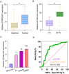
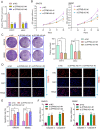
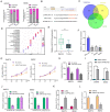
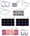
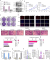
Objective: Our purpose was to study the roles and molecular mechanisms of long non-coding RNA (lncRNA) ZFPM2 Antisense RNA 1 (ZFPM2-AS1) in thyroid cancer. Methods: Firstly, the expression of ZFPM2-AS1, miR-515-5p and TUSC3 was detected in thyroid cancer tissues and cells. Secondary, their biological functions (proliferation, apoptosis, migration and invasion) were analyzed by a serious of functional experiments including cell counting kit-8 (CCK-8), clone formation, 5-Ethynyl-2'-deoxyuridine (EdU), enzyme-linked immunosorbent assay (ELISA), wound healing and Transwell assays. Thirdly, the mechanisms of STAT1/ZFPM2-AS1 and ZFPM2-AS1/miR-515-5p/TUSC were validated using chromatin immunoprecipitation (CHIP), pull-down and luciferase reporter assays. Results: ZFPM2-AS1 and TUSC were both highly expressed and miR-515-5p was down-regulated in thyroid cancer tissues as well as cells. Their knockdown weakened thyroid cancer cell growth, migration, and invasion. ZFPM2-AS1 was mainly distributed in the nucleus and cytoplasm of thyroid cancer cells. Mechanistically, up-regulation of ZFPM2-AS1 was induced by transcription factor STAT1 in line with CHIP and luciferase reporter assays. Furthermore, as a sponge of miR-515-5p, ZFPM2-AS1 decreased the ability of miR-515-5p to inhibit TUSC3 expression by pull-down, luciferase reporter and gain-and-loss assays, thereby promoting malignant progression of thyroid cancer. Conclusion: ZFPM2-AS1 acted as an oncogene in thyroid cancer, which was transcriptionally mediated by STAT1. Furthermore, ZFPM2-AS1 weakened the inhibitory effect of miR-515-5p on TUSC3. Thus, ZFPM2-AS1 could be an underlying biomarker for thyroid cancer.
Keywords: TUSC3; ZFPM2-AS1; miR-515-5p; thyroid cancer; transcription factor..
© The author(s).
Conflict of interest statement
Competing Interests: The authors have declared that no competing interest exists.
Figures




Similar articles
-
LncRNA ZFPM2-AS1 Enhances Retinoblastoma Development by Targeting the miR-3612/NT5E Signaling Axis.Crit Rev Eukaryot Gene Expr. 2022;32(6):69-82. doi: 10.1615/CritRevEukaryotGeneExpr.2022042697. Crit Rev Eukaryot Gene Expr. 2022. PMID: 35997119
-
SP1-induced lncRNA ZFPM2 antisense RNA 1 (ZFPM2-AS1) aggravates glioma progression via the miR-515-5p/Superoxide dismutase 2 (SOD2) axis.Bioengineered. 2021 Dec;12(1):2299-2310. doi: 10.1080/21655979.2021.1934241. Bioengineered. 2021. PMID: 34077295 Free PMC article.
-
YY1-induced lncRNA ZFPM2-AS1 facilitates cell proliferation and invasion in small cell lung cancer via upregulating of TRAF4.Cancer Cell Int. 2020 Apr 3;20:108. doi: 10.1186/s12935-020-1157-7. eCollection 2020. Cancer Cell Int. 2020. PMID: 32280300 Free PMC article.
-
Long noncoding RNA ZFPM2-AS1 acts as a miRNA sponge and promotes cell invasion through regulation of miR-139/GDF10 in hepatocellular carcinoma.J Exp Clin Cancer Res. 2020 Aug 14;39(1):159. doi: 10.1186/s13046-020-01664-1. J Exp Clin Cancer Res. 2020. PMID: 32795316 Free PMC article.
-
Silencing of OIP5-AS1 Protects Endothelial Cells From ox-LDL-Triggered Injury by Regulating KLF5 Expression via Sponging miR-135a-5p.Front Cardiovasc Med. 2021 Mar 12;8:596506. doi: 10.3389/fcvm.2021.596506. eCollection 2021. Front Cardiovasc Med. 2021. PMID: 33778018 Free PMC article.
Cited by
-
Interplay between JAK/STAT pathway and non-coding RNAs in different cancers.Noncoding RNA Res. 2024 Apr 7;9(4):1009-1022. doi: 10.1016/j.ncrna.2024.04.001. eCollection 2024 Dec. Noncoding RNA Res. 2024. PMID: 39022684 Free PMC article. Review.
-
MiR-221-3p Facilitates Thyroid Cancer Cell Proliferation and Inhibit Apoptosis by Targeting FOXP2 Through Hedgehog Pathway.Mol Biotechnol. 2022 Aug;64(8):919-927. doi: 10.1007/s12033-022-00473-5. Epub 2022 Mar 7. Mol Biotechnol. 2022. PMID: 35257310
-
In Silico Prioritization of STAT1 3' UTR SNPs Identifies rs190542524 as a miRNA-Linked Variant with Potential Oncogenic Impact.Noncoding RNA. 2025 Apr 29;11(3):32. doi: 10.3390/ncrna11030032. Noncoding RNA. 2025. PMID: 40407590 Free PMC article.
-
Long Non-coding RNA ZFPM2-AS1: A Novel Biomarker in the Pathogenesis of Human Cancers.Mol Biotechnol. 2022 Jul;64(7):725-742. doi: 10.1007/s12033-021-00443-3. Epub 2022 Jan 31. Mol Biotechnol. 2022. PMID: 35098483 Review.
-
lncRNA ZFPM2-AS1 promotes retinoblastoma progression by targeting microRNA miR-511-3p/paired box protein 6 (PAX6) axis.Bioengineered. 2022 Jan;13(1):1637-1649. doi: 10.1080/21655979.2021.2021346. Bioengineered. 2022. PMID: 34989314 Free PMC article.
References
-
- Seib CD, Sosa JA. Evolving Understanding of the Epidemiology of Thyroid Cancer. Endocrinol Metab Clin North Am. 2019;48:23–35. - PubMed
-
- Cabanillas ME, McFadden DG, Durante C. Thyroid cancer. Lancet. 2016;388:2783–95. - PubMed
-
- Sui F, Ji M, Hou P. Long non-coding RNAs in thyroid cancer: Biological functions and clinical significance. Mol Cell Endocrinol. 2018;469:11–22. - PubMed
-
- Wang Y, Bhandari A, Niu J, Yang F, Xia E, Yao Z. et al. The lncRNA UNC5B-AS1 promotes proliferation, migration, and invasion in papillary thyroid cancer cell lines. Hum Cell. 2019;32:334–42. - PubMed
LinkOut - more resources
Full Text Sources
Research Materials
Miscellaneous

