. 2021 Feb 25;8(9):2003535.
doi: 10.1002/advs.202003535. eCollection 2021 May.

MgFe-LDH Nanoparticles: A Promising Leukemia Inhibitory Factor Replacement for Self-Renewal and Pluripotency Maintenance in Cultured Mouse Embryonic Stem Cells

Affiliations

MgFe-LDH Nanoparticles: A Promising Leukemia Inhibitory Factor Replacement for Self-Renewal and Pluripotency Maintenance in Cultured Mouse Embryonic Stem Cells

Xiaolie He et al. Adv Sci (Weinh). .

Abstract

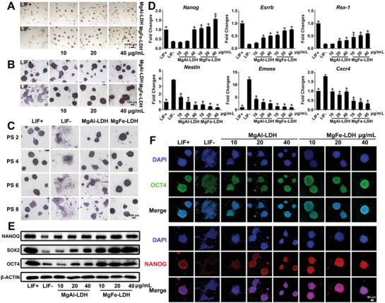
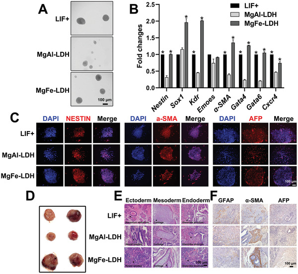
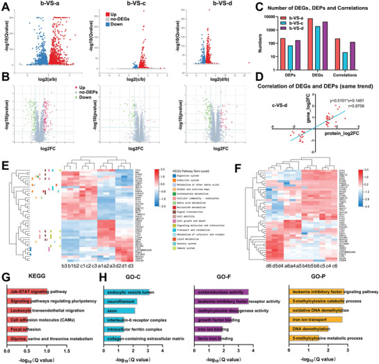
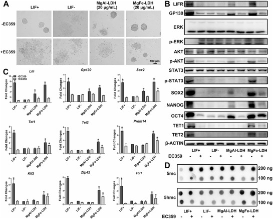
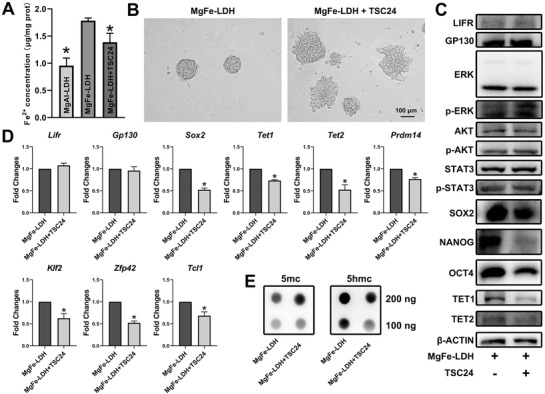
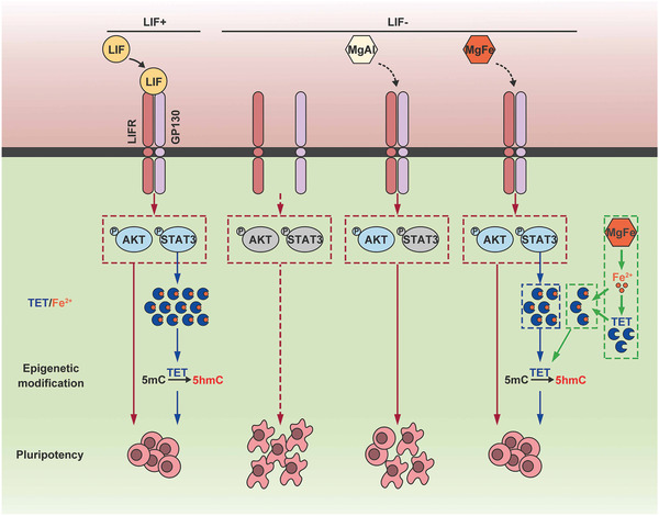
Leukemia inhibitory factor (LIF), an indispensable bioactive protein that sustains self-renewal and pluripotency in stem cells, is vital for mouse embryonic stem cell (mESC) culture. Extensive research is conducted on reliable alternatives for LIF as its clinical application in stable culture and large-scale expansion of ESCs is limited by its instability and high cost. However, few studies have sought to replace LIF with nanoparticles to provide a xeno-free culture condition. MgAl-LDH (layered double hydroxide) nanoparticles can partially replace LIF in maintaining pluripotency of mESCs; however, the requirement and tolerance for aluminum ions in mice are far lesser than those of iron ions. Hence, MgFe-LDH nanoparticles are selected for this study. MgFe-LDH is superior to MgAl-LDH in maintaining self-renewal and pluripotency of mESCs, in the absence of LIF and mouse embryonic fibroblast. Furthermore, combined transcriptomic and proteomic analysis confirms that MgFe-LDH can activate the LIF receptor (LIFR)/phosphatidylinositol 3-kinase (PI3K)/protein kinase B(AKT), LIFR/JAK/janus kinase (JAK)/signal transducer and activator of transcription 3(STAT3), and phospho-signal transducer and activator of transcription 3(p-STAT3)/ten-eleven translocation (TET) signaling pathways, while the extra Fe2+ provided by MgFe-LDH would also enhance TET1/2 abundance thus affecting the TET1/2 regulated pluripotency related marker expression and TET1/2 meditated DNA demethylation. These results suggest that MgFe-LDH nanoparticles can thus be used as an affordable and efficient replacement for LIF in mESC cultivation.

Keywords: LIFR/JAK/STAT3; MgFe‐LDH nanoparticles; combined transcriptomic and proteomic analysis; embryonic stem cells; pluripotency.


Conflict of interest statement

The authors declare no conflict of interest.

Figures

Figure 1
Figure 1
MgFe‐LDH is superior to MgAl‐LDH in supporting mESC self‐renewal. A) Morphology of clones observed via bright‐field microscopy. B) Representative images of mESCs cultured with nanoparticles and stained with ALP after 3 d of culture. C) ALP staining of mESCs treated with 20 µg mL−1 MgAl‐LDH or MgFe‐LDH at the indicated passage. D) qPCR analysis of key pluripotency genes (Nanog, Esrrb, and Rex‐1) and key differentiation genes (Nestin, Eomes, and Cxcr4). * represents p < 0.05, when compared to the LIF‐ treatment. E) Protein levels of NANOG, SOX2, and OCT4 in mESCs quantified by western blot. * represents p < 0.05, when compared to the LIF‐group. F) Representative confocal microscopy images of mESCs stained with OCT4 (green), NANOG (red), and DAPI (blue) accompanied by various treatments.
Figure 2
Figure 2
MgFe‐LDH is superior to MgAl‐LDH in supporting the pluripotency of mESCs. A) The embryoid bodies differentiated from P1 mESCs treated with nanoparticles at day eight of differentiation. B) The relative mRNA expression of three germ layer‐related genes during embryoid bodies differentiation at d8 was measured by qPCR: Nestin and Sox1 in the ectoderm; Kdr, Emoes, and α‐SMA in the mesoderm; as well as Gata4, Gata6, and Cxcr4 in the endoderm. * represents p < 0.05, when compared to the MgAl‐LDH treatment. C) The embryoid bodies were transferred to a gelatin precoated confocal dish for differentiation for another 4 d following which the presence and localization of NESTIN (ectoderm), α‐SMA (mesoderm), and AFP (endoderm) were determined by immunofluorescence. D) Teratoma formation assay for assessing pluripotency of P3 mESCs treated with nanoparticles. E) Histological analysis by HE staining of teratomas tissues derived from P3 mESCs treated with nanoparticles, to analyze the morphological features of the three germ layers. Arrowheads indicate these morphological features. F) Immunohistochemistry analysis of teratomas sections.
Figure 3
Figure 3
LIF signaling pathway, DNA demethylation, and iron ion transport differs significantly between the MgFe‐LDH and MgAl‐LDH groups, as identified via combined transcriptomic and proteomic analysis. A) Volcano plot of DEGs. B) Volcano plot of DEPs. C) The number of DEGs, DEPs, and correlations were quantified. a, b, c, and d represent the LIF+, LIF‐, MgAl‐LDH, and MgFe‐LDH groups, respectively. D) Correlation analysis of 44 DEGs and DEPs that had similar trends between c) the MgAl‐LDH and d) MgFe‐LDH groups. E) Heatmap of these 44 DEGs from correlation analysis, colors indicating relative expression. F) Clustering analysis of these 44 DEPs from the correlation analysis. G) KEGG pathway analysis of these 44 DEGs. H) GO enrichment analysis of these 44 DEGs.
Figure 4
Figure 4
MgFe‐LDH nanoparticles maintain the pluripotency of mECSs via LIFR/PI3K/AKT, LIFR/JAK/STAT3, and p‐STAT3/TET pathways. A) Bright‐field images of mESCs cultured for 3 d in media containing LIF/LIFR inhibitor EC359. B) Western blot was applied to measure protein expression changes in mESCs cultured for 3 d on gelatin, with or without EC359, subjected to the requisite treatment. C) Analysis of changes in mRNA expression following EC359 treatment via qPCR. * represents p < 0.05, when compared to the LIF‐group. D) Global 5mc and 5hmc levels detected by dot blot.
Figure 5
Figure 5
Fe2+ supplied by MgFe‐LDH enhances TET expression. A) Fe2+ concentration in the MgAl‐LDH, MgFe‐LDH, and MgFe‐LDH+TSC24 groups. B) Morphological changes in mESCs after Fe2+ chelator TSC24 was added to the MgFe‐LDH group. C) Changes in protein expression following TSC24 treatment, detected by western blot. D) Tet1/2 and Tet1/2 regulated pluripotency‐related gene expression following TSC24 administration. * represents p < 0.05, when compared to the MgFe‐LDH group. E) Detection of global 5mc and 5hmc levels following TSC24 administration.
Figure 6
Figure 6
Schematic representation of the functional mechanisms of MgFe‐LDH nanoparticles in regulating mESC pluripotency. MgFe‐LDH nanoparticles can activate the LIFR/PI3K/AKT and LIFR/JAK/STAT3 signaling pathways, resulting in downstream TET1/2 expression promoted by the activated p‐STAT3 thus controlling the expression of pluripotency‐related genes and DNA demethylation. In addition, Fe2+ released from MgFe‐LDH nanoparticles also proves beneficial in enhancing TET expression. All the above mechanisms ultimately make MgFe‐LDH an excellent substitute for LIF in maintaining mESC pluripotency.

Similar articles

Cited by

References

    1. Wang C., Yue H. B., Feng Q., Xu B. Z., Bian L. M., Shi P., ACS Appl. Mater. Interfaces 2018, 10, 29299. - PubMed
    1. Uzel S. G. M., Platt R. J., Subramanian V., Pearl T. M., Rowlands C. J., Chan V., Boyer L. A., So P. T. C., Kamm R. D., Sci. Adv. 2016, 2, 13. - PMC - PubMed
    1. Wang L., Wu W., Gu Q., Liu Z. P., Li Q. Y., Li Z. W., Fang J. H., Liu W. J., Wu J., Zhang Y., Wang L., Xu H. W., Li W., Hu B. Y., Zhou Q., Yin Z. Q., Hao J., Protein Cell 2019, 10, 455. - PMC - PubMed
    1. Liu Y., Xu H. W., Wang L., Li S. Y., Zhao C. J., Hao J., Li Q. Y., Zhao T. T., Wu W., Wang Y., Zhou Q., Qian C., Wang L., Yin Z. Q., Cell Discovery 2018, 4, 10. - PMC - PubMed
    1. Chong J. J. H., Yang X. L., Don C. W., Minami E., Liu Y. W., Weyers J. J., Mahoney W. M., Van Biber B., Cook S. M., Palpant N. J., Gantz J. A., Fugate J. A., Muskheli V., Gough G. M., Vogel K. W., Astley C. A., Hotchkiss C. E., Baldessari A., Pabon L., Reinecke H., Gill E. A., Nelson V., Kiem H. P., Laflamme M. A., Murry C. E., Nature 2014, 510, 273. - PMC - PubMed

Publication types

LinkOut - more resources