Review
. 2021 Oct;11(10):3060-3091.
doi: 10.1016/j.apsb.2021.04.023. Epub 2021 May 7.

Nanomedicine for acute respiratory distress syndrome: The latest application, targeting strategy, and rational design

Affiliations
Review

Nanomedicine for acute respiratory distress syndrome: The latest application, targeting strategy, and rational design

Qi Qiao et al. Acta Pharm Sin B. 2021 Oct.

Abstract

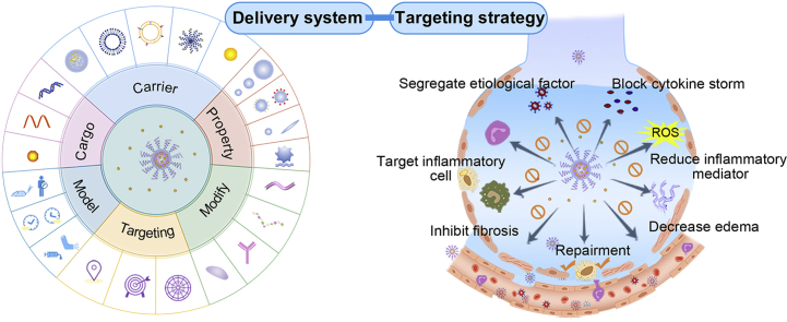
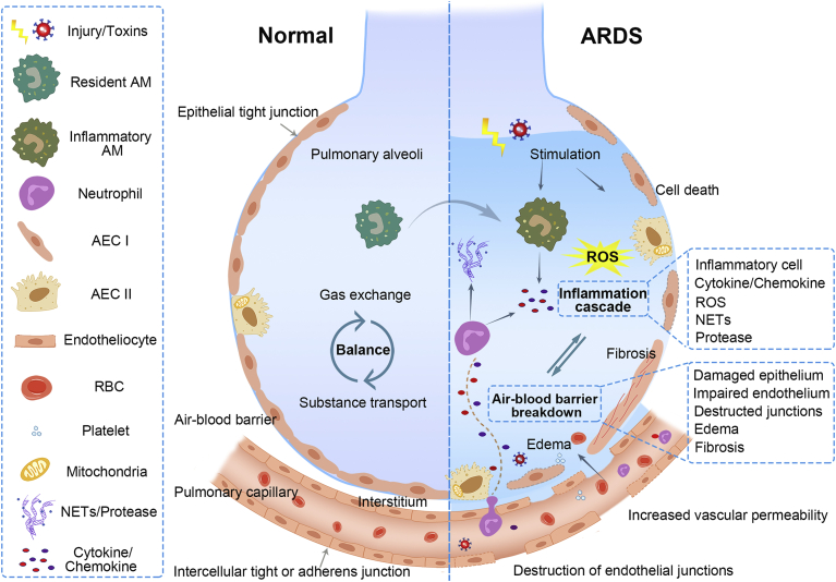
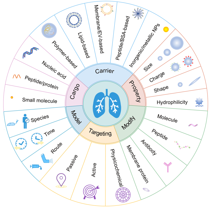
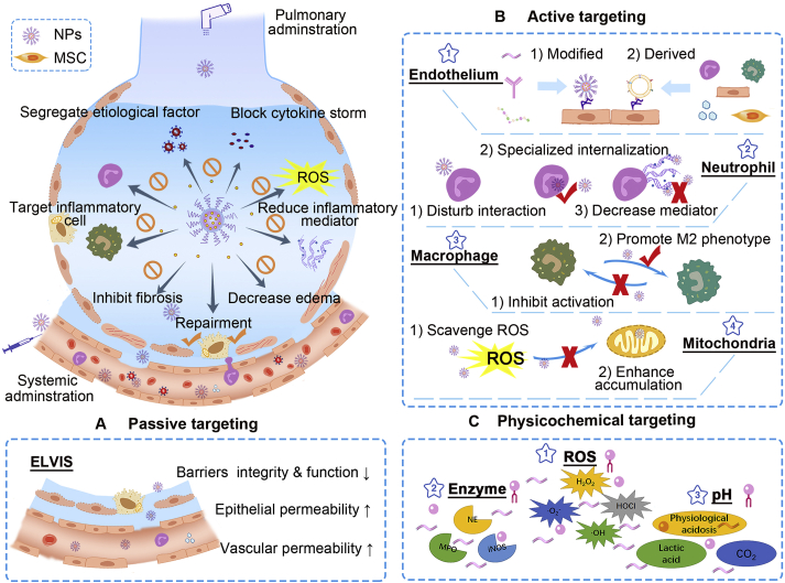
Acute respiratory distress syndrome (ARDS) is characterized by the severe inflammation and destruction of the lung air-blood barrier, leading to irreversible and substantial respiratory function damage. Patients with coronavirus disease 2019 (COVID-19) have been encountered with a high risk of ARDS, underscoring the urgency for exploiting effective therapy. However, proper medications for ARDS are still lacking due to poor pharmacokinetics, non-specific side effects, inability to surmount pulmonary barrier, and inadequate management of heterogeneity. The increased lung permeability in the pathological environment of ARDS may contribute to nanoparticle-mediated passive targeting delivery. Nanomedicine has demonstrated unique advantages in solving the dilemma of ARDS drug therapy, which can address the shortcomings and limitations of traditional anti-inflammatory or antioxidant drug treatment. Through passive, active, or physicochemical targeting, nanocarriers can interact with lung epithelium/endothelium and inflammatory cells to reverse abnormal changes and restore homeostasis of the pulmonary environment, thereby showing good therapeutic activity and reduced toxicity. This article reviews the latest applications of nanomedicine in pre-clinical ARDS therapy, highlights the strategies for targeted treatment of lung inflammation, presents the innovative drug delivery systems, and provides inspiration for strengthening the therapeutic effect of nanomedicine-based treatment.

Keywords: ACE2, angiotensin-converting enzyme 2; AEC II, alveolar type II epithelial cells; AM, alveolar macrophages; ARDS, acute respiratory distress syndrome; Acute lung injury; Acute respiratory distress syndrome; Anti-inflammatory therapy; BALF, bronchoalveolar lavage fluid; BSA, bovine serum albumin; CD, cyclodextrin; CLP, cecal ligation and perforation; COVID-19; COVID-19, coronavirus disease 2019; DOPE, phosphatidylethanolamine; DOTAP, 1-diolefin-3-trimethylaminopropane; DOX, doxorubicin; DPPC, dipalmitoylphosphatidylcholine; Drug delivery; ECM, extracellular matrix; ELVIS, extravasation through leaky vasculature and subsequent inflammatory cell-mediated sequestration; EPCs, endothelial progenitor cells; EPR, enhanced permeability and retention; EVs, extracellular vesicles; EphA2, ephrin type-A receptor 2; Esbp, E-selectin-binding peptide; FcgR, Fcγ receptor; GNP, peptide-gold nanoparticle; H2O2, hydrogen peroxide; HO-1, heme oxygenase-1; ICAM-1, intercellular adhesion molecule-1; IKK, IκB kinase; IL, interleukin; LPS, lipopolysaccharide; MERS, Middle East respiratory syndrome; MPMVECs, mouse pulmonary microvascular endothelial cells; MPO, myeloperoxidase; MSC, mesenchymal stem cells; NAC, N-acetylcysteine; NE, neutrophil elastase; NETs, neutrophil extracellular traps; NF-κB, nuclear factor-κB; Nanomedicine; PC, phosphatidylcholine; PCB, poly(carboxybetaine); PDA, polydopamine; PDE4, phosphodiesterase 4; PECAM-1, platelet-endothelial cell adhesion molecule; PEG, poly(ethylene glycol); PEI, polyetherimide; PEVs, platelet-derived extracellular vesicles; PLGA, poly(lactic-co-glycolic acid); PS-PEG, poly(styrene-b-ethylene glycol); Pathophysiologic feature; RBC, red blood cells; RBD, receptor-binding domains; ROS, reactive oxygen species; S1PLyase, sphingosine-1-phosphate lyase; SARS, severe acute respiratory syndrome; SARS-CoV-2, severe acute respiratory syndrome coronavirus 2; SDC1, syndecan-1; SORT, selective organ targeting; SP, surfactant protein; Se, selenium; Siglec, sialic acid-binding immunoglobulin-like lectin; TLR, toll-like receptor; TNF-α, tumor necrosis factor-α; TPP, triphenylphosphonium cation; Targeting strategy; YSA, YSAYPDSVPMMS; cRGD, cyclic arginine glycine-d-aspartic acid; iNOS, inducible nitric oxide synthase; rSPANb, anti-rat SP-A nanobody; scFv, single chain variable fragments.


Figures

Image 1
Graphical abstract
Figure 1
Figure 1
Pathophysiology of ARDS. Healthy lungs maintain a balance of gas change and substance transport, ascribing to the integrated structure and function of the air–blood barrier (the left sections). The air–blood barrier is mainly consisting of epithelium and endothelium barrier, where cells are lined up continuously and connected through intercellular junctions. Once injury or toxins stimulate, AM and AEC cells are initiated, secreting inflammatory cytokines and chemokines, and recruiting neutrophils to the inflamed lungs (the right sections). The activated neutrophils can release NETs and multiple proteases in response, while excessive productions can be harmful and induce lung edema. Various inflammatory cells may overproduce pro-inflammatory cytokines, ROS, and other inflammatory mediators, resulting in cytokine storms and severe lung injury. The inflammatory environment can damage the lung cells, dissolve the intercellular junctions, increase epithelium and vascular permeability, leading to the breakdown of epithelium and endothelium barrier. The impairment of lung barrier can prompt more extravasation of inflammatory cells into alveoli, which can exacerbate the inflammatory state and cause tissue damage, forming a vicious circle between inflammation cascade and air–blood barrier breakdown. Adapted with permission from Ref. . Copyright © 2017, Massachusetts Medical Society.
Figure 2
Figure 2
Parameters that affecting delivery and therapeutic efficiency of nanomedicine in ARDS. Various drugs of distinct properties are applied for ARDS treatment. Both organic and inorganic/metallic carriers have been employed for drug delivery. Physicochemical properties can be manipulated for optimal drug delivery, including particle size, charge, shape, and hydrophilicity. Through engineering approaches such as modification or conjugation with specific molecules, peptides antibody, or membrane proteins on the surface, nanocarriers can accomplish passive, active, and physicochemical targeting. The therapeutic effect of drug delivery systems was mainly conducted in animals with few studies on human lung sections. Therapeutic regimens including administration time and route can also influence drug efficiency.
Figure 3
Figure 3
Nanocarriers-mediated drug delivery for ARDS therapy. Current targeting strategies have been focused on handling overwhelming inflammations and restoring pulmonary functions by inhibiting inflammatory cells, capturing toxins and cytokines, decreasing inflammatory mediators, and recovering the air–blood barrier. Passive, active, and physicochemical targeting tactics were applied. (A) The passive targeting delivery has primarily relied on ELVIS (extravasation through leaky vasculature and subsequent inflammatory cell-mediated sequestration) effect. (B) Active targeting has been concentrated on inflamed endothelium, inflammatory neutrophils and macrophages, and impaired mitochondria. (i) targeting endothelium: nanocarriers with modification of particular molecules, antibodies, and peptides can be applied for targeting inflamed endothelium, where specific markers are highly expressed; biomimetic carriers derived from various functional cells (neutrophils, macrophages/monocytes, endothelial cells) can inherit good tropism to inflammatory endothelium; (ii) targeting neutrophils: particular nanocarriers can interfere with neutrophils to disturb their migrations to the lungs; some nanocarriers can be specifically internalized by activated neutrophils and hitchhiked to inflammatory site subsequently; nanocarriers can be employed for decreasing inflammatory mediators released by neutrophils; (iii) targeting macrophages: inhibiting pro-inflammatory M1 and promoting polarization to M2 phenotype; (iv) targeting mitochondria: scavenge ROS to protect mitochondria from damage; employing mitochondria-targeted materials for enhancing intracellular drug accumulation. (C) Physicochemical targeting: utilizing the aberrant inflammatory state such as excessive ROS, overproduced enzyme, and low pH to achieve site-specific drug delivery and stimuli-responsive release.
Figure 4
Figure 4
Targeting neutrophils by drug-loaded BSA nanoparticles. (A) The schematic of neutrophils-mediated delivery of BSA nanoparticles to reach the inflammatory site. (B) TPCA-1 concentration in plasma and BALF after TPCA-1 or TPCA-1 BSA nanoparticles injection. (C) Cell count of leukocytes and neutrophils in BALF. (D) IL-6 and (E) TNF-α concentration in BALF after drug administration (vehicle of TPCA-1 solution, 5% glucose, TPCA-1 solution, or TPCA-1 BSA nanoparticles). All data represent mean ± SD (n = 3–4, per group). Statistics were performed by a two-sample Student's t test (∗∗P < 0.01). Reprinted with the permission from Ref. . Copyright © 2015, American Chemical Society.
Figure 5
Figure 5
Long-acting DNase-I nanoparticles for COVID-19 treatment. (A) Fabrication of long-acting DNase-I nanoparticle. (B) Quantitative analysis of plasma cell-free DNA (cfDNA) level and DNase-I activity from patient with SARS-CoV-2 sepsis after free DNase-I or long-acting DNase-I treatment (n = 10). (C) NET ratio, MPO, and NE concentration after free drug or long-acting nanoparticles therapy in SARS-CoV-2 Sepsis patient’ PBMCs. (D) NF-κB p65 binding activity and plasma cytokine levels with free DNase-I or long-acting DNase-I treatment in PBMCs from SARS-CoV-2 patients. The experiment was repeated at least three times. Statistics were analyzed using a two-tailed unpaired t-test. Data are displayed as mean ± SEM. ∗∗P < 0.01, ∗∗∗P < 0.001. Reprinted with the permission from Ref. . Copyright © 2020, Elsevier Inc.
Figure 6
Figure 6
MPO and ROS dual-responsive nanoparticles for real-time imaging of inflammatory site. (A) The fabrication of luminescent materials with dual-responsive properties. (B) In vivo luminescence images of luminol or Lu-bCD nanoparticles before and after i.v. injection in ARDS mice. (C) Ex vivo fluorescence imaging of lung tissue after Lu-bCD nanoparticles treatment. (D) The luminescent intensities, MPO, and H2O2 levels, and neutrophil amounts in the lungs at different time points after LPS challenge. (E) Linear correlation analysis between luminescent intensity and neutrophil count, H2O2, and MPO level. Error bars, mean ± SD (n = 4, B and C; n = 6, D and E). Reprinted with the permission from Ref. . Copyright © 2017, Elsevier Inc.
Figure 7
Figure 7
RBC-hitchhiking for lung targeted delivery. (A) Scheme of RBC-hitchhiking: nanocarriers (NCs) were attached to RBCs, followed by the injection via an intravascular catheter, then the NCs transferred to the first downstream capillary. (B) Representative scanning electron micrographs of nanoparticles or nanogels absorbed on RBC (scale bars, 1 μm). (C) i.v. injection of RBC-hitchhiking NCs enhanced lung delivery. Mice were injected with nanogels that were uncoated (bare) or different antibodies (anti-PECAM, anti-ICAM, or IgG) coated, with or without RBC-hitchhiking. Data are plotted as % of the injected dose (%ID) per organ. Each data point represents mean ± SEM (n = 3). ∗P < 0.05, non-paired, two-tailed t-test. Reprinted with the permission from Ref. . Copyright © 2018, Nature Publishing Group.
Figure 8
Figure 8
Biomimetic drug delivery systems for virus and cytokine neutralization. (A) The scheme of cellular nanosponges preventing viruses from entering host cells through surface antigen. (B) Western blotting analysis of cell lysate, cell membrane, and cellular nanosponges obtained from epithelial cells and macrophages. Data are presented as mean ± SD. n = 3. (C) The illustration of nanodecoy preparation. The nanodecoy was derived from cell membrane vesicles of human THP-1 cells and genetically engineered 293T/ACE2. Reprinted with the permission from Ref. . Copyright © 2020, American Chemical Society and Ref. . Copyright © 2020, the Author(s).
Figure 9
Figure 9
Platelet-derived extracellular vesicles for targeting inflammatory lungs. (A) Schematic of platelet-derived EVs with TPCA-1 loaded for pneumonia therapy. The inflammatory cytokine storm was remarkably reduced, as indicated by TNF-α, IL-6, and IL-1β. (B) Ex vivo imaging and corresponding fluorescence level of main organs after i.v. injection of DiD-labeled PEVs. Data are presented as mean ± SEM (n = 3–5). Statistical significance is calculated by one-way ANOVA using Tukey's post-test. ∗∗P < 0.01; ∗∗∗P < 0.005. Reprinted with the permission from Ref. . Copyright ©2020, Elsevier Inc.
Figure 10
Figure 10
Neutrophil elastase-sensitive nanoparticles-in-microgels for particle allosteric strategy. (A) The preparation of neutrophil elastase-sensitive nanoparticles-in-microgels (N-in-M) microgels and illustration of the mechanism of NE-responsive drug release. (B) MPO activity detected by fluorescence image of lung tissue. (C) Neutrophil numbers and (D) neutrophil percentage in BALF after treatment. Statistical tests: Shapiro-Wilk followed by Kruskal-Wallis (C) and 1-way ANOVA (D), with ∗P < 0.05, ∗∗P < 0.01, and ∗∗∗P < 0.001 between groups denoted. n = 5 each group. Reprinted with the permission from Ref. . Copyright © 2019, The American Society for Clinical Investigation.

Similar articles

Cited by

References

    1. Fan E., Brodie D., Slutsky A.S. Acute respiratory distress syndrome: advances in diagnosis and treatment. J Am Med Assoc. 2018;319:698–710. - PubMed
    1. Bellani G., Laffey J.G., Pham T., Fan E., Brochard L., Esteban A. Epidemiology, patterns of care, and mortality for patients with acute respiratory distress syndrome in intensive care units in 50 Countries. J Am Med Assoc. 2016;315:788–800. - PubMed
    1. Thompson B.T., Chambers R.C., Liu K.D. Acute respiratory distress syndrome. N Engl J Med. 2017;377:562–572. - PubMed
    1. Li X., Ma X. Acute respiratory failure in COVID-19: is it "typical" ARDS? Crit Care. 2020;24:198. - PMC - PubMed
    1. Quan C., Li C.Y., Ma H., Li Y.S., Zhang H.L. Immunopathogenesis of coronavirus-induced acute respiratory distress syndrome (ARDS): potential infection-associated hemophagocytic lymphohistiocytosis. Clin Microbiol Rev. 2021;34 e00074−20. - PMC - PubMed