

doi: 10.1096/fba.2020-00138.
eCollection 2021 May.
Emerging roles for the autophagy machinery in extracellular vesicle biogenesis and secretion
Affiliations
- PMID: 33977236
- PMCID: PMC8103724
- DOI: 10.1096/fba.2020-00138
Item in Clipboard
Emerging roles for the autophagy machinery in extracellular vesicle biogenesis and secretion
FASEB Bioadv.
.
Abstract
Autophagy classically functions to maintain cell health during stressful conditions by targeting cytosolic components for degradation and recycling via lysosomal pathways. However, accumulating evidence also supports roles for autophagy-related genes (ATGs) in non-degradative processes including cellular secretion. Here, we review emerging roles for the autophagy machinery in regulating extracellular vesicle loading and secretion and discuss how functional coupling of these pathways may impact normal physiology and disease.
© 2021 The Authors. FASEB BioAdvances published by the Federation of American Societies for Experimental Biology.
Conflict of interest statement
JD is the member of the Scientific Advisory Board of Vescor Therapeutics, LLC.
Figures
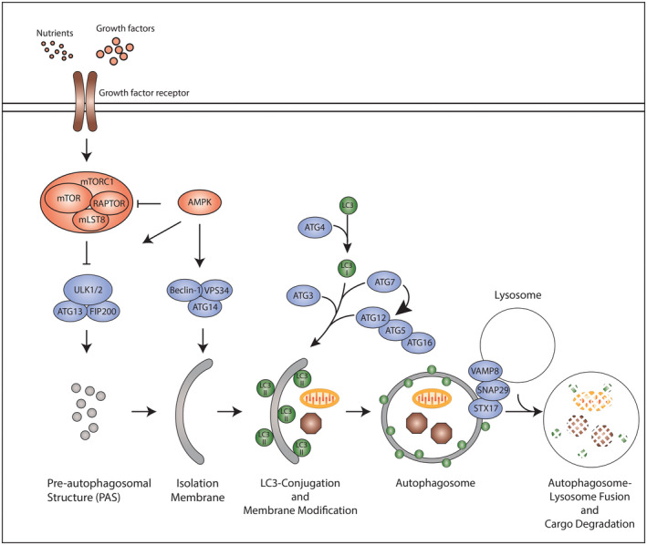
The classical autophagy pathway. Signals are integrated by the core autophagy machinery, which hierarchically regulate individual steps within the autophagy pathway. Formation of the pre‐autophagosomal structure (PAS), the first step in the autophagy pathway, is controlled by the ULK1‐ATG13‐FIP200 kinase complex. Nutrients and growth factors (e.g. amino acids, IGF‐1) trigger signaling through growth factor receptors. Ultimately, these signals converge upon the mammalian target of rapamycin complex 1 (mTORC1) comprised of mTOR, RAPTOR and mLST8, as well as AMPK, which reciprocally modify the ULK complex to regulate its functions in PAS formation. The PAS is subsequently modified by the Beclin‐1‐ATG14‐VPS34 complex to mediate formation of the isolation membrane. Expansion of the isolation membrane is associated with two ubiquitin‐like reactions involving ATG7, ATG5, ATG12, ATG16 and ATG3 which ultimately conjugate phosphatidylethanolamine (PE) to microtubule‐associated protein 1 light chain 3 (MAP1LC3B; also known as LC3) and other ATG8 family proteins. LC3‐PE targets LC3 to autophagosomal membranes where it facilitates membrane expansion and cargo sequestration. Finally, the autophagosome double‐membrane is sealed and captured cargo targeted to the lysosome through autophagosome‐lysosome fusion. In this schematic, arrows indicate activating signals, whereas blunt‐end lines represent inhibitory signals
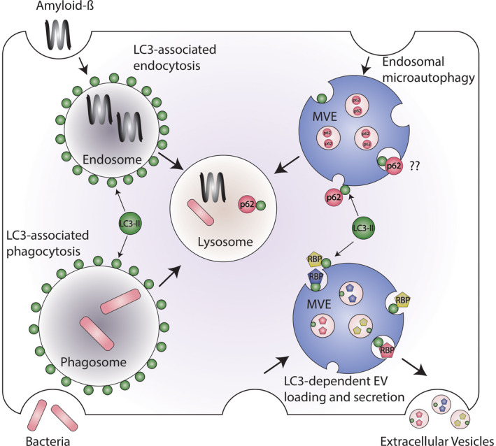
Emerging autophagy‐related pathways that employ LC3‐conjugation to single‐membrane endocytic and phagocytic vesicles. Components of the autophagy conjugation machinery, in addition to regulating classical autophagy, facilitate the delivery of microtubule‐associated protein 1 light chain 3 (MAP1LC3B; also known as LC3) and other ATG8 family proteins to single‐membrane vesicles including phagosomes, endosomes, and multivesicular endosomes (MVEs). During LC3‐associated phagocytosis (LAP) and LC3‐associated endocytosis (LANDO), LC3 delivery to the surface of phagosomes and endosomes, respectively, controls the trafficking and lysosomal degradation of material engulfed from the extracellular space such as bacteria or amyloid‐β. In contrast, LC3 is targeted to discrete subdomains at the limiting membrane of MVEs during endosomal microautophagy (eMI) and LC3‐dependent EV loading and secretion (LDELS), which undergo budding to form small intraluminal vesicles. LC3 facilitates the packaging of RNA‐binding proteins (RBPs) into these intraluminal vesicles, which subsequently are released as extracellular vesicles in the LDELS pathway. LC3 may also mediate the packaging of autophagy cargo receptors such as p62/SQSTM1 into intraluminal vesicles during endosomal microautophagy, but it remains unclear whether such pathways contribute to EV secretion
References
-
- van Niel G, D'Angelo G, Raposo G. Shedding light on the cell biology of extracellular vesicles. Nat Rev Mol Cell Biol. 2018;19:213‐228. - PubMed
-
- Pegtel DM, Gould SJ. Exosomes. Annu Rev Biochem. 2019;88:487‐514. - PubMed
-
- Mathieu M, Martin‐Jaular L, Lavieu G, Théry C. Specificities of secretion and uptake of exosomes and other extracellular vesicles for cell‐to‐cell communication. Nat Cell Biol. 2019;21:9‐17. - PubMed
Grants and funding
LinkOut - more resources
Full Text Sources
Other Literature Sources