. 2021 Mar 21;3(2):fcab047.
doi: 10.1093/braincomms/fcab047. eCollection 2021.

Immune modulation attenuates infantile neuronal ceroid lipofuscinosis in mice before and after disease onset

Affiliations

Immune modulation attenuates infantile neuronal ceroid lipofuscinosis in mice before and after disease onset

Janos Groh et al. Brain Commun. .

Abstract

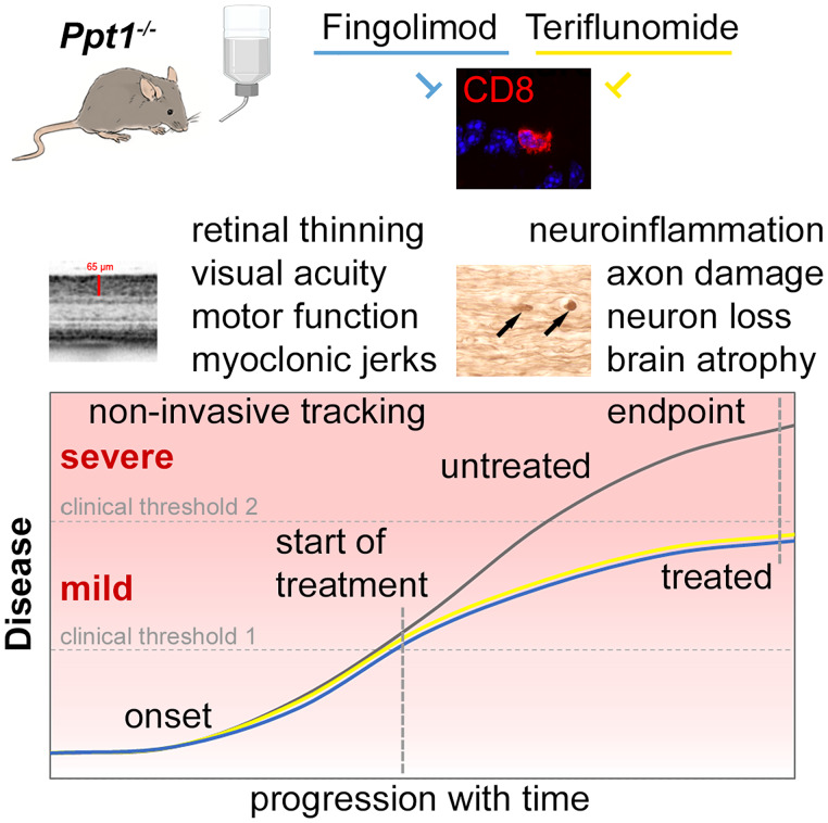
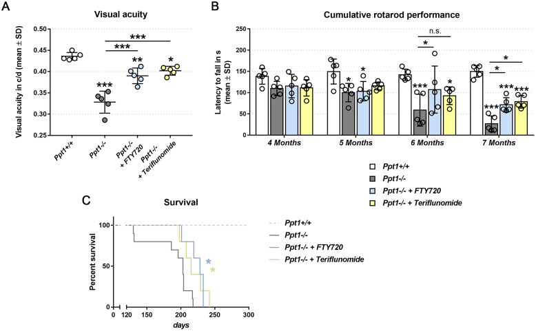
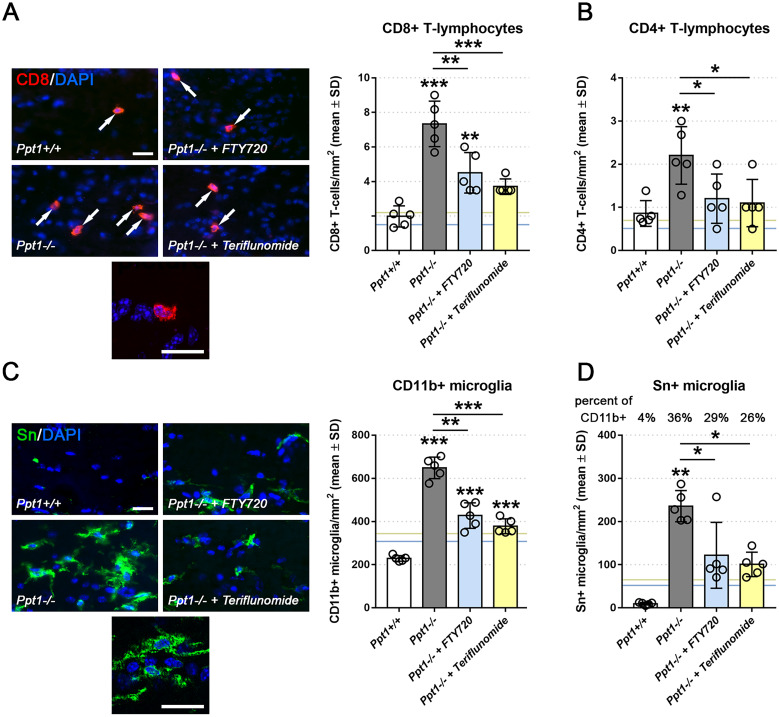
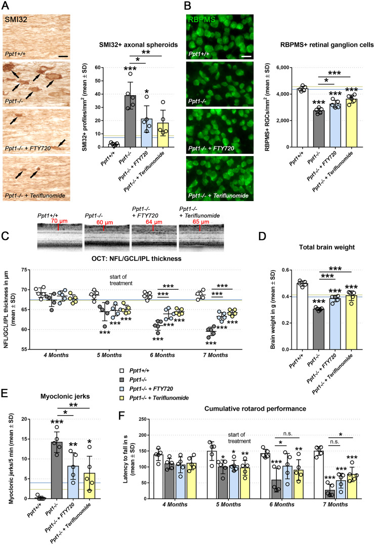
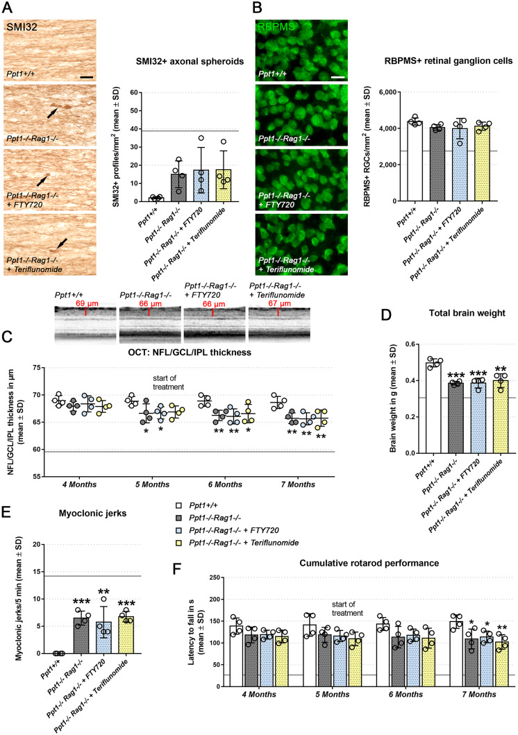
Targeting neuroinflammation in models for infantile and juvenile forms of neuronal ceroid lipofuscinosis (NCL, CLN disease) with the clinically established immunomodulators fingolimod and teriflunomide significantly attenuates the neurodegenerative phenotype when applied preventively, i.e. before the development of substantial neural damage and clinical symptoms. Here, we show that in a mouse model for the early onset and rapidly progressing CLN1 form, more complex clinical phenotypes like disturbed motor coordination and impaired visual acuity are also ameliorated by immunomodulation. Moreover, we show that the disease outcome can be attenuated even when fingolimod and teriflunomide treatment starts after disease onset, i.e. when neurodegeneration is ongoing and clinical symptoms are detectable. In detail, treatment with either drug led to a reduction in T-cell numbers and microgliosis in the CNS, although not to the same extent as upon preventive treatment. Pharmacological immunomodulation was accompanied by a reduction of axonal damage, neuron loss and astrogliosis in the retinotectal system and by reduced brain atrophy. Accordingly, the frequency of myoclonic jerks and disturbed motor coordination were attenuated. Overall, disease alleviation was remarkably substantial upon therapeutic treatment with both drugs, although less robust than upon preventive treatment. To test the relevance of putative immune-independent mechanisms of action in this model, we treated CLN1 mice lacking mature T- and B-lymphocytes. Immunodeficient CLN1 mice showed, as previously reported, an improved neurological phenotype in comparison with genuine CLN1 mice which could not be further alleviated by either of the drugs, reflecting a predominantly immune-related therapeutic mechanism of action. The present study supports and strengthens our previous view that repurposing clinically approved immunomodulators may alleviate the course of CLN1 disease in human patients, even though diagnosis usually occurs when symptoms have already emerged.

Keywords: T-lymphocytes; attenuation of disease; immunomodulation; infantile neuronal ceroid lipofuscinosis; neurodegeneration; neuroinflammation; preventive treatment.


Figures

Graphical Abstract
Graphical Abstract
Figure 1
Figure 1
Preventive fingolimod and teriflunomide treatment improves complex clinical phenotypes in CLN1 mice. (A) Automated optokinetic response analysis of visual acuity (c/d, cycles per degree) of 6-month-old Ppt1+/+, Ppt1/, fingolimod-treated Ppt1/ (+ FTY720) and teriflunomide-treated Ppt1/ (+ Teriflunomide) mice (circle = mean value of one mouse; n = 5 mice per group, one-way ANOVA and Tukey’s post hoc test). (B) Longitudinal analysis of cumulative Rotarod performance in Ppt1+/+, Ppt1/, fingolimod-treated Ppt1/ and teriflunomide-treated Ppt1/ mice (circle = mean value of five consecutive runs of one mouse; n = 5 mice per group, two-way ANOVA and Tukey’s post hoc test). (C) Kaplan-Meier survival analysis of Ppt1+/+, Ppt1/, fingolimod-treated Ppt1/ and teriflunomide-treated Ppt1/ mice (n = 5 to 10 mice per group, Log-rank test). *P < 0.05, **P < 0.01, ***P < 0.001.
Figure 2
Figure 2
Therapeutic fingolimod and teriflunomide treatment attenuate neuroinflammation in CLN1 mice. (A) Representative immune fluorescence microscopy and quantification of CD8+ and (B) CD4+ T-lymphocytes in longitudinal optic nerve sections from 7-month-old Ppt1+/+, Ppt1/, fingolimod-treated Ppt1/ (+ FTY720) and teriflunomide-treated Ppt1/ (+ Teriflunomide) mice (circle = mean value of one mouse; n = 5 mice per group, one-way ANOVA and Tukey’s post hoc test). Scale bars: 20 µm. (C) Representative immune fluorescence microscopy and quantification of CD11b+ and (D) Sn+ microglia in longitudinal optic nerve sections (n = 5 mice per group, one-way ANOVA and Tukey’s post hoc test). Blue and yellow lines indicate mean values from previously published preventive (from 1 to 6 months of age) fingolimod and teriflunomide treatment approaches, respectively . Scale bars: 20 µm. *P < 0.05, **P < 0.01, ***P < 0.001.
Figure 3
Figure 3
Therapeutic fingolimod and teriflunomide treatment improves histopathological and behavioural readout measures in CLN1 mice. (A) Representative light microscopy and quantification of SMI32+ axonal spheroids in optic nerves and (B) immune fluorescence microscopy and quantification of RBPMS+ retinal ganglion cells in retinae from 7-month-old Ppt1+/+, Ppt1/, fingolimod-treated Ppt1/ (+ FTY720) and teriflunomide-treated Ppt1/ (+ Teriflunomide) mice (circle = mean value of one mouse; n = 5 mice per group, one-way ANOVA and Tukey’s post hoc test). Scale bars: 20 µm. (C) Representative peripapillary OCT circle scans at 7 months and longitudinal analysis of NFL/GCL/IPL thickness (n = 5 mice per group, one-way ANOVA and Tukey’s post hoc test). (D) Total brain weights of 7-month-old Ppt1+/+, Ppt1/, fingolimod-treated Ppt1/ and teriflunomide-treated Ppt1/ mice (n = 5 mice per group, one-way ANOVA and Tukey’s post hoc test). (E) Quantification of myoclonic jerks in 7-month-old Ppt1+/+, Ppt1/, fingolimod-treated Ppt1/ and teriflunomide-treated Ppt1/ mice (circle = mean value of one mouse; n = 5 mice per group, one-way ANOVA and Tukey’s post hoc test). (F) Longitudinal analysis of cumulative Rotarod performance (circle = mean value of five consecutive runs of one mouse; n = 5 mice per group, two-way ANOVA and Tukey’s post hoc test). Blue and yellow lines (in b–e) indicate mean values from previously published preventive (from 1 to 6 months of age) fingolimod and teriflunomide treatment approaches, respectively. *P < 0.05, **P < 0.01, ***P < 0.001.
Figure 4
Figure 4
Treatment effects of fingolimod and teriflunomide are predominantly immunomodulatory. (A) Representative light microscopy and quantification of SMI32+ axonal spheroids in optic nerves and (B), immune fluorescence microscopy and quantification of RBPMS+ retinal ganglion cells in retinae from 7-month-old Ppt1+/+, Ppt1/Rag1/, fingolimod-treated Ppt1/Rag1/ (+ FTY720) and teriflunomide-treated Ppt1/Rag1/ (+ Teriflunomide) mice (circle = mean value of one mouse; n = 4 mice per group, one-way ANOVA and Tukey’s post hoc test). Scale bars: 20 µm. (C) Representative peripapillary OCT circle scans at 7 months and longitudinal analysis of NFL/GCL/IPL thickness (n = 4 mice per group, one-way ANOVA and Tukey’s post hoc test). (D) Total brain weights of 7-month-old Ppt1+/+, Ppt1/Rag1/, fingolimod-treated Ppt1/Rag1/ and teriflunomide-treated Ppt1/Rag1/ mice (n = 4 mice per group, one-way ANOVA and Tukey’s post hoc test). (E) Quantification of myoclonic jerks in 7-month-old Ppt1+/+, Ppt1/Rag1/, fingolimod-treated Ppt1/Rag1/ and teriflunomide-treated Ppt1/Rag1/ mice (circle = mean value of one mouse; n = 4 mice per group, one-way ANOVA and Tukey’s post hoc test). (F) Longitudinal analysis of cumulative Rotarod performance (circle = mean value of five consecutive runs of one mouse n = 4 mice per group, two-way ANOVA and Tukey’s post hoc test). Note that none of the investigated parameters was further improved by either of the immunomodulators in comparison to untreated Ppt1/Rag1/ mice. Grey horizontal lines in all graphs indicate mean values of immune competent Ppt1/Rag1+/+ mice. *P < 0.05, **P < 0.01, ***P < 0.001.

References

    1. Kohlschutter A, Schulz A, Bartsch U, Storch S.. Current and emerging treatment strategies for neuronal ceroid lipofuscinoses. Cns Drugs. 2019;33(4):315–325. - PMC - PubMed
    1. Mole SE, Anderson G, Band HA, et al.Clinical challenges and future therapeutic approaches for neuronal ceroid lipofuscinosis. Lancet Neurol. 2019;18(1):107–116. - PubMed
    1. Santavuori P. Neuronal ceroid-lipofuscinoses in childhood. Brain Dev. 1988;10(2):80–83. - PubMed
    1. Vesa J, Hellsten E, Verkruyse LA, et al.Mutations in the palmitoyl protein thioesterase gene causing infantile neuronal ceroid lipofuscinosis. Nature. 1995;376(6541):584–587. - PubMed
    1. Gupta P, Soyombo AA, Atashband A, et al.Disruption of PPT1 or PPT2 causes neuronal ceroid lipofuscinosis in knockout mice. Proc Natl Acad Sci U S A. 2001;98(24):13566–13571. - PMC - PubMed

LinkOut - more resources