

Effects of uncomplicated Descemet membrane endothelial keratoplasty on the central retinal thickness
- PMID: 33977320
- PMCID: PMC8380572
- DOI: 10.1007/s00417-021-05203-2
Effects of uncomplicated Descemet membrane endothelial keratoplasty on the central retinal thickness
Abstract
Purpose: To determine retinal thickness (RT) changes and the incidence of macular edema after uncomplicated Descemet membrane endothelial keratoplasty (DMEK-ME) in patients without ME risk factors.
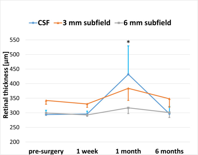
Methods: In this retrospective study, 107 pseudophakic eyes of 74 patients with Fuchs endothelial dystrophy (FED) (79.4%) or bullous keratopathy (BK) (20.6%) underwent DMEK surgery between 2016 and 2019 at the Department of Ophthalmology, RWTH Aachen University. Patients with intra- or postoperative complications as well as pre-existing risk factors for ME were excluded. Macular spectral-domain optical coherence tomography (SD-OCT) and best spectacle-corrected visual acuity (BSCVA) measurements were performed before, 1 week, 1 month, and 6 months after surgery. Retinal thickness (RT) was analyzed in the central foveal 1 mm (CSF), parafoveal 3 mm and 6 mm subfield.
Results: Eight eyes (7.5%) developed DMEK-ME 1 month after surgery. Six DMEK-ME eyes (75%) were rebubbled, compared with 31.3% (31 of 99; P = 0.02) of the non DMEK-ME eyes. DMEK-ME eyes had a significantly thicker CSF 1 month after surgery (432.0 ± 97.6 μm) compared with non-DMEK-ME eyes (283.7 ± 22.2 μm; P = 0.01). The other subfields and time points showed no significant RT changes. DMEK-ME significantly impaired BSCVA (0.38 ± 0.92 logMAR) only 1 month after surgery in comparison to the non DMEK-ME eyes (0.23 ± 0.87 logMAR, P = 0.015).
Conclusion: Excluding systemic and surgery-related risk factors, rebubbling increases the risk of DMEK-ME. Performing a CSF scan 1 month after surgery, particularly in rebubbled eyes, efficiently detects DMEK-ME and allows the prompt initiation of treatment, e.g., topical corticosteroid and non-steroidal (NSAID) eye drops.
Keywords: Descemet membrane endothelial keratoplasty; Lamellar corneal surgery; Macular edema; Retinal thickness.
© 2021. The Author(s).
Conflict of interest statement
The authors report no conflicts of interest.
All authors certify that they have no affiliations with or involvement in any organization or entity with any financial interest (such as honoraria; educational grants; participation in speakers’ bureaus; membership, employment, consultancies, stock ownership, or other equity interest; and expert testimony or patent-licensing arrangements), or non-financial interest (such as personal or professional relationships, affiliations, knowledge or beliefs) in the subject matter or materials discussed in this manuscript.
Figures


References
-
- Goldich Y, Showail M, Avni-Zauberman N, Perez M, Ulate R, Elbaz U, Rootman DS. Contralateral eye comparison of descemet membrane endothelial keratoplasty and descemet stripping automated endothelial keratoplasty. Am J Ophthalmol. 2015;159(1):155–159.e151. doi: 10.1016/j.ajo.2014.10.009. - DOI - PubMed
MeSH terms
LinkOut - more resources
Full Text Sources
Other Literature Sources
Research Materials
