. 2021 Jun 7;60(11):8123-8134.
doi: 10.1021/acs.inorgchem.1c00769. Epub 2021 May 12.

Os(II)-Bridged Polyarginine Conjugates: The Additive Effects of Peptides in Promoting or Preventing Permeation in Cells and Multicellular Tumor Spheroids

Affiliations

Os(II)-Bridged Polyarginine Conjugates: The Additive Effects of Peptides in Promoting or Preventing Permeation in Cells and Multicellular Tumor Spheroids

Karmel S Gkika et al. Inorg Chem. .

Abstract

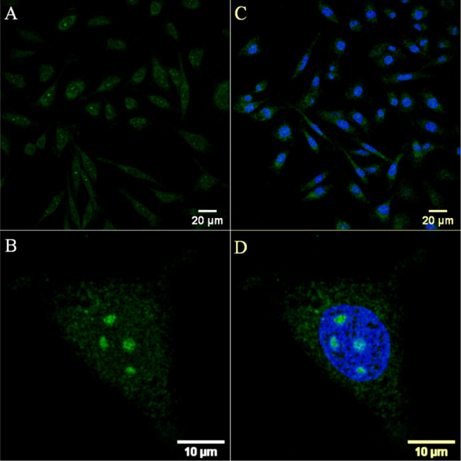
The preparation of two polyarginine conjugates of the complex Os(II) [bis-(4'-(4-carboxyphenyl)-2,2':6',2″-terpyridine)] [Os-(Rn)2]x+ (n = 4 and 8; x = 10 and 18) is reported, to explore whether the R8 peptide sequence that promotes cell uptake requires a contiguous amino acid sequence for membrane permeation or if this can be accomplished in a linearly bridged structure with the additive effect of shorter peptide sequences. The conjugates exhibit NIR emission centered at 754 nm and essentially oxygen-insensitive emission with a lifetime of 89 ns in phosphate-buffered saline. The uptake, distribution, and cytotoxicity of the parent complex and peptide derivatives were compared in 2D cell monolayers and a three-dimensional (3D) multicellular tumor spheroid (MCTS) model. Whereas, the bis-octaarginine sequences were impermeable to cells and spheroids, and the bis-tetraarginine conjugate showed excellent cellular uptake and accumulation in two 2D monolayer cell lines and remarkable in-depth penetration of 3D MCTSs of pancreatic cancer cells. Overall, the data indicates that cell permeability can be promoted via non-contiguous sequences of arginine residues bridged across the metal centre.


Conflict of interest statement

The authors declare no competing financial interest.

Figures

Figure 1
Figure 1
(A) General chemical structure of Os(II)-terpyridine-based polyarginine conjugates following amide coupling of Rn (n = 4 and 8) to [Os(tpybenzCOOH)2]2+ parent complex. (B) Absorbance and normalised emission spectra of [Os-(R4)2]10+ (30 μM/PBS buffer pH 7.4) under aerated and de-aerated conditions with λexc 490 nm and excitation and emission slit widths of 10 nm.
Figure 2
Figure 2
Uptake and co-localization studies of [Os-(R4)2]10+ in live A549 cells where the osmium channel is shown in green. Cells were incubated in the absence of light with 30 μM [Os-(R4)2]10+ for (A) 24 h and for (B,C) 48 h. Co-localization studies at 48 h with Lysotracker Green (50 nM) confirmed lysosomal confinement evident by the overlap of the (D) osmium channel with (E) Lysotracker Green (orange) in the (F) overlay image (Pearson’s coefficient = 0.69). A 490 nm white light laser was used to excite the conjugate, and emission was collected between 650 and 800 nm. The LysoTracker Green dye was excited at 504 nm and emission was collected at 511 nm.
Figure 3
Figure 3
Confocal luminescence images of [Os-(R4)2]10+ in CHO cells. Live cells were incubated with 30 μM [Os-(R4)2]10+ for 24 h in the absence of light and co-stained with DRAQ7. (A,C) The distribution of the conjugate (in green) is shown in a group of cells co-stained with DRAQ7 (in blue). (B,D) Closed-up image of a single A549 cell shows nucleoli staining. The 633 nm laser was used to excite DRAQ 7 and emission was collected between 635 and 750 nm.
Figure 4
Figure 4
Luminescence lifetime imaging [Os-(R4)2]10+ at 30 μM in live A549 cells. (A) Confocal image of a single cell following conjugate uptake at 24 h and (B) lifetime distribution in the expanded cytoplasmic region of the cell. (C) PLIM acquired following uptake at 48 h. The PLIM images were acquired using the 405 nm excitation laser line. The PLIM images of the entire cell of (B) and (C) and corresponding emission decays are shown in the Supporting Information (Figures S19–S20).
Figure 5
Figure 5
3D reconstruction depth coding images of whole live HPAC spheroids treated with [Os-(R4)2]10+ at (A) 30 μM/24 h and (B) 100 μM/24 h. Confocal images were acquired at different planes in the z direction throughout the spheroids (from the bottom to above each spheroid). A 490 nm white light laser was used to excite the conjugate, and emission was collected between 650 and 800 nm. Scale bar reads 100 μm.
Figure 6
Figure 6
Z-stack images of a single live HPAC spheroid pre-treated with [Os-(R4)2]10+ (100 μM/48 h) and co-stained with DAPI (10 μM). Each image corresponds to the cross section from the bottom to the upper part along the z-axis. Representative cross sections are shown using bright-field contrast as the background. Scale bar reads 100 μm. A 490 nm white light laser was used to excite the conjugate, and emission was collected between 650 and 800 nm. The 405 nm excitation laser was used to excite DAPI, and emission was collected between 423 and 580 nm.
Figure 7
Figure 7
Confocal images (2D projection) of HPAC spheroid regions treated with Os-(R4)2 at (A) 30 μM/24 h, (B) 100 μM/24 h, and (C) 100 μM/48 h at 37 °C. The spheroids were co-stained with DAPI (10 μM) and (D–F) overlay images with DAPI channel. A 490 nm white light laser was used to excite the conjugate, and emission was collected between 650 and 800 nm. The 405 nm excitation laser was used to excite DAPI, and emission was collected between 423 and 580 nm (40× obj.).
Figure 8
Figure 8
Confocal images of a single live HPAC spheroid treated with [Os-(R4)2]10+ (100 μM/48 h). A 490 nm white light laser was used for excitation, and emission was collected between (A) 650 and 800 nm; Os(II) channel and (B) 500–570 nm; autofluorescence window. (C) Os(II)/autofluorescence channel overlay.

References

    1. Dolan C.; Burke C. S.; Byrne A.; Keyes T. E.. Cellular Uptake and Sensing Capability of Transition Metal Peptide Conjugates. In Inorganic and Organometallic Transition Metal Complexes with Biological Molecules and Living Cell; Elsevier, 2017, pp 55–89.
    1. Lo K. K.-W. Luminescent Rhenium(I) and Iridium(III) Polypyridine Complexes as Biological Probes, Imaging Reagents, and Photocytotoxic Agents. Acc. Chem. Res. 2015, 48, 2985–2995. 10.1021/acs.accounts.5b00211. - DOI - PubMed
    1. Sowell J.; Strekowski L.; Patonay G. DNA and Protein Applications of Near-Infrared Dyes. J. Biomed. Opt. 2002, 7, 571.10.1117/1.1502262. - DOI - PubMed
    1. Hemmer E.; Benayas A.; Légaré F.; Vetrone F. Exploiting the Biological Windows: Current Perspectives on Fluorescent Bioprobes Emitting above 1000 Nm. Nanoscale Horiz. 2016, 1, 168–184. 10.1039/C5NH00073D. - DOI - PubMed
    1. Welsher K.; Sherlock S. P.; Dai H. Deep-Tissue Anatomical Imaging of Mice Using Carbon Nanotube Fluorophores in the Second near-Infrared Window. Proc. Natl. Acad. Sci. U. S. A. 2011, 108, 8943–8948. 10.1073/pnas.1014501108. - DOI - PMC - PubMed

MeSH terms

LinkOut - more resources