Review
. 2021 Jun 7;218(6):e20210518.
doi: 10.1084/jem.20210518. Epub 2021 May 12.

Ferroptosis in infection, inflammation, and immunity

Affiliations
Review

Ferroptosis in infection, inflammation, and immunity

Xin Chen et al. J Exp Med. .

Abstract

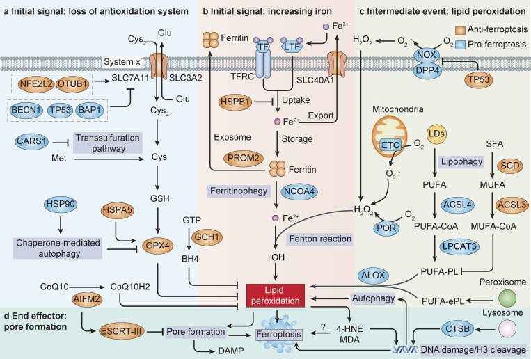
Ferroptosis is a type of regulated necrosis that is triggered by a combination of iron toxicity, lipid peroxidation, and plasma membrane damage. The upstream inducers of ferroptosis can be divided into two categories (biological versus chemical) and activate two major pathways (the extrinsic/transporter versus the intrinsic/enzymatic pathways). Excessive or deficient ferroptotic cell death is implicated in a growing list of physiological and pathophysiological processes, coupled to a dysregulated immune response. This review focuses on new discoveries related to how ferroptotic cells and their spilled contents shape innate and adaptive immunity in health and disease. Understanding the immunological characteristics and activity of ferroptotic death not only illuminates an intersection between cell death and immunity but may also lead to the development of novel treatment approaches for immunopathological diseases.


Conflict of interest statement

Disclosures: G. Kroemer reported grants from Daiichi Sankyo, Eleor, Kaleido, Lytix Pharma, PharmaMar, Samsara, Sanofi, Vascage, and Vasculox outside the submitted work; additionally, G. Kroemer is on the Board of Directors of the Bristol Myers Squibb Foundation France and is a scientific co-founder of everImmune, Samsara Therapeutics, and Therafast Bio. No other disclosures were reported.

Figures

Figure 1.
Figure 1.
Core mechanisms of ferroptosis. (a and b) The initiation of ferroptosis requires two key signals, namely the inhibiting antioxidant SLC7A11-GSH-GPX4 system and the accumulation of free iron. This process is further regulated by epigenetic, transcriptional, and posttranslational mechanisms. For example, the expression and activity of SLC7A11 is regulated by protein–protein interaction (e.g., by BECN1), gene expression (e.g., by NFE2L2, TP53, and BAP1), and protein stabilization (e.g., by OTUB1), whereas autophagy promotes ferroptosis partly by degrading GPX4 or ferritin. Alternatively, CoQ10 or tetrahydrobiopterin (BH4) inhibits ferroptosis independently of GSH. (c) The generation of polyunsaturated phospholipid (by ACSL4 and LPCAT3) or PUFA-ePL (by peroxisomal enzymes) and subsequent activation of ALOX have a main role in promoting lipid peroxidation. This process requires hydrogen peroxide (H2O2) production from an iron-mediated Fenton reaction or the activation of POR, NOX, or mitochondria electron transport chain pathways. (d) At the final step of ferroptosis, lipid peroxidation or its secondary products (e.g., 4-HNE and MDA) directly or indirectly induce pore formation in plasma or organelle membrane, which eventually triggers cell death. The lysosomal enzyme CTSB mediates ferroptosis by transporting to the nucleus and inducing DNA damage or histone H3 cleavage. In contrast, endosomal sorting complex required for transport-III (ESCRT-III)–mediated membrane repair prevents ferroptotic cell death and/or DAMP release.
Figure 2.
Figure 2.
Models of ferroptosis in innate immune cells. (a) Ferroptosis in macrophages. The activation of the SLC7A110-GSH-GPX4 axis or an increase in iron storage protein ferritin by nuclear receptor coactivator 4–mediated ferritinophagy prevents lipid peroxidation in macrophages. In contrast, erythrophagocytosis or exogenous ferric ions can trigger iron-dependent ferroptosis in macrophages. Compared with M2 microglia/macrophages, M1 cells are resistant to ferroptosis due to the loss of ALOX15 activity by NOS2-mediated NO production. The released DAMPs (e.g., HMGB1, KRASG12D, and 8-OHG) by ferroptotic cancer cells cause inflammation-related immunosuppression through AGER- or STING1-mediated macrophage polarization. (b) Ferroptosis in neutrophils. The activation of NOX and PADI4 is essential for NET formation in neutrophils. The release of myeloperoxidase (MPO) by neutrophils through NET induces lipid peroxidation and subsequent ferroptosis in glioblastoma cells.
Figure 3.
Figure 3.
Models of ferroptosis in adaptive immune cells. (a) Ferroptosis in T cells. SLC7A11, GPX4, or AIFM2 inhibits, whereas ACSL4 and CD36 promotes, ferroptosis in CD8+ T cells. IFNG released by CD8+ T cells induces ferroptotic tumor cell death by activating STAT3-dependent down-regulation of system xc subunit (SLC3A2 and SLC7A11) expression. In addition, ferroptotic cancer cells activate CD8+ T cell–mediated immunogenic cell death by releasing DAMPs (e.g., HMGB1 and ATP). (b) Ferroptosis in B cells. The depletion of GPX4 induces ferroptotic cell death and impairs IgM antibody responses in B1 and MZB cells. CD36 increases the absorption of fatty acids and the sensitivity to ferroptosis of B1 and MZB cells, and the degradation of lipid droplets (LDs) caused by lipophagy may promote ferroptosis by increasing the concentration of free fatty acids within cells. Lipid peroxidation promotes human peripheral blood mononuclear cell proliferation and differentiation into B cells and natural killer cells by inhibiting BMPs.
Figure 4.
Figure 4.
Models of DAMPs and PRRs in ferroptosis. (a) TLR4 pathway in ferroptosis. TLR4 plays a central role in innate immunity by signaling to adaptor MYD88 or TICAM1/TRIF to induce proinflammatory cytokines. The activation of TLR4 induces type I IFN signaling in vascular endothelial cells, triggering neutrophil recruitment and subsequent ferroptosis-mediated cardiac damage. TLR4-dependent NF-κB activation promotes ferroptosis-related inflammation through the production of proinflammatory cytokines (cytokines C-C motif chemokine ligand 2 [CCL2], TNF, and TGFB1) during rhabdomyolysis-associated kidney damage. Moreover, lipid oxidation products (e.g., 4-HNE and oxPLs) can trigger inflammation partly through activating TLR4 signaling. The interaction of TLR4 and NOX4 may enhance ferroptotic cell death–mediated inflammation. (b) AGER signaling in ferroptosis. AGER recognizes various DAMPs, leading to the activation of signaling pathways, including the protein kinase C [PKC], JAK-STAT, phophatidylinositol 3-kinase [PI3K]–protein kinase B, and MAPK–NF-κB pathways. In addition, AGER is responsible for HMGB1- or KRASG12D-mediated macrophage activation and polarization in response to ferroptotic cancer cells. (c) STING1 pathway in ferroptosis. In this pathway, 4-HNE inhibits STING1 activity by the carbonylation of STING1, whereas the release of 8-OHG from ferroptotic cancer cells activates the STING1-dependent inflammatory pathway in macrophages by the DNA sensor CGAS. STING1 directly promotes ferroptosis by increasing MAP1LC3 lipidation, thereby activating autophagy-dependent cell death caused by zalcitabine-induced mitochondrial DNA (mtDNA) damage and CTSB-mediated genomic DNA damage.

References

    1. Amaral, E.P., Conceição E.L., Costa D.L., Rocha M.S., Marinho J.M., Cordeiro-Santos M., D’Império-Lima M.R., Barbosa T., Sher A., and Andrade B.B.. 2016. N-acetyl-cysteine exhibits potent anti-mycobacterial activity in addition to its known anti-oxidative functions. BMC Microbiol. 16:251. 10.1186/s12866-016-0872-7 - DOI - PMC - PubMed
    1. Amaral, E.P., Costa D.L., Namasivayam S., Riteau N., Kamenyeva O., Mittereder L., Mayer-Barber K.D., Andrade B.B., and Sher A.. 2019. A major role for ferroptosis in Mycobacterium tuberculosis-induced cell death and tissue necrosis. J. Exp. Med. 216:556–570. 10.1084/jem.20181776 - DOI - PMC - PubMed
    1. Anthonymuthu, T.S., Tyurina Y.Y., Sun W.Y., Mikulska-Ruminska K., Shrivastava I.H., Tyurin V.A., Cinemre F.B., Dar H.H., VanDemark A.P., Holman T.R., et al. . 2021. Resolving the paradox of ferroptotic cell death: Ferrostatin-1 binds to 15LOX/PEBP1 complex, suppresses generation of peroxidized ETE-PE, and protects against ferroptosis. Redox Biol. 38:101744. 10.1016/j.redox.2020.101744 - DOI - PMC - PubMed
    1. Baba, Y., Higa J.K., Shimada B.K., Horiuchi K.M., Suhara T., Kobayashi M., Woo J.D., Aoyagi H., Marh K.S., Kitaoka H., and Matsui T.. 2018. Protective effects of the mechanistic target of rapamycin against excess iron and ferroptosis in cardiomyocytes. Am. J. Physiol. Heart Circ. Physiol. 314:H659–H668. 10.1152/ajpheart.00452.2017 - DOI - PMC - PubMed
    1. Badgley, M.A., Kremer D.M., Maurer H.C., DelGiorno K.E., Lee H.J., Purohit V., Sagalovskiy I.R., Ma A., Kapilian J., Firl C.E.M., et al. . 2020. Cysteine depletion induces pancreatic tumor ferroptosis in mice. Science. 368:85–89. 10.1126/science.aaw9872 - DOI - PMC - PubMed

Publication types