

Persisting Salivary IgG Against SARS-CoV-2 at 9 Months After Mild COVID-19: A Complementary Approach to Population Surveys
- PMID: 33978762
- PMCID: PMC8244549
- DOI: 10.1093/infdis/jiab256
Persisting Salivary IgG Against SARS-CoV-2 at 9 Months After Mild COVID-19: A Complementary Approach to Population Surveys
Erratum in
-
Correction to: Persisting Salivary IgG Against SARS-CoV-2 at 9 Months After Mild COVID-19: A Complementary Approach to Population Surveys.J Infect Dis. 2023 Feb 14;227(4):603. doi: 10.1093/infdis/jiac121. J Infect Dis. 2023. PMID: 35859351 Free PMC article. No abstract available.
Abstract
Background: Declining humoral immunity in coronavirus disease 2019 (COVID-19) patients and possible reinfection have raised concern. Mucosal immunity, particularly salivary antibodies, may be short lived although long-term studies are lacking.
Methods: Using a multiplex bead-based array platform, we investigated antibodies specific to severe acute respiratory syndrome coronavirus 2 (SARS-CoV-2) proteins in 256 saliva samples from convalescent patients 1-9 months after symptomatic COVID-19 (n = 74, cohort 1), undiagnosed individuals with self-reported questionnaires (n = 147, cohort 2), and individuals sampled prepandemic (n = 35, cohort 3).
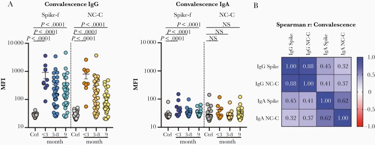
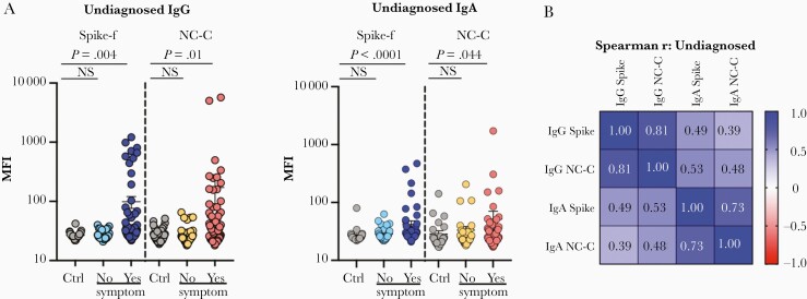
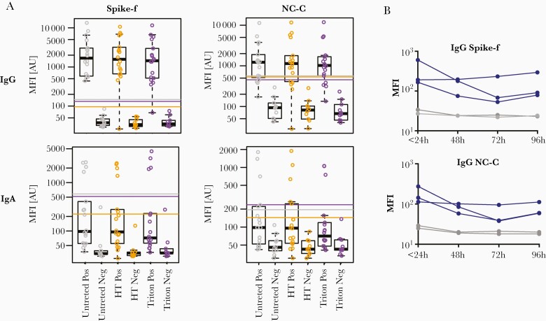
Results: Salivary IgG antibody responses in cohort 1 (mainly mild COVID-19) were detectable up to 9 months postrecovery, with high correlations between spike and nucleocapsid specificity. At 9 months, IgG remained in blood and saliva in most patients. Salivary IgA was rarely detected at this time point. In cohort 2, salivary IgG and IgA responses were significantly associated with recent history of COVID-19-like symptoms. Salivary IgG tolerated temperature and detergent pretreatments.
Conclusions: Unlike SARS-CoV-2 salivary IgA that appeared short lived, specific saliva IgG appeared stable even after mild COVID-19, as for blood serology. This noninvasive saliva-based SARS-CoV-2 antibody test with home self-collection may be a complementary alternative to conventional blood serology.
Keywords: COVID-19; antibody; convalescence; immunoassay; saliva; serology.
© The Author(s) 2021. Published by Oxford University Press for the Infectious Diseases Society of America.
Figures
Similar articles
-
A Multiplex Noninvasive Salivary Antibody Assay for SARS-CoV-2 Infection and Its Application in a Population-Based Survey by Mail.Microbiol Spectr. 2021 Oct 31;9(2):e0069321. doi: 10.1128/Spectrum.00693-21. Epub 2021 Sep 15. Microbiol Spectr. 2021. PMID: 34523986 Free PMC article.
-
COVID-19 Serology at Population Scale: SARS-CoV-2-Specific Antibody Responses in Saliva.J Clin Microbiol. 2020 Dec 17;59(1):e02204-20. doi: 10.1128/JCM.02204-20. Print 2020 Dec 17. J Clin Microbiol. 2020. PMID: 33067270 Free PMC article.
-
Salivary and serum IgA and IgG responses to SARS-CoV-2-spike protein following SARS-CoV-2 infection and after immunization with COVID-19 vaccines.Allergy Asthma Proc. 2022 Sep 1;43(5):419-430. doi: 10.2500/aap.2022.43.220045. Allergy Asthma Proc. 2022. PMID: 36065108 Free PMC article.
-
Salivary diagnostics of the novel coronavirus SARS-CoV-2 (COVID-19).Oral Dis. 2022 Apr;28 Suppl 1(Suppl 1):867-877. doi: 10.1111/odi.13729. Epub 2020 Dec 4. Oral Dis. 2022. PMID: 33211392 Free PMC article. Review.
-
The Use of Saliva as a Biosample in the Light of COVID-19.Diagnostics (Basel). 2021 Sep 26;11(10):1769. doi: 10.3390/diagnostics11101769. Diagnostics (Basel). 2021. PMID: 34679467 Free PMC article. Review.
Cited by
-
Persistence of salivary antibody responses after COVID-19 vaccination is associated with oral microbiome variation in both healthy and people living with HIV.Front Immunol. 2023 Jan 10;13:1079995. doi: 10.3389/fimmu.2022.1079995. eCollection 2022. Front Immunol. 2023. PMID: 36703980 Free PMC article. Clinical Trial.
-
Blood and saliva SARS-CoV-2 antibody levels in self-collected dried spot samples.Med Microbiol Immunol. 2022 Aug;211(4):173-183. doi: 10.1007/s00430-022-00740-x. Epub 2022 Jun 13. Med Microbiol Immunol. 2022. PMID: 35697945 Free PMC article.
-
Guardians of the oral and nasopharyngeal galaxy: IgA and protection against SARS-CoV-2 infection.Immunol Rev. 2022 Aug;309(1):75-85. doi: 10.1111/imr.13118. Epub 2022 Jul 11. Immunol Rev. 2022. PMID: 35815463 Free PMC article. Review.
-
Differences in systemic and mucosal SARS-CoV-2 antibody prevalence in a prospective cohort of Dutch children.Front Immunol. 2022 Sep 9;13:976382. doi: 10.3389/fimmu.2022.976382. eCollection 2022. Front Immunol. 2022. PMID: 36159841 Free PMC article.
-
Multiplex Antibody Analysis of IgM, IgA and IgG to SARS-CoV-2 in Saliva and Serum From Infected Children and Their Close Contacts.Front Immunol. 2022 Jan 27;13:751705. doi: 10.3389/fimmu.2022.751705. eCollection 2022. Front Immunol. 2022. PMID: 35154094 Free PMC article.
References
-
- Johns Hopkins University. Coronavirus resource center. https://coronavirus.jhu.edu/map.html. Accessed 13 March 2021.
-
- Krammer F. SARS-CoV-2 vaccines in development. Nature 2020; 586:516–27. - PubMed
Publication types
MeSH terms
Substances
Grants and funding
LinkOut - more resources
Full Text Sources
Other Literature Sources
Medical
Miscellaneous
