Review
. 2021 Nov;51(12):2284-2302.
doi: 10.1007/s00247-020-04867-y. Epub 2021 May 12.

Contrast-enhanced ultrasound of transplant organs - liver and kidney - in children

Affiliations
Review

Contrast-enhanced ultrasound of transplant organs - liver and kidney - in children

Doris Franke et al. Pediatr Radiol. 2021 Nov.

Abstract

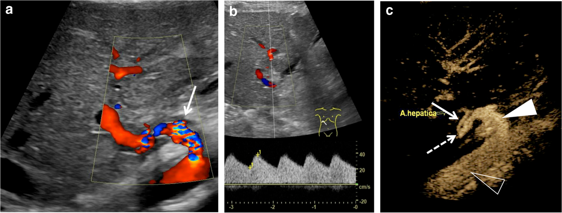
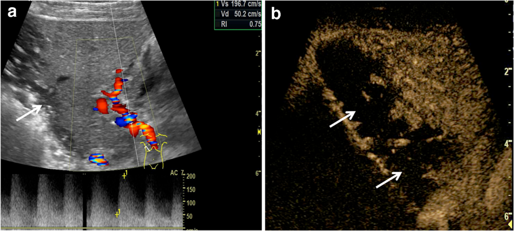
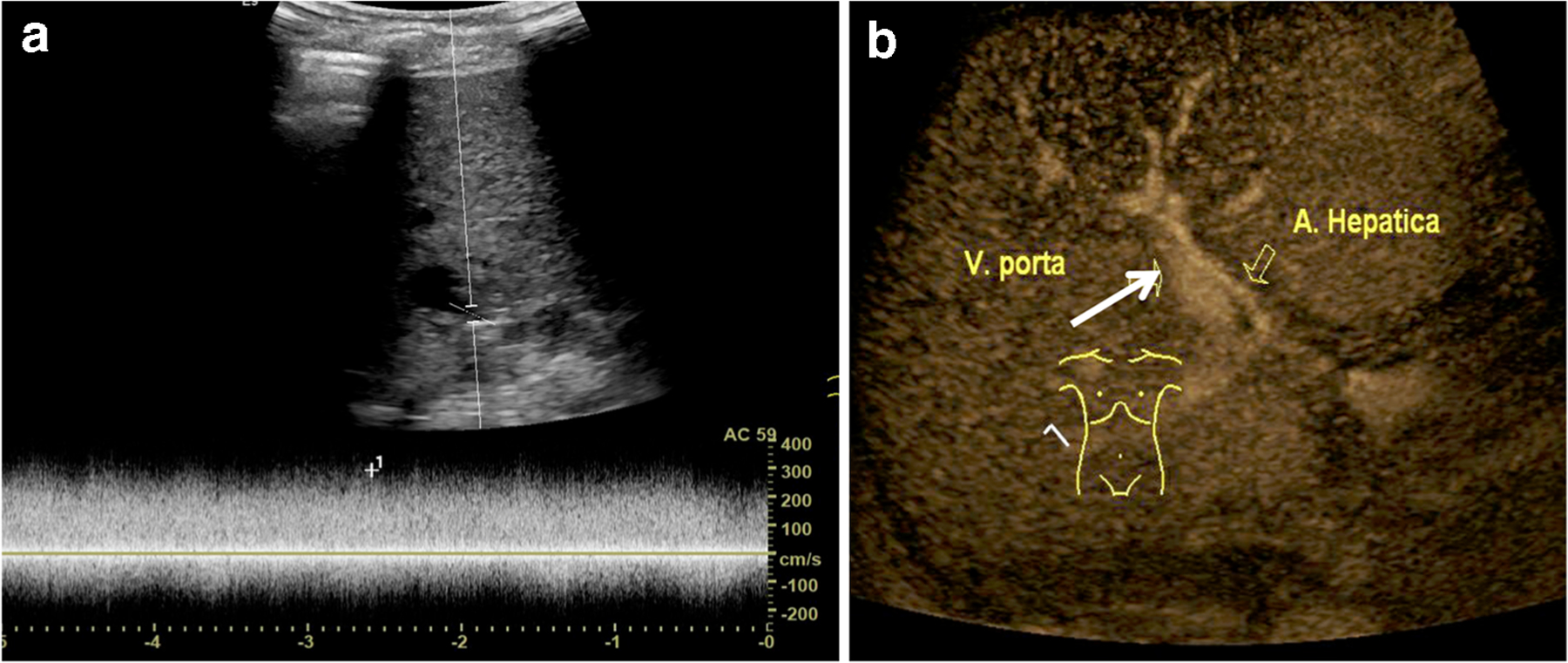
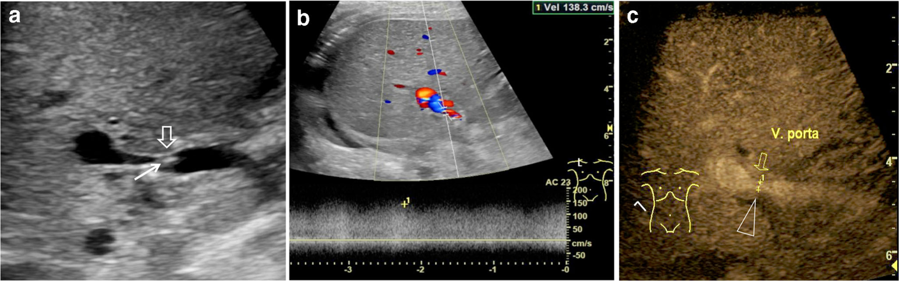
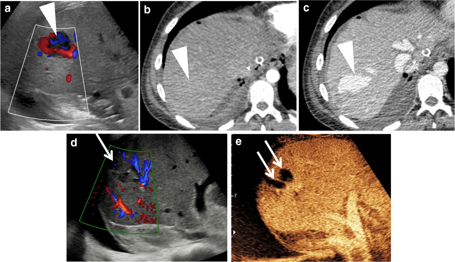
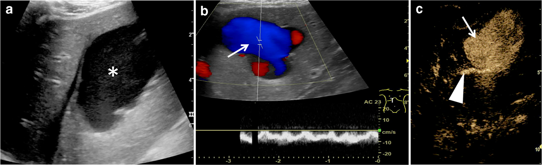
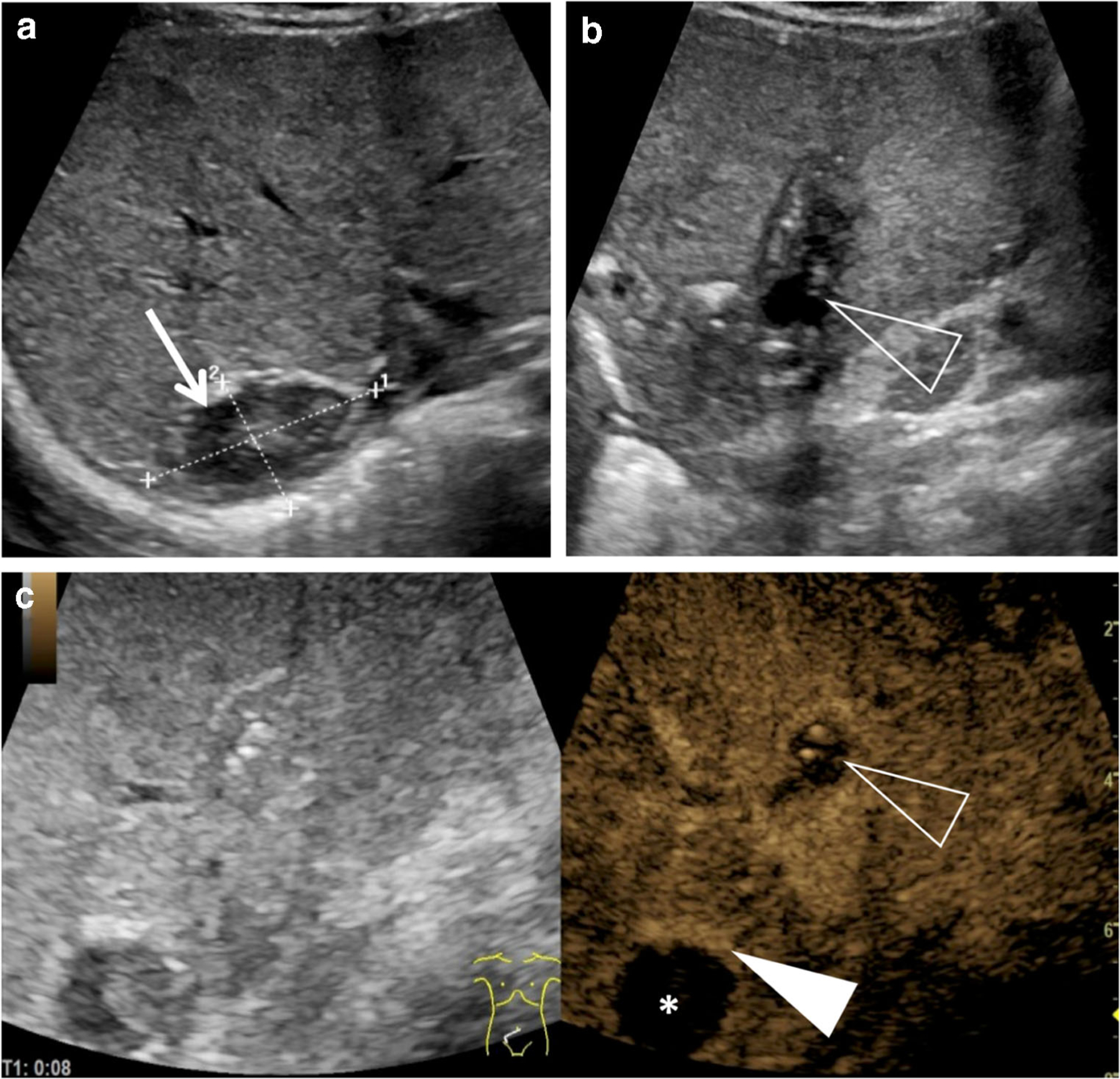
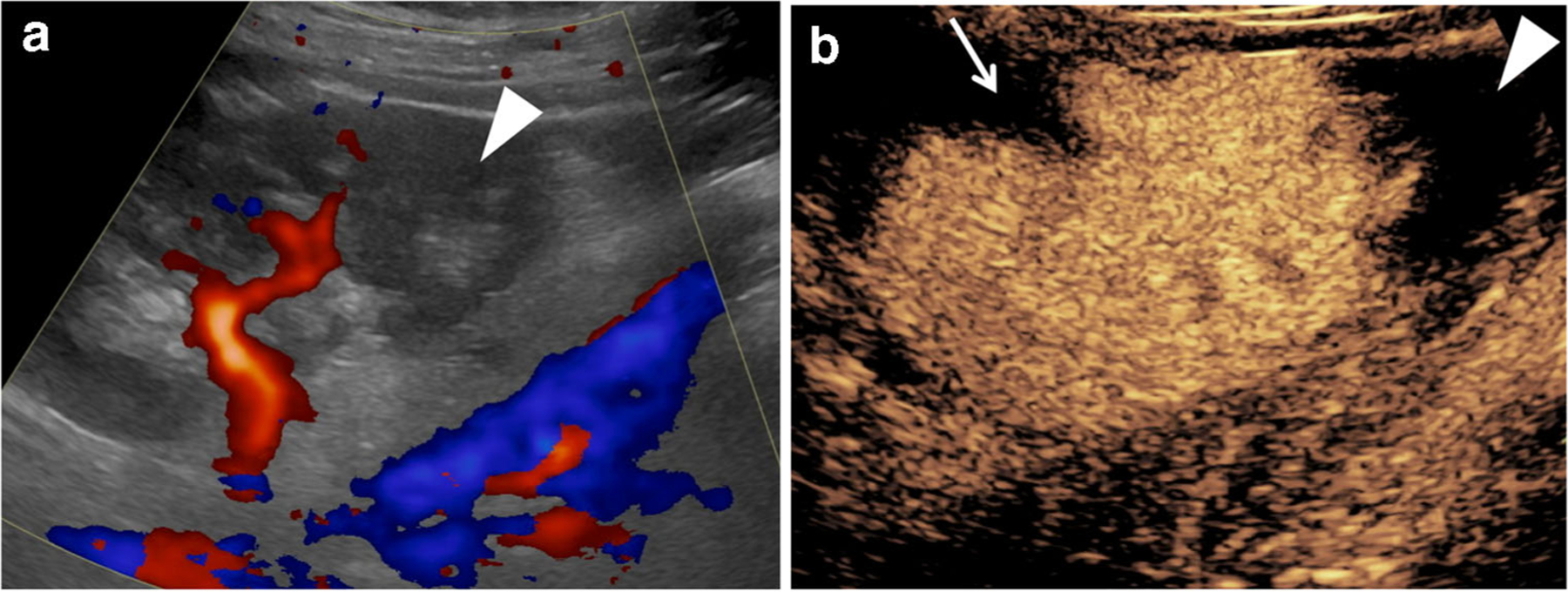
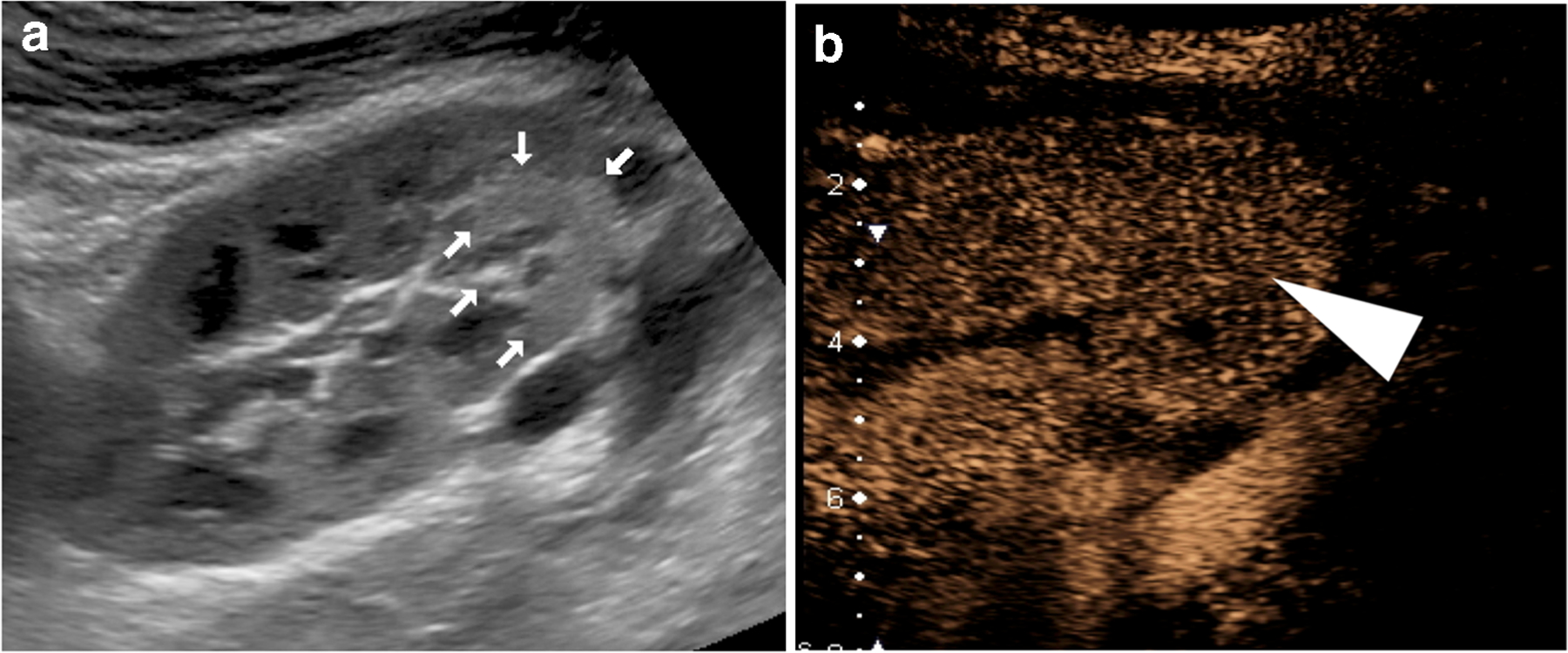
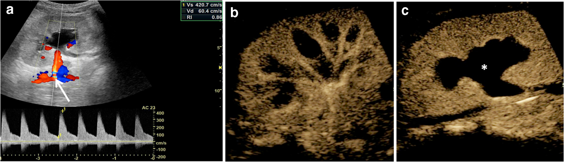
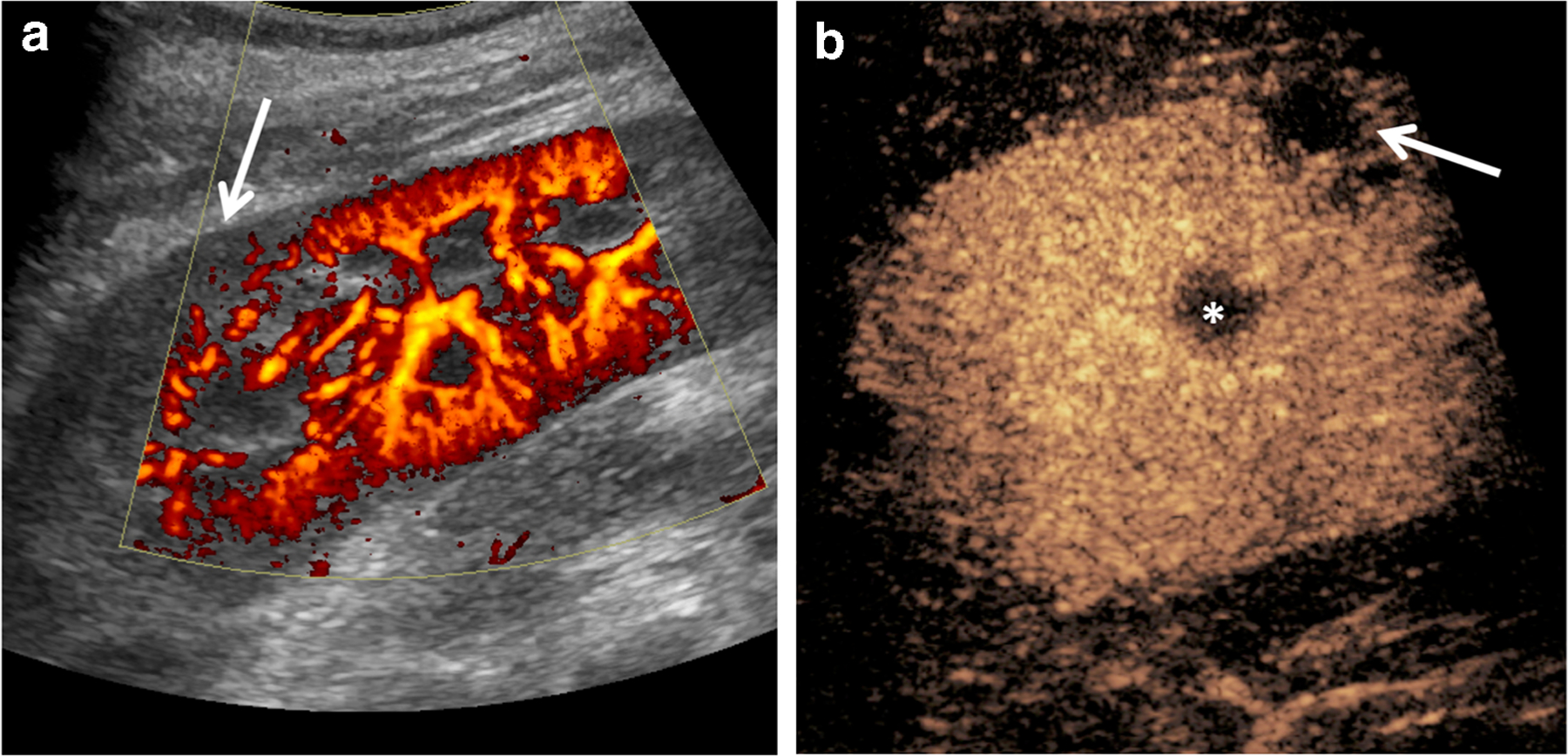
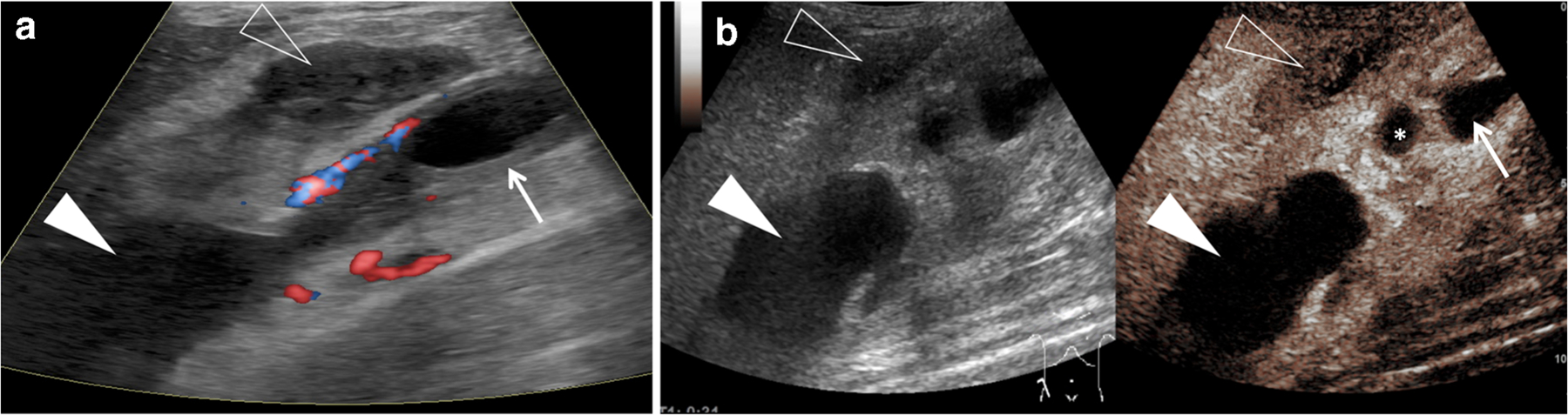
Ultrasound (US) is the first-line imaging tool for evaluating liver and kidney transplants during and after the surgical procedures. In most patients after organ transplantation, gray-scale US coupled with color/power and spectral Doppler techniques is used to evaluate the transplant organs, assess the patency of vascular structures, and identify potential complications. In technically difficult or inconclusive cases, however, contrast-enhanced ultrasound (CEUS) can provide prompt and accurate diagnostic information that is essential for management decisions. CEUS is indicated to evaluate for vascular complications including vascular stenosis or thrombosis, active bleeding, pseudoaneurysms and arteriovenous fistulas. Parenchymal indications for CEUS include evaluation for perfusion defects and focal inflammatory and non-inflammatory lesions. When transplant rejection is suspected, CEUS can assist with prompt intervention by excluding potential underlying causes for organ dysfunction. Intracavitary CEUS applications can evaluate the biliary tract of a liver transplant (e.g., for biliary strictures, bile leak or intraductal stones) or the urinary tract of a renal transplant (e.g., for urinary obstruction, urine leak or vesicoureteral reflux) as well as the position and patency of hepatic, biliary and renal drains and catheters. The aim of this review is to present current experience regarding the use of CEUS to evaluate liver and renal transplants, focusing on the examination technique and interpretation of the main imaging findings, predominantly those related to vascular complications.

Keywords: Children; Complications; Contrast-enhanced ultrasound; Intracavitary; Intravenous; Kidney; Liver; Transplant; Ultrasound; Ultrasound contrast agents.


Conflict of interest statement

Conflicts of interest None

Figures

Fig. 1
Fig. 1
A 13-year-old girl with a full-liver graft from a deceased donor. Transverse contrast-enhanced ultrasound (CEUS) of the liver graft, contrast-only mode in gray-scale display, shows echogenic microbubbles within the patent portal vein (arrowhead) and patent hepatic artery (arrow)
Fig. 2
Fig. 2
Transient vasospasm of the right hepatic artery in a 3-year-old boy with full-liver graft from a deceased donor. a, b Transverse intraoperative spectral Doppler US of the liver graft shows decreased flow velocities with a tardus parvus waveform in the right hepatic artery (RHA) (a). Velocities and waveform in the left hepatic artery (LHA) are within normal limits (b). c, d Contrast-enhanced ultrasound (CEUS) performed immediately after Doppler US in contrast-only mode. Transverse views of the liver graft show contrast microbubbles within the RHA (solid arrowhead) and LHA (open arrowhead), suggestive of patency (c). There is homogeneous opacification of the liver parenchyma (asterisk) with no evidence of focal perfusion defects to suggest infarct (d)
Fig. 3
Fig. 3
Biliary atresia and full-liver graft from a deceased donor in a 7-month-old girl. a Contrast-enhanced ultrasound (CEUS) with simultaneous display of gray-scale (left) and contrast-enhanced (right) modes. Transverse view of the liver graft 11 s post contrast injection shows enhancement of the portal vein (arrow). Absence of enhancement is evident in the expected location of the hepatic artery (arrowhead). There is no enhancement of the liver parenchyma. b Transverse CEUS, contrast-only mode at 1 min 41 s post contrast injection, shows enhancement of the portal vein (arrow) with persistent absence of enhancement in the anatomical location of the hepatic artery (arrowhead), indicating hepatic artery thrombosis. A large area of parenchymal non-enhancement (asterisk) is evident within the right lobe of the transplant in keeping with an infarct
Fig. 4
Fig. 4
Thrombosis of the hepatic artery and portal vein in a 1-year-old girl with liver failure of unknown origin after full-liver graft from a deceased donor. Transverse contrast-enhanced ultrasound (CEUS) with simultaneous display of gray-scale (left) and contrast-enhanced (right) modes of the liver graft, 43 s post contrast injection, shows that the three hepatic veins are patent (arrows) by demonstrating enhancement. This is reduced to nearly absent enhancement of the liver parenchyma (asterisk), indicating liver failure
Fig. 5
Fig. 5
Full-liver graft from a deceased donor in a 14-year-old girl with progressive familial intrahepatic cholestasis Type II. a Color Doppler US of the liver graft in sagittal plane shows aliasing effect within the hepatic artery (arrow). b Spectral Doppler US in oblique sagittal plane shows tardus parvus waveform in a peripheral branch of the left hepatic artery that is highly suspicious for a high-grade stenosis proximally. c Contrast-enhanced ultrasound (CEUS), contrast-only mode in sagittal plane during early arterial phase (16 s after contrast injection). Note enhancement of the aorta (open arrowhead) and of the celiac trunk (solid arrowhead). The hepatic artery (solid arrow) is patent, as confirmed by opacification of its lumen. However, it demonstrates a sharp curve in its course (dashed arrow), with reduced caliber of its lumen distally. This finding explains the spectral Doppler waveform alterations
Fig. 6
Fig. 6
Abnormally high liver enzymes postoperatively in a 7-month-old boy after a right-reduced full-liver graft. a Spectral Doppler US of the liver graft in oblique sagittal plane shows heterogeneous reflectivity at the margins of the split-liver graft (arrow). Aliasing effect is evident within the hepatic artery, which also demonstrates increased flow velocities of nearly 200 cm/s (normal range 70–120 cm/s in school-age children, even lower in infants [61]). b Contrast-enhanced ultrasound (CEUS) in contrast-only mode, oblique sagittal plane, demonstrates an extensive area of non-enhancement (arrows) confined at the resection site of the liver graft, indicative of ischemia
Fig. 7
Fig. 7
High-grade hepatic artery and portal vein stenosis of the liver graft in a 7-month-old girl. a Spectral Doppler US of the liver graft, transverse view, shows increased flow velocity in the portal vein measuring up to 250 cm/s (normal range in healthy-term neonates 24.0±1.3 cm/s, fasting, to 35.9±2.4 cm/s, within 15 min after feeding [61]). b Contrast-enhanced ultrasound (CEUS) in contrast-only mode, transverse view, shows contrast microbubbles within the hepatic artery and portal vein, suggestive of patency. However, a focal narrowing (arrow) of the lumen of the portal vein is suggestive of portal vein stenosis. There is normal perfusion of the liver graft
Fig. 8
Fig. 8
Left lateral split-liver graft in a 10-month old boy with biliary atresia. a Baseline gray-scale US of the liver graft in transverse plane shows a focal narrowing at the anastomotic site of the portal vein, where a highly reflective linear structure (arrows) is seen, presumably representing the suture material. b Spectral Doppler US, transverse plane, shows increased flow velocity within the portal vein, with mean velocity measured at 138 cm/s (normal range in healthy term neonates 24.0±1.3 cm/s, fasting, to 35.9±2.4 cm/s, within 15 min after feeding [61]). c Contrast-enhanced ultrasound (CEUS) in contrast-only mode, transverse view of the liver graft. The portal vein is normally opacified with echogenic microbubbles. CEUS confirms the focal narrowing (arrowhead, arrow) of the portal vein at the anastomotic site. The lumen of the portal vein has been reduced to 2 mm in diameter at the level of the narrowing. Normal diameter of the portal vein is noted proximally and distally to that level. There is normal perfusion of the liver graft
Fig. 9
Fig. 9
Full-liver graft from a deceased donor in an 18-year-old woman with primary biliary cirrhosis. a Color Doppler US of the liver graft, transverse view. Routine post-transplantation US reveals a vascular abnormality (arrowhead) adjacent to the right hepatic vein. b, c CT was subsequently performed for further evaluation. Select axial CT images in arterial (b) and venous (c) phases confirm the vascular origin of the lesion (arrowheads), which demonstrates enhancement only in the venous phase, indicating venous ectasia. Laboratory testing was unremarkable and the woman was managed conservatively. d Follow-up color Doppler US on postoperative Day 5, transverse view. The area of concern (arrow) in the right liver lobe appears significantly smaller in size and hypoechoic centrally, without any color Doppler vascularity. e Contrast-enhanced ultrasound (CEUS), contrast-only mode, transverse view. Corresponding to the area of concern in the right liver lobe, CEUS reveals two small areas (arrows) with complete absence of enhancement in all contrast phases, in keeping with small infarcts from thrombosis of the previously noted venous ectasia. The remaining liver parenchyma enhances homogeneously
Fig. 10
Fig. 10
Large arteriovenous fistula after liver transplant biopsy in a 3-year-old boy. The boy had left lateral split-liver graft from a deceased donor due to acute liver failure of unknown origin. a Baseline gray-scale US of the liver graft performed 4 weeks after liver transplant biopsy, sagittal plane, shows a large anechoic lesion (asterisk) within the liver transplant. b Spectral Doppler US, sagittal plane, shows a mixed-vascularity lesion with presence of a large draining vein centrally (arrow). Imaging findings are in keeping with a large arteriovenous fistula. c Contrast-enhanced ultrasound (CEUS), contrast-only mode, sagittal plane. Examination was performed to better evaluate the feeding and draining vessels of the arteriovenous fistula prior to intervention. Ten seconds post contrast injection, the feeding artery (arrowhead) enhances first and more intensely compared to the larger draining vein (arrow). The arteriovenous fistula spontaneously thrombosed on the day of the planned catheter intervention, leaving the hepatic artery and portal vein of the transplant patent
Fig. 11
Fig. 11
Biliary complications after early hepatic artery thrombosis in an 8-month-old girl with full-liver graft from a deceased donor. The girl was on the waiting list for re-transplantation for more than 3 weeks. a, b Baseline gray-scale US, transverse views of the liver graft. A heterogeneously hypoechoic lesion (arrow in a) is evident in the posterior aspect of the right lobe of the graft. The adjacent bile ducts are dilated and contain echogenic material (arrowhead in b). Imaging findings are suspicious for cholangitis with possible abscess formation. c Contrast-enhanced ultrasound (CEUS) with simultaneous display of gray-scale (left) and contrast-enhanced (right) modes, transverse view. CEUS was performed as a problem-solving imaging modality to clarify the nature of the lesions. The lesion in the posterior right liver lobe demonstrates absence of enhancement centrally (asterisk) with a peripheral rim of enhancement (solid arrowhead), in keeping with an abscess. Note the adjacent dilated bile ducts (open arrowhead) that do not enhance
Fig. 12
Fig. 12
Infarct in a 15-year-old girl on Day 2 post right lower quadrant transplant allograft kidney. a Color Doppler US of the transplant kidney, sagittal plane, shows an area of decreased perfusion (arrowhead) in the lower pole of the transplant kidney that is highly suspicious for infarct. b Contrast-enhanced ultrasound (CEUS), contrast-only mode in sagittal plane, performed on postoperative Day 3. Parenchymal phase of enhancement. Corresponding to the area of concern, there is lack of contrast enhancement in the lower pole of the renal transplant (arrowhead), in keeping with an infarct. A similar second wedge-shaped defect is seen in the middle aspect (arrow) of the transplant, also representing an infarct
Fig. 13
Fig. 13
A 15-year-old boy 2 days after renal transplantation with satisfactory onset of renal transplant function presented for routine scan. a Baseline gray-scale US of the transplant kidney, sagittal view, shows an ill-defined, slightly echogenic area in the lower pole of the transplant kidney (arrows). The allograft contains two renal arteries, one of which could not be visualized on color Doppler postoperatively (not presented here). An early parenchymal infarct in the lower pole could not be ruled out. b Contrast-enhanced ultrasound (CEUS) was performed for further evaluation. CEUS in contrast-only mode, sagittal view, shows homogeneous enhancement in the lower pole of the renal transplant (arrowhead). No evidence of ischemic infarct or other focal lesion is identified
Fig. 14
Fig. 14
First episode of urosepsis 4 weeks after renal transplantation in a 4-year-old girl with Mayer–Rokitansky–Küster–Hauser syndrome. Vascular reconstruction was performed with anastomosis of the graft renal artery to the recipient common iliac artery. a Baseline pulsed-wave Doppler US of the transplant kidney, sagittal view, shows aliasing effect at the vascular anastomosis site and markedly accelerated flow velocities of the graft renal artery (arrow), with a mean peak systolic flow velocity measured at 420 cm/s and a mean diastolic flow velocity of 60 cm/s (normal range in toddlers for peak systolic flow velocity 71.3±13.5 cm/s and for diastolic flow velocity 20.3±6.0 cm/s [67]). The resistive index is elevated at 0.86 (normal range for toddlers 0.71±0.008). b, c Contrast-enhanced ultrasound (CEUS), contrast-only mode, sagittal views of the transplant kidney 8 s (b) and 18 s (c) after injection of the contrast agent show homogeneous enhancement of the cortex and medulla. There is no evidence of parenchymal infarcts. The dilated urinary pelvis (asterisk) and proximal ureter show no enhancement
Fig. 15
Fig. 15
Urosepsis, elevated C-reactive protein (CRP) of 350 mg/L (normal range up to 5 mg/L), high fever and low blood pressure in a 10-year-old girl with bilateral dysplastic native kidneys and a right renal transplant. a Power Doppler US of the transplant kidney in sagittal plane reveals an equivocal area of decreased corticomedullary differentiation in the middle aspect of the renal transplant, with reduced vascularity in the region of concern (arrow). b Contrast-enhanced ultrasound (CEUS), contrast-only mode in transverse view, 41 s after contrast injection. Corresponding to the US findings, a poorly demarcated wedge-shaped lesion (arrow) of reduced enhancement is noted in the periphery of the cortex in the middle aspect of the transplant. Given the clinical context, this lesion is in keeping with an abscess. The small non-enhancing structure (asterisk) central within the renal transplant corresponds to the partially visualized renal pelvis. There is normal homogeneous enhancement of the remaining renal parenchyma
Fig. 16
Fig. 16
Urinary tract dilatation and urosepsis in a 14-year-old boy with a history of posterior urethral valves and renal transplant. a Baseline color Doppler US of the transplant kidney, oblique sagittal view, shows marked thickening of the uroepithelium of the dilated proximal ureter (arrow). There are a large anechoic lesion in the upper pole of the renal transplant (solid arrowhead) and a second, smaller hypoechoic lesion (open arrowhead) along the lateral aspect of the transplant. Neither lesion demonstrates internal vascularity on color Doppler US. b Contrast-enhanced US with simultaneous display of gray-scale (left) and contrast-enhanced (right) modes, oblique sagittal view. Corresponding to the US findings, there is hypoenhancement of both lesions (arrowheads) with a subtle ill-defined peripheral rim of enhancement around the smaller lateral lesion. These findings are in keeping with renal abscesses. The renal abscesses were thought to originate from the renal cortex and extend into the perirenal space laterally. The proximal ureter (arrow) and a lower pole calyx (asterisk) remain non-enhancing

Similar articles

Cited by

References

    1. Piardi T, Lhuaire M, Bruno O et al. (2016) Vascular complications following liver transplantation: a literature review of advances in 2015. World J Hepatol 8:36–57 - PMC - PubMed
    1. Nixon JN, Biyyam DR, Stanescu L et al. (2013) Imaging of pediatric renal transplants and their complications: a pictorial review. Radiographics 33:1227–1251 - PubMed
    1. Friedewald SM, Molmenti EP, Friedewald JJ et al. (2005) Vascular and nonvascular complications of renal transplants: sonographic evaluation and correlation with other imaging modalities, surgery, and pathology. J Clin Ultrasound 33:127–139 - PubMed
    1. Camacho JC, Coursey-Moreno C, Telleria JC et al. (2015) Nonvascular post-liver transplantation complications: from US screening to cross-sectional and interventional imaging. Radiographics 35:87–104 - PubMed
    1. Kim N, Juarez R, Levy AD (2018) Imaging non-vascular complications of renal transplantation. Abdom Radiol 43:2555–2563 - PubMed

LinkOut - more resources