Review
. 2021 Nov;51(12):2214-2228.
doi: 10.1007/s00247-020-04868-x. Epub 2021 May 12.

Contrast-enhanced ultrasound of the pediatric bowel

Affiliations
Review

Contrast-enhanced ultrasound of the pediatric bowel

Ami Gokli et al. Pediatr Radiol. 2021 Nov.

Abstract

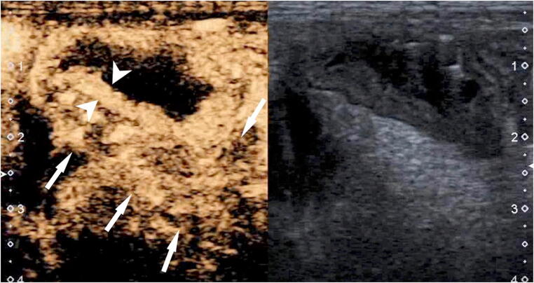
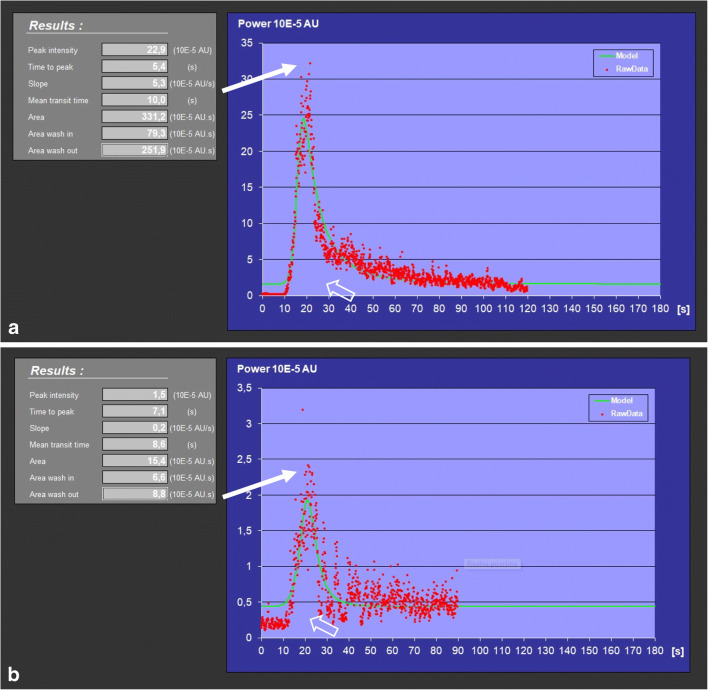
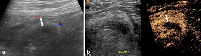
Contrast-enhanced ultrasound (CEUS) has emerged as a valuable modality for bowel imaging in adults and children. CEUS enables visualization of the perfusion of the bowel wall and of the associated mesentery in healthy and disease states. In addition, CEUS images can be used to make quantitative measurements of contrast kinetics, allowing for objective assessment of bowel wall enhancement. Bowel CEUS is commonly applied to evaluate inflammatory bowel disease and to monitor treatment response. It has also been applied to evaluate necrotizing enterocolitis, intussusception, appendicitis and epiploic appendagitis, although experience with these applications is more limited. In this review article, we present the current experience using CEUS to evaluate the pediatric bowel with emphasis on inflammatory bowel disease, extrapolating the established experience from adult studies. We also discuss emerging applications of CEUS as an adjunct or problem-solving tool for evaluating bowel perfusion.

Keywords: Bowel; Children; Contrast-enhanced ultrasound; Crohn disease; Inflammatory bowel disease; Ultrasound; Ultrasound contrast agents.


Conflict of interest statement

Dr. Dillman receives grant funding from Bracco Diagnostics.

Figures

Fig. 1
Fig. 1
Crohn disease and sigmoid wall thickening on baseline US examination in an 11-year-old girl. Contrast-enhanced ultrasound (CEUS) examination of the bowel. Dual image with simultaneous side-by-side display of the contrast-enhanced (left) and gray-scale (right) modes. Longitudinal still image captured from a CEUS video clip demonstrates significant mural thickening (arrowheads) with transmural hyperenhancement and marked enhancement of the surrounding mesentery (arrows), consistent with active disease
Fig. 2
Fig. 2
Quantitative contrast-enhanced ultrasound (CEUS). a–c Time-intensity curves from three children with Crohn disease and variable severity of inflammatory bowel changes: a 13-year-old boy (a), an 11-year-old girl (b) and a 10-year-old girl (c). All bowel CEUS examinations were performed on the same US unit and the log-converted time-intensity curves were generated off-line using the same quantification contrast software. Regions of interest were placed within the thickened bowel wall, which was imaged in longitudinal plane. The vertical axis of the time-intensity curves graph represents the signal intensity of contrast enhancement expressed in a logarithmic scale. The horizontal axis represents the time interval (in seconds) since the administration of contrast agent, which occurs at 0 s. The height of the upward slope (solid arrows) represents the peak enhancement. The area under the curve (open arrows) corresponds to the regional blood volume. There is a progressive increase of the peak enhancement and also of the area under the curve in mild (a), moderate (b) and severe (c) grades of inflammatory disease. Peak enhancement and area under the curve can provide an objective measurement for treatment monitoring
Fig. 3
Fig. 3
Anti-tumor necrosis factor alpha (anti-TNFα) treatment in a 14-year-old boy with known Crohn disease. Sigmoid colon stenosis was detected on routine US examination and later confirmed on colonoscopy. Treatment with a corticosteroid was initiated. a, b Contrast-enhanced ultrasound (CEUS) examination of this bowel segment was performed to evaluate the activity of the inflammation. Dual image with simultaneous side-by-side display of the contrast-enhanced (left) and gray-scale (right) modes. Longitudinal views. CEUS at the initial presentation (a). The stenotic bowel segment (arrowheads) and the pre-stenotic dilation (arrows) are clearly demonstrated. The stenotic segment appears thickened and demonstrates moderate homogeneous wall enhancement. Mild enhancement of the submucosal layer in the pre-stenotic segment is also noted. Follow-up CEUS 3 weeks after the initiation of corticosteroid treatment (b). There is significant improvement of the enhancement in the stenotic and pre-stenotic segments, indicative of a good response to the treatment. No more signs of active inflammation are noted
Fig. 4
Fig. 4
Inadequate response to azathioprine and enteral diet in a 13-year-old boy with relapse of Crohn disease. Change of therapy was decided. a, b Quantitative contrast-enhanced ultrasound (CEUS) of the bowel with time-intensity curve analysis was performed before (a) and after (b) two cycles of anti-tumor necrosis factor alpha (anti-TNFα, infliximab) therapy. Regions of interest were placed in the thickened bowel wall, which was imaged in longitudinal plane. The vertical axis of the graph represents the signal intensity and the horizontal axis of the graph represents the time interval (in seconds) since the injection of the contrast medium that was performed at 0 s. The height of the upward slope (solid arrows) represents the peak enhancement. The area under the curve (open arrows) corresponds to the regional blood flow and volume. There is considerable decrease in the height of the upward slope and in the area under the curve, indicative of good response to treatment. Note the units in the longitudinal axis on the initial study are ×10 higher
Fig. 5
Fig. 5
A fulminant course of Crohn disease in a 10-year-old girl. a, b Longitudinal contrast-enhanced ultrasound (CEUS) examination of the sigmoid and descending colon. Dual image with simultaneous side-by-side display of the contrast-enhanced (left) and gray-scale (right) modes. CEUS examination performed at the time of diagnosis before introduction of anti-TNFα medication treatment (a). There is markedly thickened bowel wall with intense transmural hyper-enhancement and surrounding mesenteric enhancement indicative of severe disease activity. Time-intensity curve demonstrates high values for peak enhancement and area under the curve. Follow-up CEUS examination 2.5 months later (b) demonstrates similar findings as the baseline CEUS examination, indicating no significant response to therapy
Fig. 6
Fig. 6
Crohn disease of the terminal ileum in a 12-year-old girl. Longitudinal contrast-enhanced ultrasound (CEUS) examination of the terminal ileum, dual image with simultaneous side-by-side display of the contrast-enhanced (left) and gray-scale (right) modes. Approximately 10 cm from the ileocecal valve, there is a short segment of bowel stenosis (arrowheads) with pre-stenotic dilatation. CEUS shows a mild degree of enhancement of the bowel wall near stenosis, but the stenotic part is not enhancing, presumably because of dominant fibrosis. In addition, next to the stenotic segment, another small bowel segment (arrow) is closely opposed and remained attached to the stenotic segment throughout the CEUS examination. This was a highly suspicious sign for presence of entero-enteric fistula
Fig. 7
Fig. 7
Contrast-enhanced ultrasound (CEUS) in an 8-year-old boy who presented with right lower quadrant pain and diagnosis of appendicitis. a Color Doppler US image in longitudinal plane demonstrates the blind-ending tip of the appendix (arrow), which is dilated with a thick wall but shows no significant hyperemia. There is increased echogenicity of the surrounding mesentery. Apart from the lack of pronounced wall hyperemia, the combination of the other findings is radiologically suggestive of appendicitis. b CEUS of the appendix was subsequently performed to increase the diagnostic confidence. Longitudinal dual image with simultaneous side-by-side display of the gray-scale (left) and contrast-enhanced (right) modes. The appendix demonstrates wall thickening and dilation, as well as significant hyperenhancement (arrow), with no surrounding fluid collection. There is also hyperenhancement of the surrounding inflammed mesenteric fat. Subsequent appendectomy confirmed acute suppurative non-perforated appendicitis with serositis and periappendicitis
Fig. 8
Fig. 8
Acute epiploic appendagitis in a 78-year-old man. a Gray-scale US of the sigmoid colon in transverse plane shows a homogeneous hyperechoic mesenteric mass (arrows) located near the sigmoid colon (S). b Contrast-enhanced ultrasound (CEUS) in transverse plane shows mixed enhancement of the mesenteric mass with a central non-enhancing region surrounded by an oval region of hyperenhancement (arrows). c Axial CT shows a fat-density lesion (arrows) adjacent to the sigmoid colon, demonstrating a central hyperdense dot and surrounded by a thin high-density rim. There is inflammatory stranding of the associated mesentery and thickening of the adjacent peritoneal lining. Imaging findings are in keeping with epiploic appendagitis. Reprinted from [7] with permission
Fig. 9
Fig. 9
Preterm 1-day-old girl who presented with concern for duodenal atresia based on a prenatal US that was performed to evaluate for bowel perfusion because of theorized in utero volvulus. a Transverse postnatal baseline US scan shows a cluster of featureless echogenic aperistaltic bowel loops (arrows) without significant wall thickening. There is a small amount of free fluid with mild complexity manifested as several septations and echoes (asterisk). There was a normal orientation of the superior mesenteric artery and vein. b Transverse postnatal contrast-enhanced ultrasound (CEUS). Dual image with simultaneous side-by-side display of the gray-scale (left) and contrast-enhanced (right) modes. There is a complete lack of enhancement of the multiple bowel walls (arrows), consistent with diffuse bowel necrosis. No swirl sign was evident. Exploratory laparotomy was subsequently performed. The entirety of the small bowel was found to be necrotic, with parts nearly liquefying. This was a nonsurvivable finding that suggested that this process had been going on since before birth. Because the intestine was congealed together, adequate visualization of the ligament of Treitz and assessment of the intestinal rotation could not be determined intraoperatively. The family declined an autopsy. Based on the prenatal US findings and the operative findings, in utero volvulus was theorized

Similar articles

Cited by

References

    1. Medellin A, Merrill C, Wilson SR. Role of contrast-enhanced ultrasound in evaluation of the bowel. Abdom Radiol. 2018;43:918–933. doi: 10.1007/s00261-017-1399-6. - DOI - PMC - PubMed
    1. Pecere S, Holleran G, Ainora ME, et al. Usefulness of contrast-enhanced ultrasound (CEUS) in inflammatory bowel disease (IBD) Dig Liver Dis. 2018;50:761–767. doi: 10.1016/j.dld.2018.03.023. - DOI - PubMed
    1. Ripolles T, Martinez-Perez MJ, Blanc E, et al. Contrast-enhanced ultrasound (CEUS) in Crohn's disease: technique, image interpretation and clinical applications. Insights Imaging. 2011;2:639–652. doi: 10.1007/s13244-011-0124-1. - DOI - PMC - PubMed
    1. Serafin Z, Bialecki M, Bialecka A, et al. Contrast-enhanced ultrasound for detection of Crohn's disease activity: systematic review and meta-analysis. J Crohns Colitis. 2016;10:354–362. doi: 10.1093/ecco-jcc/jjv196. - DOI - PMC - PubMed
    1. Ma X, Li Y, Jia H, et al. Contrast-enhanced ultrasound in the diagnosis of patients suspected of having active Crohn's disease: meta-analysis. Ultrasound Med Biol. 2015;41:659–668. doi: 10.1016/j.ultrasmedbio.2014.09.013. - DOI - PubMed

LinkOut - more resources