. 2021 Jul;142(1):87-115.
doi: 10.1007/s00401-021-02316-0. Epub 2021 May 12.

Multiple system atrophy-associated oligodendroglial protein p25α stimulates formation of novel α-synuclein strain with enhanced neurodegenerative potential

Affiliations

Multiple system atrophy-associated oligodendroglial protein p25α stimulates formation of novel α-synuclein strain with enhanced neurodegenerative potential

Nelson Ferreira et al. Acta Neuropathol. 2021 Jul.

Abstract

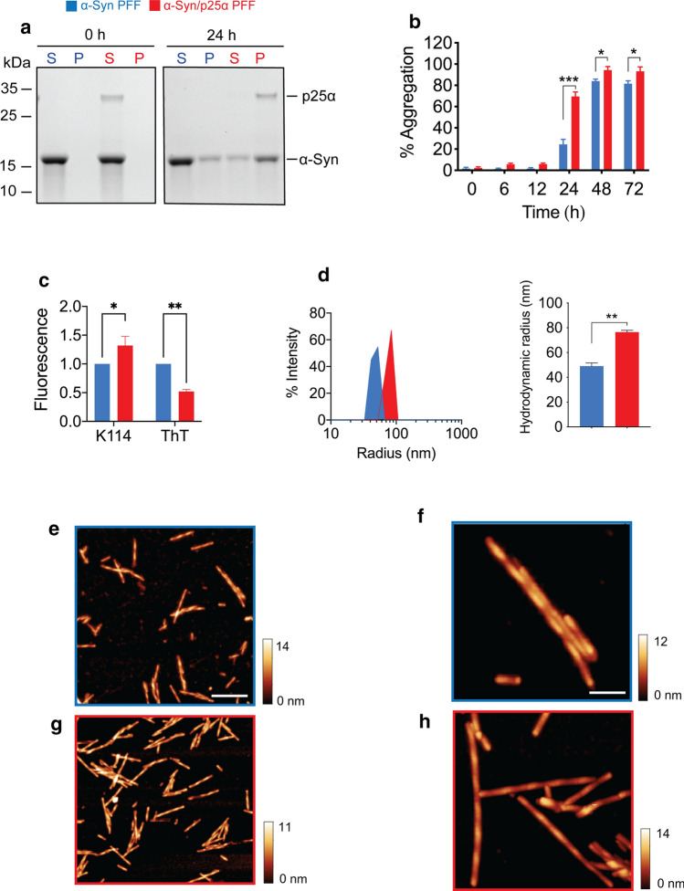
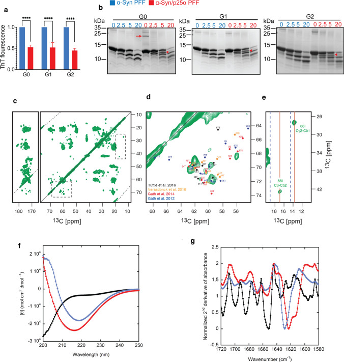
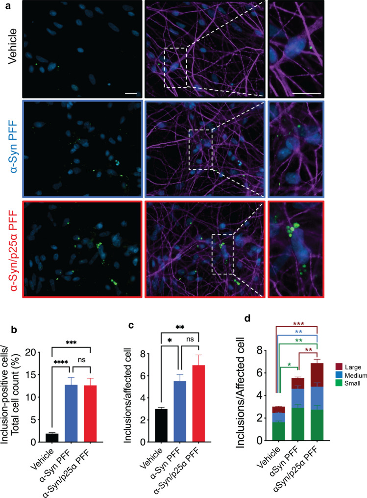
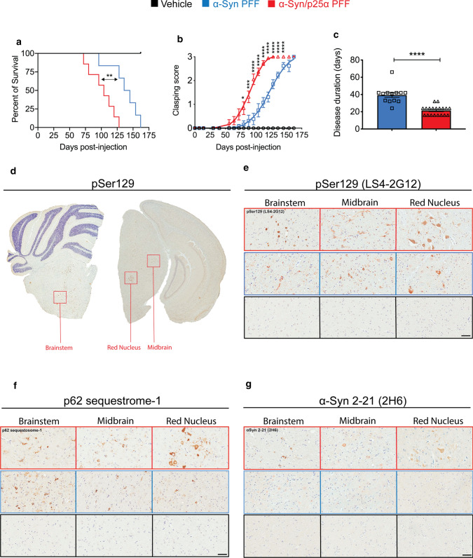
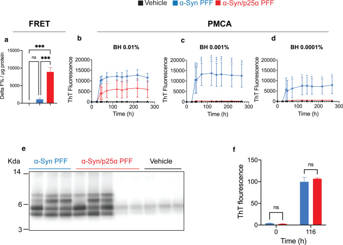
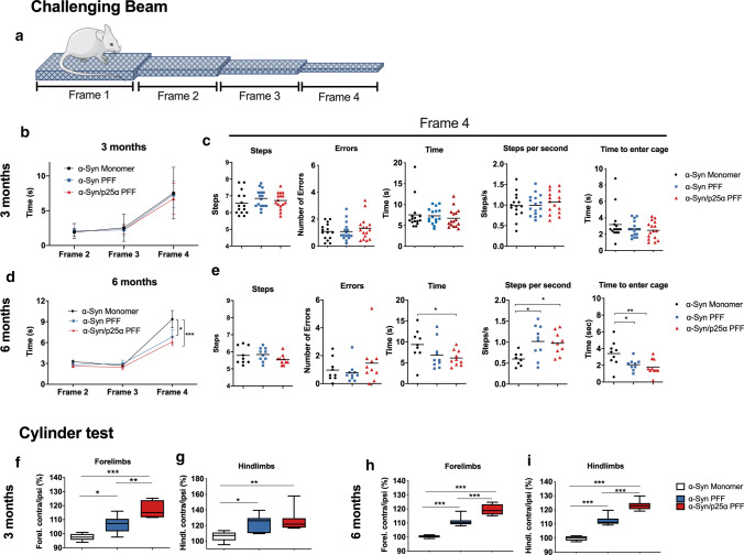
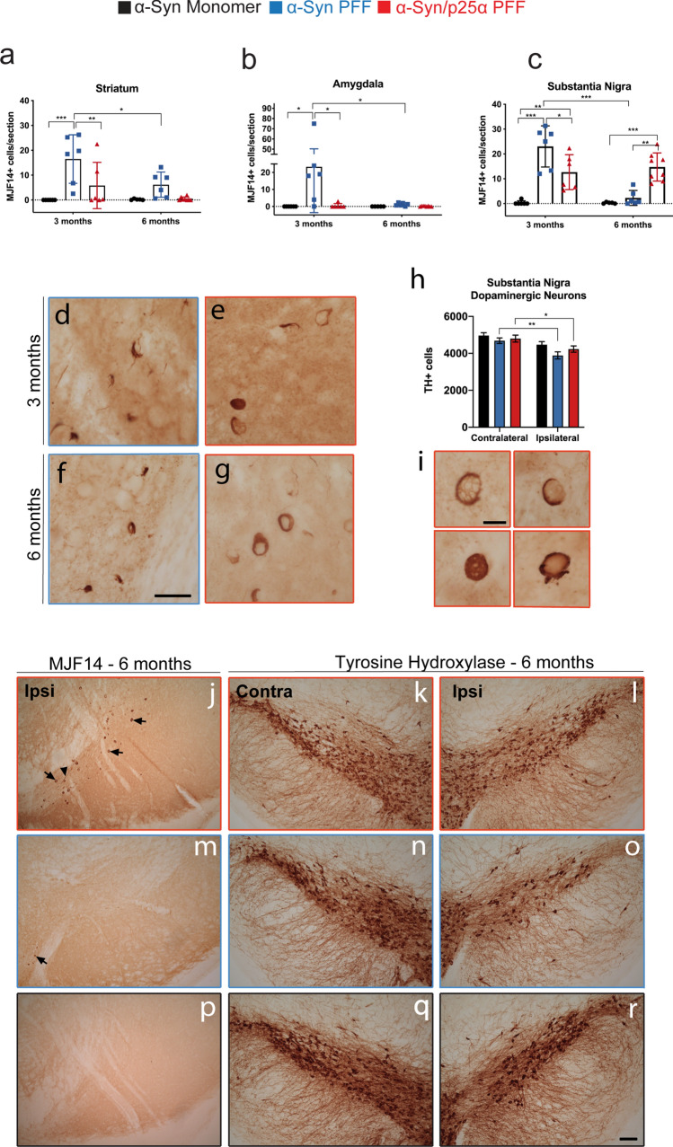
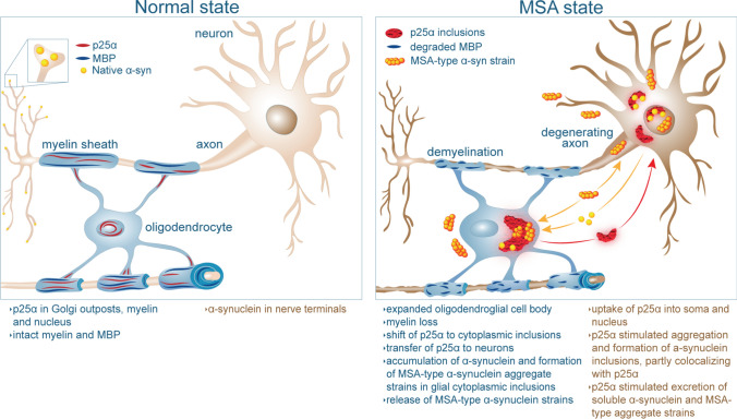
Pathology consisting of intracellular aggregates of alpha-Synuclein (α-Syn) spread through the nervous system in a variety of neurodegenerative disorders including Parkinson's disease, dementia with Lewy bodies, and multiple system atrophy. The discovery of structurally distinct α-Syn polymorphs, so-called strains, supports a hypothesis where strain-specific structures are templated into aggregates formed by native α-Syn. These distinct strains are hypothesised to dictate the spreading of pathology in the tissue and the cellular impact of the aggregates, thereby contributing to the variety of clinical phenotypes. Here, we present evidence of a novel α-Syn strain induced by the multiple system atrophy-associated oligodendroglial protein p25α. Using an array of biophysical, biochemical, cellular, and in vivo analyses, we demonstrate that compared to α-Syn alone, a substoichiometric concentration of p25α redirects α-Syn aggregation into a unique α-Syn/p25α strain with a different structure and enhanced in vivo prodegenerative properties. The α-Syn/p25α strain induced larger inclusions in human dopaminergic neurons. In vivo, intramuscular injection of preformed fibrils (PFF) of the α-Syn/p25α strain compared to α-Syn PFF resulted in a shortened life span and a distinct anatomical distribution of inclusion pathology in the brain of a human A53T transgenic (line M83) mouse. Investigation of α-Syn aggregates in brain stem extracts of end-stage mice demonstrated that the more aggressive phenotype of the α-Syn/p25α strain was associated with an increased load of α-Syn aggregates based on a Förster resonance energy transfer immunoassay and a reduced α-Syn aggregate seeding activity based on a protein misfolding cyclic amplification assay. When injected unilaterally into the striata of wild-type mice, the α-Syn/p25α strain resulted in a more-pronounced motoric phenotype than α-Syn PFF and exhibited a "tropism" for nigro-striatal neurons compared to α-Syn PFF. Overall, our data support a hypothesis whereby oligodendroglial p25α is responsible for generating a highly prodegenerative α-Syn strain in multiple system atrophy.

Keywords: Multiple system atrophy (MSA); P25α; Protein aggregation; Strains; Tubulin polymerisation-promoting protein (TPPP); Α-Synuclein.


Conflict of interest statement

The authors declare no competing interests.

Figures

Fig. 1
Fig. 1
p25α accelerates α-Syn aggregation and binds to α-Syn aggregates. Soluble monomeric α-Syn (346 μM) was assembled in the absence (labelled blue) and presence of p25α (17 μM) (labelled red) into PFF by incubation at 37 °C with continuous shaking for up to  72 h. During the incubation, samples were removed and analysed for solubility by centrifugation and amyloid formation by K114 and ThT fluorescence. a Representative Coomassie Blue stained SDS–polyacrylamide gel of supernatant (S) and pellet (P) fractions at the beginning of incubation and after 24 h. b Time course for development of insolubility based on densitometric analysis of gels as in panel a. Y-axis demonstrates the percentage of aggregation as calculated by [P/(P + S)] × 100]. Bars represent as mean ± SEM of four experiments. *P < 0.05; ***P < 0.001 based on two-way ANOVA followed by Tukey’s multiple comparisons test. c K114 and ThT fluorescence of α-Syn and α-Syn/p25α PFF measured at end-stage plateau of aggregation experiment. Y-axis represents arbitrary units normalised to the fluorescence of the control α-Syn fibrils. Bars represented as mean ± SEM of three experiments normalized to α-Syn signal. *P < 0.05; **P < 0.01. d Dynamic light scattering (DLS) analysis of α-Syn and α-Syn/p25α PFF aggregates after sonication to shear the filaments. Left panel demonstrates the scattering intensity on the Y-axis and the hydrodynamic radius on the log scaled X-axis. The right panel displays the hydrodynamic radius of the two fibril populations displayed as mean ± SEM of four experiments. **P < 0.01 based on two-tailed paired t test. e–h Atomic force microscopy (AFM) characterisation of α-Syn fibrils (e, f, outlined in blue) and α-Syn/p25α fibrils (g, h, outlined in red). Scale bar in e = 500 nm also applies to g and scale bar in f =  100 nm also applies to h
Fig. 2
Fig. 2
α-Syn strains show different structural organisation. De novo generated α-Syn and α-Syn/p25α parental PFF (G0) were used to seed second- and third-generation fibrils (G1-G2) by seeding of 69 µM α-Syn monomers with 3.5 µM parental α-Syn or α-Syn/p25α PFF as seeds. a ThT fluorescence of G0-G2 of the α-Syn and α-Syn/p25α PFF (blue and red, respectively. Values are normalized to the ThT fluorescence signal of α-Syn PFF from the same generation. Asterisks indicate ****P < 0.0001 based on two-way ANOVA followed by Sidak's multiple comparisons test. b α-Syn and α-Syn/p25α PFF (blue and red, respectively) incubated with increasing concentrations of proteinase K, indicated in μg/ml above the gels, and resolved by SDS-PAGE followed and Coomassie Blue staining. Molecular size standards in kDa are indicated to the left of each gel. The p25α present in the parental α-Syn/p25α G0 strain is indicated by red arrow and a prominent α-Syn/p25α strain-specific proteinase K fragment that is templated from G0 to G2 is indicated by red arrow head. c Solid-state NMR analysis of α-Syn/p25α PFF. d, e Represents the regions marked with dashed line in panel c. Published chemical shift sets from different structures are inserted in different colours: orange [107], black [103], red [28] and blue [29] presented in d. f Circular dichroism (CD) spectra and g Fourier-transformed infrared spectroscopy (FTIR) spectra of 0.4 mg/ml α-Syn monomer (black line), α-Syn PFF (blue line) or α-Syn/p25α PFF (red line)
Fig. 3
Fig. 3
α-Syn/p25α PFF template more and larger α-Syn inclusions in human iPSCs-derived neurons. a Human iPSCs-derived neurons were differentiated from neuronal stem cells for 45 days. After 38 days of differentiation, neurons were supplemented with vehicle (upper panels) or G1 generation of α-Syn PFF (middle panels, outlined in blue) and α-Syn/p25α PFF (lower panels, outlined in red) (14 µg/ml) for 24 h followed by media shift to remove excess PFF. They were kept in culture for an additional 7 days in fresh medium before fixation in 4% PFA. Cells were immunostained for microtubule-associated protein 2 (MAP2) (purple) to label neuronal cell bodies and pS129 α-Syn (green) to label inclusions, and the nuclei were stained with DAPI (blue). Pictures were taken randomly with a X63 objective on a Zeiss Observer Z1 microscope. Scalebar = 20 µm. b Columns demonstrate percentage of total MAP2-positive neurons carrying inclusions. c Columns demonstrate number of pS129-α-Syn-positive inclusions/MAP2 neuron carrying inclusions. d Bar graph illustrating inclusion size distribution categorized into small (25–100 pixel2, green), medium (100–300 pixel2, blue), and large (> 300 pixel2, maroon) inclusions for the cells analysed in c. In the two differentiations performed, a total of n = 308 inclusion-bearing cells were analysed for the α-Syn/p25α PFF group, n = 267 cells for the α-Syn PFF group and n = 189 cells for the untreated group. Bars represent mean ± SEM from six different cultures derived from two independent differentiations. *P < 0.05, **P < 0.01, ***P < 0.001 based on one-way ANOVA
Fig. 4
Fig. 4
Intramuscular injection of α-Syn/p25α PFF induces earlier and more severe disease than α-Syn PFF in a human A53T α-Syn-transgenic mouse model. Human A53T α-Syn-expressing M83+/− mice were bilaterally injected with α-Syn or α-Syn/p25α PFF (10 μg/leg) or PBS (vehicle control) into the hindlimb gastrocnemius muscle muscle. a Kaplan–Meier survival plot shows decreased survival time for age-matched M83± mice injected with α-Syn/p25α PFF (red line, n = 19) compared to α-Syn PFF (blue line, n = 13) and PBS (black line, n = 7). **P < 0.01 as determined by log-rank (Mantel-Cox) test. b Hindlimb clasping was scored on a scale from 0 to 3 as a function of days after injection in the mice displayed in panel a and displayed as mean ± s.e.m. The PBS-injected control mice did not develop clasping. Asterisks indicate P < 0.05. ***P < 0.001. ****P < 0.0001 based on two-way ANOVA followed by Tukey's multiple comparisons test. c Duration of disease (from onset of clasping behaviour until death) for individual mice displayed in panel a injected with α-Syn/p25α PFF and α-Syn PFF. PBS-injected control mice did not develop disease. Error bars indicate mean ± SEM and ****P < 0.001 as determined by one-way ANOVA test. d Low-magnification coronal section of a α-Syn/p25α PFF-injected mouse stained with a pSer129 α-Syn-directed antibody LS4-2G12. Boxes indicate locations of brainstem, midbrain, and red nucleus regions analysed in age-matched M83± mice injected with α-Syn/p25α PFF (n = 7), α-Syn PFF (n = 7), and PBS (n = 7). Representative images are displayed of tissue immunohistochemically stained with antibodies reactive to: e pSer129 α-Syn (LS4-2G12), f p62/sequestosome-1 staining as a marker of general inclusions, and g N-terminal of α-Syn using the 2H6 monoclonal antibody against α-Syn 2–21. Scale bar = 50 µm applies to panels e–g
Fig. 5
Fig. 5
Detection of α-Syn aggregates in mouse brain samples by immuno- and PMCA assay and proteinase K digestion and thioflavin T analyses of amplified aggregates. a Brain homogenates of mice injected with α-Syn/p25α PFF contain more aggregated α-Syn than mice injected with α-Syn PFF. Aggregated α-Syn was quantified in brain homogenates using an FRET immune-based Cisbio assay with three technical replicates per homogenate. The level of aggregated α-Syn in the samples is expressed as delta F%/ug protein. This represents the ratio of emission at 665 nm/620 nm of the sample compared to the negative assay control normalized to the protein content (vehicle, n = 3, mice #19–21; α-Syn PFF, n = 4, mice #3–6; α-Syn/p25α PFF n = 4, mice mice #12–15). Neuropathological information of mice samples is available in Supplementary Table 2, online resource. BCA protein measurements determined the total protein concentration of the homogenates. One-way ANOVA followed by Tukey’s post hoc. Bars show mean ± SEM ***P < 0.001. b-d Brain homogenates of mice injected with α-Syn/p25α PFF contain less templating active seeds than mice injected with α-Syn PFF. Brain samples from mouse inoculated with α-Syn PFF (blue line, n = 3, mice #1–3), α-Syn/p25 PFF (red line, n = 5, mice #7–11), and vehicle (PBS, pH 7.4, black line, n = 3, mice #16–18) were homogenized at 10% w/v. Brain samples (at final concentrations of 0.01, 0.001 and 0.0001%) were analysed in a 96-well plate α-Syn-PMCA assay. The extent of aggregation was monitored by the increase in ThT fluorescence by a spectrofluorometer using an λex = 435 nm and an λem = 485 nm. Neuropathological information of mice samples is available in Supplementary Table 2, online resource. The experiments were carried out in duplicates, and error bars indicate mean ± s.e.m. Asterisks indicate *P < 0.05. **P < 0.01. ***P < 0.001 based on two-way ANOVA followed by Tukey's multiple comparisons test. e PMCA-amplified α-Syn aggregates (G0) were incubated with proteinase K (1 mg/ml) at 37 °C for 2 h. Proteins were separated on 12% Bis–Tris gel and immunoblotted with anti-alpha synuclein Syn-1 to visualise cleavage patterns. Molecular weight markers are indicated on the left in kilo-Daltons (KDa). f G1 of PMCA-amplified α-Syn aggregates were generated by seeding of 69 µM α-Syn monomers with 3.5 µM parental aggregated PMCA-amplified α-Syn material (G0). ThT fluorescence was measured at beginning (0 h) and end-stage plateau (116 h) of the G1 re-amplification experiment. Bars represented as mean of signal from α-Syn PFF-injected mice (blue, n = 3) or α-Syn/p25α PFF-injected mice (red, n = 3,) ± s.e.m analysed by two-way ANOVA followed by Sidak’s multiple comparisons test. ns not significant
Fig. 6
Fig. 6
α-Syn/p25α PFF induces abnormal, progressive motor hyperactivity in wild-type mice. Wild-type mice were injected with α-Syn or α-Syn/p25α PFF (10 μg) or monomer α-Syn (10 μg) as negative control in their right striatum and were analysed for motor function by the challenging beam and cylinder test at 3 and 6 months after injection. a The challenging beam test measures the time spent, steps taken, and errors in crossing a progressively thinner beam and to enter a cage after the last frame 4. b-c No significant changes were observed between the three groups at 3 months after injection (α-Syn monomeric, n = 15; α-Syn PFF, n = 16; α-Syn/p25α PFF, n = 16), d 6 months after injection, both α-Syn PFF and α-Syn/p25α PFF-injected animals transversed frame 4 faster than α-Syn monomeric animals (α-Syn monomeric, n = 9; α-Syn PFF, n = 10; α-Syn/p25α PFF, n = 10). Two-way ANOVA followed by Tukey’s post hoc. Error bars indicate mean ± SEM. *P < 0.05. ***P < 0.001. e The faster crossing of frame 4 for both groups was due to more steps/s than the α-Syn monomeric animals and they also used less time to enter the home cage. One-way ANOVA followed by Tukey’s post hoc. Error bars indicate mean ± SEM. *P < 0.05. **P < 0.01. The cylinder test revealed an early and persistent side-bias hyperactivity of PFF-injected animals that at 3 month fg where animals injected with both PFF polymorphs used the contralateral limbs more than α-Syn monomeric-injected mice, and with the α-Syn/p25α PFF even more strongly affecting the forelimb than in the α-Syn PFF mice (α-Syn monomeric, n = 6; α-Syn PFF, n = 8; α-Syn/p25α PFF, n = 8). One-way ANOVA followed by Tukey’s post hoc. Error bars indicate mean ± s.e.m. *P < 0.05. **P < 0.01. ***P < 0.001. h, i This difference between the α-Syn PFF and the α-Syn/p25α PFF mice became more obvious at 6 months after injection, where there also was a difference in hindlimb use. (α-Syn monomeric, n = 9; α-Syn PFF, n = 10; α-Syn/p25α PFF, n = 10). One-way ANOVA followed by Tukey’s post hoc. Bars show min and max. ***P < 0.001
Fig. 7
Fig. 7
α-Syn/p25α PFF induces preferential α-Syn aggregation in nigro-striatal neurons. Progression of the α-Syn pathology and dopaminergic neurodegeneration in mice injected with α-Syn monomer, α-Syn PFF or α-Syn/p25α PFF was assessed by immunohistochemistry and stereology. ac Brain sections were immunostained with an antibody against aggregated α-Syn (MJF-14); the labelled cell body structures were counted in striatum, amygdala, and substantia nigra (SN), and the average number of cells per coronal section in each group was calculated at 3 and 6 months after injection. dg Representative images of cellular structures immunostained with MJF-14 antibody in the SN. Notice that MJF-14-labelled structures observed in the ventral midbrain cells of α-Syn PFF-injected animals extended into neurites and preferentially were segregated into parts of the perinuclear space (d, f); that contrasts with the circular perinuclear staining found in the mice injected with α-Syn/p25α PFF (e, g). h Stereological quantification of tyrosine hydroxylase (TH)-positive cells in SN showed a significant decrease of neurons in the ipsilateral side of the α-Syn PFF-injected group and in the α-Syn/p25α PFF-injected group, while no changes were found after monomeric α-Syn injections. In i, details of the different cells found in SN of the α-Syn/p25α PFF animals at 6 months are presented. j, m, p Representative images of SN immunostained with MJF-14 antibody in all three groups at 6 months. Notice that MJF-14+ structures observed in the α-Syn PFF-injected animals were normally located in the ventral medial midbrain in the border with VTA (m), while cells (arrows in j) and fibres (arrowhead in j) were located in the SN compacta in the α-Syn/p25α PFF animals. k, l, n, o, q, r Representative images of the contralateral SN and its correspondent ipsilateral side from sections immunostained with TH antibody in all three groups at 6 months. Notice the decrease in the number of cell bodies in the ipsilateral side of the α-Syn PFF-injected mice in o and the α-Syn/p25α PFF-injected mice in (l). Data are average ± SD. ac or SEM. h Two-way ANOVA followed by Sidak post hoc. * P < 0.05. ** P < 0.01. *** P < 0.001. Scale in f = 25 µm applies to dg, scale in i = 10 µm and applies to insets and scale in t: 100 µm applies to (jr)
Fig. 8
Fig. 8
Hypothetical mechanism for the generation of an MSA-associated α-Syn strain with increased neurotoxicity induced by oligodendroglial protein p25α/TPPP. Left panel: p25α/TPPP is expressed in mature oligodendrocytes and is vital for reorganisation and stabilisation myelin. In the physiological state, p25α/TPPP can be found in Golgi outposts in myelin outside the cell body and in the nucleus. The myelin basic protein (MBP) is present in intact myelin. Right panel: In MSA, the oligodendroglial cell body expands, and MBP is degraded leading to demyelination. p25α/TPPP retracts from myelin and exits the nucleus forming early cytoplasmic inclusions. Low levels of α-Syn monomers are then recruited to the inclusion where P25α/TPPP nucleate them into MSA-type α-Syn aggregate strains completing the formation of mature glial cytoplasmic inclusions. P25α/TPPP is released from oligodendrocytes and taken into neurons where it exists in nuclear and extranuclear inclusions in the absence and presence of α-Syn and induce formation of MSA-type α-Syn polymorphs that accumulate in neurites and nuclei as neuronal nuclear and cytoplasmic inclusions causing neuronal degeneration. The P25α/TPPP facilitates the spreading of the disease process by stimulating atypical secretion of α-Syn aggregates

Similar articles

Cited by

References

    1. Abdelmotilib H, Maltbie T, Delic V, Liu Z, Hu X, Fraser KB, et al. alpha-Synuclein fibril-induced inclusion spread in rats and mice correlates with dopaminergic Neurodegeneration. Neurobiol Dis. 2017;105:84–98. doi: 10.1016/j.nbd.2017.05.014. - DOI - PMC - PubMed
    1. Anderson JP, Walker DE, Goldstein JM, de Laat R, Banducci K, Caccavello RJ, et al. Phosphorylation of Ser-129 is the dominant pathological modification of alpha-synuclein in familial and sporadic Lewy body disease. J Biol Chem. 2006;281:29739–29752. doi: 10.1074/jbc.M600933200. - DOI - PubMed
    1. Aoki N, Boyer PJ, Lund C, Lin WL, Koga S, Ross OA, et al. Atypical multiple system atrophy is a new subtype of frontotemporal lobar degeneration: frontotemporal lobar degeneration associated with alpha-synuclein. Acta Neuropathol. 2015;130:93–105. doi: 10.1007/s00401-015-1442-z. - DOI - PMC - PubMed
    1. Asi YT, Simpson JE, Heath PR, Wharton SB, Lees AJ, Revesz T, et al. Alpha-synuclein mRNA expression in oligodendrocytes in MSA. Glia. 2014;62:964–970. doi: 10.1002/glia.22653. - DOI - PMC - PubMed
    1. Badiola N, de Oliveira RM, Herrera F, Guardia-Laguarta C, Goncalves SA, Pera M, et al. Tau enhances alpha-synuclein aggregation and toxicity in cellular models of synucleinopathy. PLoS ONE. 2011;6:e26609. doi: 10.1371/journal.pone.0026609. - DOI - PMC - PubMed

Publication types

LinkOut - more resources