. 2021 May 11;35(6):109105.
doi: 10.1016/j.celrep.2021.109105.

Efficient generation of isogenic primary human myeloid cells using CRISPR-Cas9 ribonucleoproteins

Affiliations

Efficient generation of isogenic primary human myeloid cells using CRISPR-Cas9 ribonucleoproteins

Joseph Hiatt et al. Cell Rep. .

Abstract

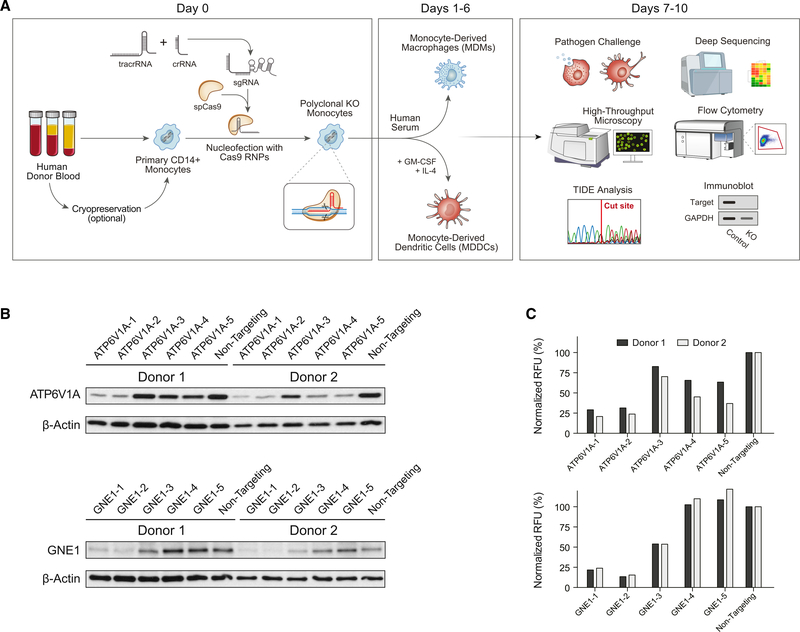
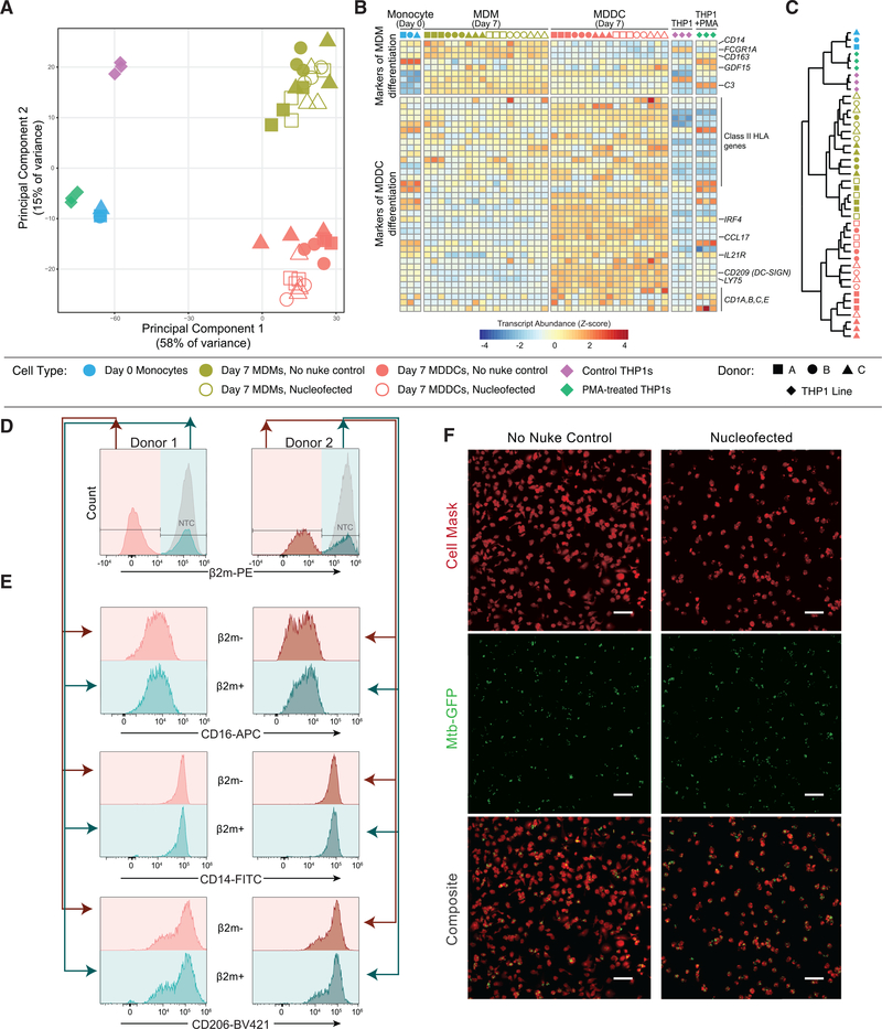
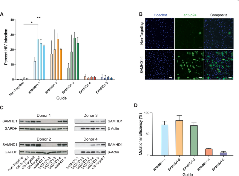
Genome engineering of primary human cells with CRISPR-Cas9 has revolutionized experimental and therapeutic approaches to cell biology, but human myeloid-lineage cells have remained largely genetically intractable. We present a method for the delivery of CRISPR-Cas9 ribonucleoprotein (RNP) complexes by nucleofection directly into CD14+ human monocytes purified from peripheral blood, leading to high rates of precise gene knockout. These cells can be efficiently differentiated into monocyte-derived macrophages or dendritic cells. This process yields genetically edited cells that retain transcript and protein markers of myeloid differentiation and phagocytic function. Genetic ablation of the restriction factor SAMHD1 increased HIV-1 infection >50-fold, demonstrating the power of this system for genotype-phenotype interrogation. This fast, flexible, and scalable platform can be used for genetic studies of human myeloid cells in immune signaling, inflammation, cancer immunology, host-pathogen interactions, and beyond, and could facilitate the development of myeloid cellular therapies.

Keywords: CRISPR; Cas9; dendritic cells; electroporation; host-pathogen interactions; knockout; macrophages; monocytes; myeloid cells; ribonculeoproteins (RNPs).


Conflict of interest statement

Declaration of interests The authors declare competing interests: T.L.R. is a co-founder of Arsenal Biosciences. A.M. is a compensated co-founder, member of the boards of directors, and a member of the scientific advisory boards of Spotlight Therapeutics and Arsenal Biosciences. A.M. was a compensated member of the scientific advisory board at PACT Pharma and was a compensated advisor to Juno Therapeutics and Trizell. A.M. owns stock in Arsenal Biosciences, Spotlight Therapeutics, and PACT Pharma. A.M. has received honoraria from Merck and Vertex, a consulting fee from AlphaSights, and is an investor in and informal advisor to Offline Ventures. The Marson lab has received research support from Juno Therapeutics, Epinomics, Sanofi, GlaxoSmithKline, Gilead, and Anthem. A.M., T.L.R., and E.S. are holders of patents pertaining to, but not resulting from, this work. The Krogan laboratory has received research support from Vir Biotechnology and F. Hoffmann-La Roche.

Figures

Figure 1.
Figure 1.. A flexible platform for CRISPR editing of human myeloid-lineage cells
(A) A generalized schematic of the platform. Human CD14+ monocytes are isolated from blood by density gradient separation of PBMCs followed by magnetic negative selection. Either PBMCs or monocytes may be cryopreserved for later editing (Figure S1B). Cells are then nucleofected with preformed CRISPR-Cas9 RNPs and immediately put into differentiating culture under MDM- or MDDC-generating conditions. After allowing for 6–7 days of differentiation and washout of the targeted gene product, cells can be subjected to a wide variety of functional, phenotypic, and genotypic studies to assess the knockout efficiency and function of the targeted gene product. (B) Guide sequence-dependent knockout of targeted genes leads to loss of gene products. CD14+ monocytes were nucleofected with RNPs containing 1 of 5 distinct guide sequences against the indicated gene or a scrambled non-targeting control, cultured under MDM-generating conditions, and then lysed for immunoblot analysis. Blots show targeted gene protein product and untargeted housekeeping gene product β-actin protein levels in cells from 2 blood donors. GNE1 and ATP6V1A ran at their expected sizes of 79 and 69 kDa, respectively. (C) Knockout was quantified by digital densitometry and normalized on a per-sample basis in relative fluorescence units (RFUs) to untargeted housekeeping control protein β-actin. See also Figure S1.
Figure 2.
Figure 2.. CRISPR-Cas9-mediated gene knockout preserves key aspects of differentiation and function in targeted myeloid cells
(A) Principal-component analysis of RNA sequencing (RNA-seq) from the indicated cell types. (B) Normalized transcript abundance (Z score) for selected markers of MDM or MDDC differentiation (Lehtonen et al., 2007). (C) Dendrogram of hierarchical clustering of the data in (B) by Euclidean distance. (D and E) Among cells subjected to CRISPR-Cas9 RNP nucleofection, cell surface protein levels of CD16, CD14, and CD206 were compared between the cells that bear the desired β2 m knockout (pink) and those that do not (teal) by flow cytometry after 7 days of MDM differentiation. (D) shows gating, while (E) shows the expression of the indicated markers. (F) Representative images of unperturbed (left) and RNP-nucleofected (right) MDMs infected with GFP-expressing M. tuberculosis (Mtb-GFP) show that CRISPR-Cas9-targeted cells remain competent to phagocytose living pathogens. Top, membrane staining with Cell Mask Far Red; CENTER, Mtb-GFP; bottom, composite. Scale bars represent 100 μm. See also Figure S2.
Figure 3.
Figure 3.. Generation of isogenic monocyte-derived macrophages for functional evaluation of an HIV-1 host restriction factor
(A) SAMHD1-targeted or non-targeting control MDMs from 4 independent, HIV blood donors were infected with HIV-1. The plot displays the percentage of cells productively infected after a 48-h exposure. Guides that most efficiently ablated the gene caused statistically significant increases in infection as assessed by 1-way ANOVA followed by Dunnett’s test. *p < 0.05, **p < 0.01. See also Figure S3E. (B) Representative images of HIV-1 infection from donor 3 comparing cells nucleofected with control non-targeting RNPs (top) to cells nucleofected with RNPs made from guide SAMHD1–1 (bottom). Left, Hoechst; center, staining of the HIV-1 antigen p24; right, composite. Scale bars represent 100 μm. For representative images of all of the guides, see Figure S3D. (C and D) Quantification of SAMHD1 knockout by immunoblot (C) and sequencing (D). No protein sample was available for guide SAMHD1–1 in donors 3 and 4; Sanger sequencing was analyzed for mutational efficiency by TIDE, bars represent means ± SDs for at least 3 biological replicates. See also Figure S3.

References

    1. Auffray C, Sieweke MH, and Geissmann F (2009). Blood monocytes: development, heterogeneity, and relationship with dendritic cells. Annu. Rev. Immunol 27, 669–692. - PubMed
    1. Barkal AA, Weiskopf K, Kao KS, Gordon SR, Rosental B, Yiu YY, George BM, Markovic M, Ring NG, Tsai JM, et al. (2018). Engagement of MHC class I by the inhibitory receptor LILRB1 suppresses macrophages and is a target of cancer immunotherapy. Nat. Immunol 19, 76–84. - PMC - PubMed
    1. Brinkman EK, Chen T, Amendola M, and van Steensel B (2014). Easy quantitative assessment of genome editing by sequence trace decomposition. Nucleic Acids Res. 42, e168. - PMC - PubMed
    1. Chao MP, Takimoto CH, Feng DD, McKenna K, Gip P, Liu J, Volkmer J-P, Weissman IL, and Majeti R (2020). Therapeutic Targeting of the Macrophage Immune Checkpoint CD47 in Myeloid Malignancies. Front. Oncol 9, 1380. - PMC - PubMed
    1. Engblom C, Pfirschke C, and Pittet MJ (2016). The role of myeloid cells in cancer therapies. Nat. Rev. Cancer 16, 447–462. - PubMed

Publication types

Substances