Review
. 2022 Jan:78:104-123.
doi: 10.1016/j.semcancer.2021.05.007. Epub 2021 May 9.

Thorny ground, rocky soil: Tissue-specific mechanisms of tumor dormancy and relapse

Affiliations
Review

Thorny ground, rocky soil: Tissue-specific mechanisms of tumor dormancy and relapse

Andrea R Lim et al. Semin Cancer Biol. 2022 Jan.

Abstract

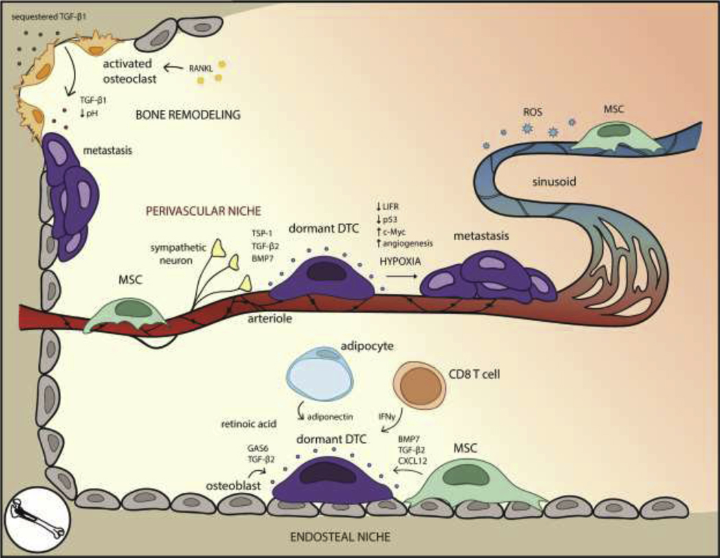
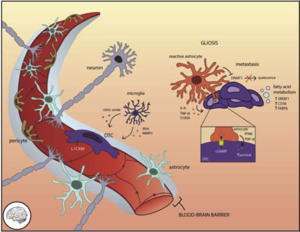
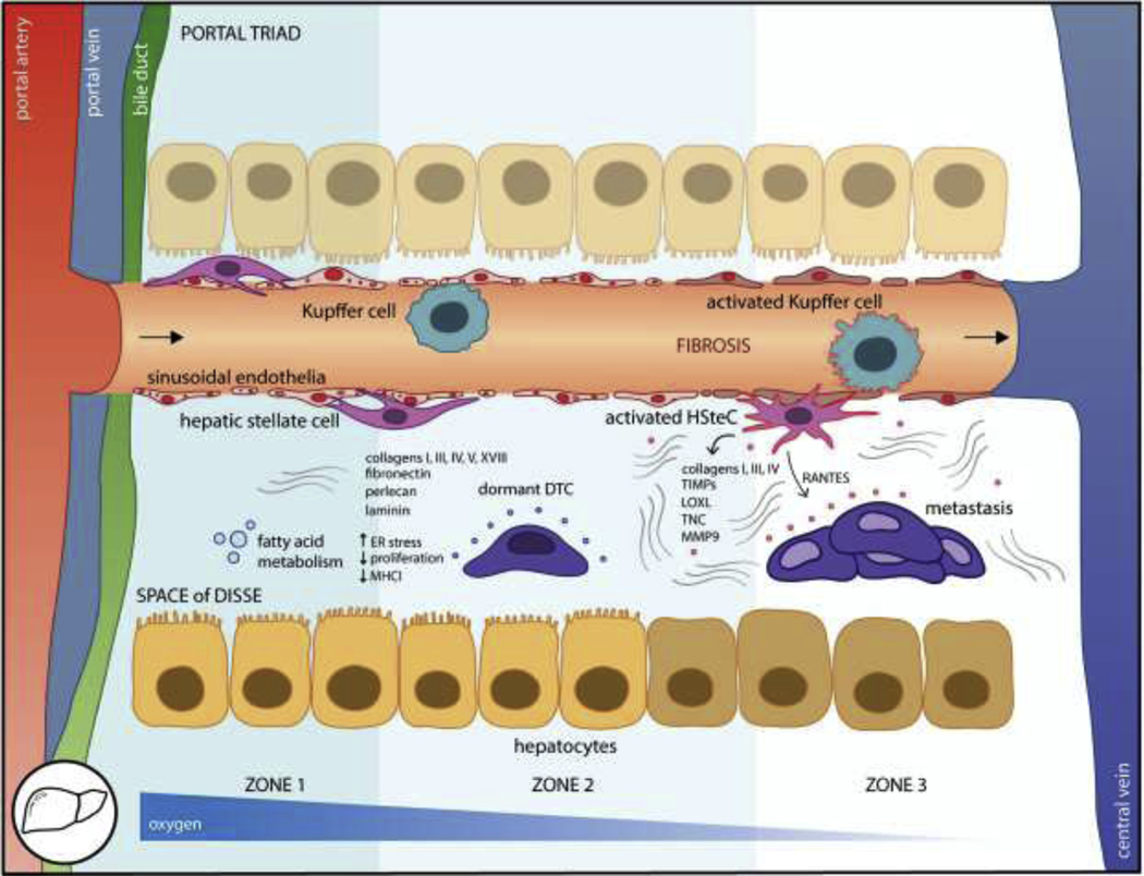
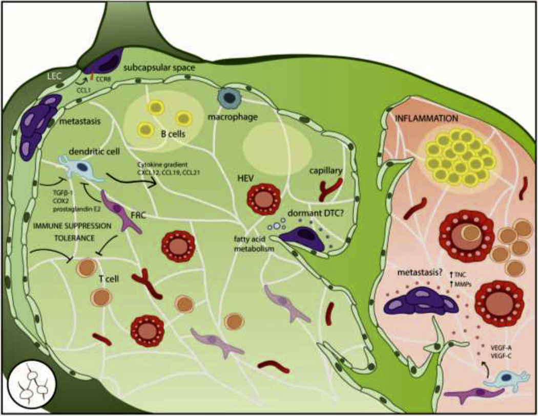
Disseminated tumor cells (DTCs) spread systemically yet distinct patterns of metastasis indicate a range of tissue susceptibility to metastatic colonization. Distinctions between permissive and suppressive tissues are still being elucidated at cellular and molecular levels. Although there is a growing appreciation for the role of the microenvironment in regulating metastatic success, we have a limited understanding of how diverse tissues regulate DTC dormancy, the state of reversible quiescence and subsequent awakening thought to contribute to delayed relapse. Several themes of microenvironmental regulation of dormancy are beginning to emerge, including vascular association, co-option of pre-existing niches, metabolic adaptation, and immune evasion, with tissue-specific nuances. Conversely, DTC awakening is often associated with injury or inflammation-induced activation of the stroma, promoting a proliferative environment with DTCs following suit. We review what is known about tissue-specific regulation of tumor dormancy on a tissue-by-tissue basis, profiling major metastatic organs including the bone, lung, brain, liver, and lymph node. An aerial view of the barriers to metastatic growth may reveal common targets and dependencies to inform the therapeutic prevention of relapse.

Keywords: Disseminated tumor cell dormancy; Dormant niche; Metastasis; Microenvironment; Quiescence.


Conflict of interest statement

Conflict of Interest statement

The authors declare that there are no conflicts of interest.

Figures

Figure 1.
Figure 1.. The bone microenvironment for dormant DTCs is well-defined due to established mechanisms that regulate HSC quiescence and bone resorption.
Within the endosteal niche, MSCs, osteoblasts, adipocytes, and CD8+ T cells secrete factors that preserve DTC quiescence. High levels of retinoic acid within the bone marrow induce cell cycle arrest. Awakening can occur as the result of environmental changes brought on by RANKL-activated osteoclasts and bone resorption. In the perivascular niche, arterioles and perivascular cells promote DTC quiescence through basement membrane factors such as TSP-1, although hypoxic regions near the vessels promote angiogenesis, which correlates with metastasis. The leakier sinusoids, by contrast, experience ROS on the basal side of the vessel, possibly inducing DNA damage and stress pathways associated with metastasis.
Figure 2.
Figure 2.. The lung microenvironment for dormant DTCs includes the perivascular and epithelial niches.
In the perivascular niche, deposited basement membrane components like TSP-1 induce cell cycle arrest. The lungs are rich in BMP signaling and BMP4 specifically promotes DTC quiescence, which is antagonized by the BMP inhibitor Coco. The epithelial niche induces SFRP2 in DTCs through an unknown factor. Environmental insults such as tobacco smoke or bacterial LPS elicit inflammation, leading to pro-metastatic angiogenesis, fibrosis, and ECM alterations such as cleavage of laminin-111 by activated neutrophil proteases. These changes are associated with metastatic outgrowth of dormant DTCs.
Figure 3.
Figure 3.. The brain microenvironment requires DTCs to adapt in order to survive.
Access to the brain parenchyma is restricted by the blood-brain barrier (BBB), a tightly reinforced layer including endothelia and concentric layers of supporting pericytes, astrocytes, and neurons. DTCs that manage to extravasate through the BBB rely on L1CAM-mediated spreading on the vasculature to trigger YAP-dependent survival. Astrocytes maintain the integrity of the vasculature and repel invasive cells through Fas ligand but undergo gliosis as an injury response. Reactive astrocytes secrete pro-metastatic cytokines such as IL-6, TNF-α, and S100a and exchange factors directly with DTCs through gap junctions, which elicits paracrine survival signaling (IFNα, TNF-α) back to the DTC. Microglia eliminate tumor cells through nitric oxide but also contribute pro-invasive Wnts and MMP3. The unique lipid composition of the brain necessitates DTCs adapt their fatty acid metabolism through SREBF1, CD36, or FABP6 for successful colonization.
Figure 4.
Figure 4.. The liver is the most common major metastatic organ.
DTCs traffic from the portal triad vessels to the fenestrated sinusoids and into the subendothelial space of Disse where they encounter stromal cells including liver sinusoidal endothelial cells (LSEC), hepatic stellate cells (HSteC), Kupffer cells, and hepatocytes. Distinct zones occur along the portal triad to central vein axis following oxygen and functional gradients. The loosely organized fibers and proteoglycans basement membrane include collagens, fibronectin, perlecan, and laminin which impose quiescence. Metabolic adaptation slows DTC proliferation, although it also coincides with downregulation of MHCI and immune evasion. However, upon inflammation or injury, the liver become fibrotic which is associated with metastatic outgrowth. Activated HSteCs deposit vast quantities of ECM fibers and other pro-metastatic factors such as RANTES, endothelia undergo capillarization, and hepatocytes lose their microvilli. These changes to the liver microenvironment alter the niche to foster metastasis.
Figure 5.
Figure 5.. The lymph node (LN) is the most common site of all metastases but its role as a dormancy niche is unknown.
The LN is highly compartmentalized to support its primary function in peripheral immunity. In addition to resident and migrating immune cells, the LN stroma includes lymphatic endothelial cells (LECs) that form the outer capsule and draining sinusoids, blood endothelial cells (BECs) that form capillaries and specialized high endothelial venules (HEVs), and fibroblastic reticular cells (FRCs) that produce an ECM conduit network to facilitate antigen sampling. DTCs frequently access the LN by lymphatic or hematogenous routes, but must adapt to the LN microenvironment through metabolic pathways and co-opting signals that guide immune cells into the LN parenchyma such as CCL1-CCR8 signaling at the afferent lymphatic vessel and cytokine gradients of CXCL12, CCL19, and CCL21; otherwise, they risk remaining confined to the subcapsular space. LN DTCs may benefit from immune suppressive and tolerogenic functions of FRCs and LECs. However, the stromal components that contribute to a dormancy niche have not been formally described. During inflammation, the LN activates into a proliferative environment including angiogenesis and lymphangiogenesis; these changes to the stroma could conceivably induce outgrowth of dormant DTCs, but must be more fully investigated.

References

    1. Spencer W, Celsus de Medicina. The American Journal of the Medical Sciences, 1936. 191(3): p. 424.
    1. Hulsbergen AFC, et al., Subtype switching in breast cancer brain metastases: a multicenter analysis. Neuro Oncol, 2020. 22(8): p. 1173–1181. - PMC - PubMed
    1. Priedigkeit N, et al., Intrinsic Subtype Switching and Acquired ERBB2/HER2 Amplifications and Mutations in Breast Cancer Brain Metastases. JAMA Oncol, 2017. 3(5): p. 666–671. - PMC - PubMed
    1. Lluch A, et al., Dynamic clonal remodelling in breast cancer metastases is associated with subtype conversion. European Journal of Cancer, 2019. 120: p. 54–64. - PubMed
    1. Nikbakht H, et al., Latency and interval therapy affect the evolution in metastatic colorectal cancer. Scientific Reports, 2020. 10(1): p. 581. - PMC - PubMed

Publication types