. 2021 Nov 1;106(11):2971-2985.
doi: 10.3324/haematol.2020.278252.

The interactome of the N-terminus of band 3 regulates red blood cell metabolism and storage quality

Affiliations

The interactome of the N-terminus of band 3 regulates red blood cell metabolism and storage quality

Aaron Issaian et al. Haematologica. .

Abstract

Band 3 (anion exchanger 1; AE1) is the most abundant membrane protein in red blood cells, which in turn are the most abundant cells in the human body. A compelling model posits that, at high oxygen saturation, the N-terminal cytosolic domain of AE1 binds to and inhibits glycolytic enzymes, thus diverting metabolic fluxes to the pentose phosphate pathway to generate reducing equivalents. Dysfunction of this mechanism occurs during red blood cell aging or storage under blood bank conditions, suggesting a role for AE1 in the regulation of the quality of stored blood and efficacy of transfusion, a life-saving intervention for millions of recipients worldwide. Here we leveraged two murine models carrying genetic ablations of AE1 to provide mechanistic evidence of the role of this protein in the regulation of erythrocyte metabolism and storage quality. Metabolic observations in mice recapitulated those in a human subject lacking expression of AE11-11 (band 3 Neapolis), while common polymorphisms in the region coding for AE11-56 correlate with increased susceptibility to osmotic hemolysis in healthy blood donors. Through thermal proteome profiling and crosslinking proteomics, we provide a map of the red blood cell interactome, with a focus on AE11-56 and validate recombinant AE1 interactions with glyceraldehyde 3-phosphate dehydrogenase. As a proof-of-principle and to provide further mechanistic evidence of the role of AE1 in the regulation of redox homeo stasis of stored red blood cells, we show that incubation with a cell-penetrating AE11-56 peptide can rescue the metabolic defect in glutathione recycling and boost post-transfusion recovery of stored red blood cells from healthy human donors and genetically ablated mice.


Figures
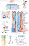
Figure 1.
Figure 1.
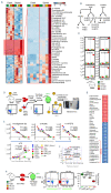
The N-terminus of band 3 controls red blood cell metabolism during storage under blood bank conditions. (A) Human and mouse N-terminal band 3 sequences are slightly different. (B) We leveraged humanized AE1 mice, and compared them to wild-type (WT) mice (C57BL6), or humanized mice lacking residues 1-11 (deletion of the high affinity binding region for glycolytic enzyme: HA Del) or 12-23 (hemoglobin binding site knockout: BS KO). (C) Red blood cells (RBC) from these mice were stored under refrigerated conditions for 12 days, prior to metabolomics analyses. (D, E) Storage and genotypes had a significant impact on RBC metabolism, as determined by unsupervised principal component analysis (D) and hierarchical clustering analysis of significant metabolites by analysis of variance (E). Specifically, strain-specific differences in fresh and stored RBC were noted in glycolysis, the pentose phosphate pathway (PPP) and glutathione homeostasis. (F) To further investigate the impact on glycolysis and the PPP, RBC from the four mouse strains were incubated with 1,2,3-C3-glucose upon stimulation with methylene blue (MB). (G, H) Rescue experiments were also performed by incubating RBC with a recombinantly expressed N-terminus AE1 peptide (residues 1-56), prior to determination of the ratios of lactate isotopologues +3 and +2, deriving from glycolysis and the PPP, respectively. (I) MB activated the PPP in all mouse strains except for the HA Del mice. Supplementation of the AE11-56 peptide increased PPP activation and decreased glycolysis in HA Del mice and further exacerbated responses in all the other strains.±
Figure 2.
Figure 2.
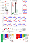
Metabolic alterations in band 3 knockout mice correlate with poor post-transfusion recovery. (A-C) Analyses of fresh and stored red blood cells (RBC) from wild-type (WT) (C57BL6) mice [green], humanized band 3 mice (HuB3) [white] and mice lacking amino acids 1-11 (HA Del) [yellow] or 12-23 (BS KO) [orange] of band 3 showed strain-specific differences in several pathways in fresh and stored RBC. Above all, stored RBC from the KO mice were characterized by increased levels of carboxylic acid and lipid peroxidation products (A), especially metabolites of the arachidonate metabolism (B), including prostaglandins, eicosanoids, hydroxyeicosatetraenoates (HETE) and hydroxyoctadecenoates (HODE) (C); the full, vectorial version of this list is provided in Online Supplementary Table S1). (D-F) Metabolomics analyses on fresh and stored RBC from 13 different mouse strains (D) highlight these metabolites as significant correlates with poor post-transfusion recovery (E), as highlighted by the metabolite set enrichment analysis in (F) and the ranked top positive and negative correlates with post-transfusion recovery reported in (G). (H) Determination of end-of-storage post-transfusion recovery of RBC from the four mouse strains showed significant decreases in recovery in the HA Del and BS KO mice.
Figure 3.
Figure 3.
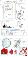
Metabolic impact of AE11-11 deletion in human red blood cells and alterations of osmotic fragility of stored red blood cells from donors with polymorphisms in AE11-56. (A, B) Lack of amino acid residues 1-11 of AE1 in humans, known as band 3 Neapolis, (A) results in increased activation of glycolysis and decreases in glutathione pools (B). (C) Tracing experiments with 1,2,3-C3-glucose showed impaired responses to methylene blue (MB)-induced activation of the pentose phosphate pathway (PPP) compared to glycolysis in band 3 Neapolis, a phenotype that is partially rescued in vitro by supplementation of a recombinant AE11-56 peptide. (D) However, band 3 Neapolis RBC were also characterized by higher levels of oxylipins, comparable to those observed in stored RBC from mice lacking AE1 amino acids 12-23 (BS KO). (E) Genome-wide association studies on 13,806 healthy donor volunteers revealed increased osmotic fragility in subjects carrying a polymorphism in the region coding for band 3 (gene name SLC4A1) residues 1-56 and neighboring introns. CTRL: control; GSH: reducd glutathione; GSSG: oxidized glutathione; HETE: hydroxyeicosatetraenoates; HODE: hydroxyoctadecenoates; GWAS: genome-wide association studies, SNP: single nucleotide polymorphisms.
Figure 4.
Figure 4.
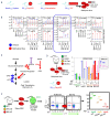
Thermal proteome profiling and crosslinking proteomics of recombinant peptide 1-56 of band 3 in red blood cell lysates. (A) Thermal proteome profiling experiments were performed by incubating red blood cell (RBC) lysates with a recombinantly expressed peptide coding for the amino acids 1-56 in the N-terminus of band 3 at a gradient of temperatures from 37°C to 67ºC, labeling with ten different tandem mass tags (TMT10), pooling and analysis via nano-ultrahigh performance liquid chromatography tandem mass spectrometry (nanoUHPLC-MS/MS). (B) Proteins were ranked as a function of their alterations in the temperature at which their solubility decreases and precipitation is observed (ΔTm). (C) A few representative melting curves for the top hits for proteins stabilized or destabilized by the presence of the band 3 peptide 1-56 (red) compared to untreated controls (blue) are provided. (D) A peptide coding for amino acids 1-56 of the N-terminus of band 3 was recombinantly expressed with a SUMO-tag and/or a His-Flag tag at either the N- or C-terminus of the peptide, prior to incubation with plasma, RBC cytosols and membrane in independent experiments, enrichment in nickel columns, pull-down against the SUMO tag, and crosslinking (XLINK) with disuccinimidyl sulfoxide (DSSO) or 4-(4,6-dimetoxy-1,3,5-triazin-2-yl)-4-metylmorpholinium (DMTMM), prior to protein digestion, fractionation of crosslinked peptides and nanoUHPLC-MS/MSbased identification of band 3 interacting partners via MS2/MS3 analyses. (E, F) Top interactors for RBC membrane and cytosol interactors are listed in (E) and (F), respectively, divided by pathway.
Figure 5.
Figure 5.
The interactome of AE11-56, as gleaned from crosslinking mass spectrometry. A network view of the interactome of AE11-56, as determined by merging the data from all the crosslinking proteomics studies. The network shows direct and indirect interactors with AE11-56 and the residues on band 3 with which the proteins were identified to crosslink (XLINK).
Figure 6.
Figure 6.
Structural studies of recombinantly expressed GAPDH and its interaction with the N-terminus of band 3 (residues 1-56). (A) Glyceraldehyde 3-phosphate dehydrogenase (GAPDH) was recombinantly expressed in E.coli prior to purification and interaction studies with the recombinantly expressed band 3 peptide (residues 1-56). (B-E) These studies included isothermal titration calorimetry (B), nuclear magnetic resonance of band 3 (AE11-56) with 100 or 200 μM GAPDH (C) and derived calculation of chemical shift perturbations (CSP) (D), and in silico modeling of band 3 (1-56) structure based on NMR data (E). Following these studies, crosslinking proteomics analyses were performed in vitro by co-incubating GAPDH and AE11-56 in the presence of disuccinimidyl sulfoxide (DSSO) or 4-(4,6-dimetoxy- 1,3,5-triazin-2-yl)-4-metylmorpholinium (DMTMM). (F, G) A representative spectrum (F) from one of the most abundant crosslinks, comprehensively mapped in the circos plot (light blue for intramolecular crosslinks, red for intermolecular ones) (G). (H) Results were thus mapped against the GAPDH monomeric structure (blue: all lysine residue on GAPDH that were experimentally found to crosslink to band 3 aspartyl or glutamyl side chains; yellow: the lysines that were available for crosslinking but were not found to face band 3 acidic residues within the reach of the crosslinker of ~20 Å). (H-K) Nuclear magnetic resonance and crosslinking proteomics data were used to build a model of the band 3 (1-56) interaction with GAPDH with the software Rosetta, resulting in the active site pocket of GAPDH being exposed to residues 1-15 of band 3.
Figure 7.
Figure 7.
Cell membrane-permeable band 31-56 peptides reverse the defect in the glutathione recycling capacity of red blood cells from band 3 knockout mice. (A) Three membrane permeable versions of the band 3 (AE11-56) peptide were generated through addition of a poly arginine (polyArg), internalization sequence or TAT sequence at the C-terminus of the peptide. Human red blood cells (RBC) were thus incubated with a control (scramble), a band 3 (B3) 1-56 peptide (non-penetrating) or the three penetrating peptides in the presence of 1,2,3-C3-glucose and methylene blue (MB) stimulation to activate the pentose phosphate pathway (PPP). (B) C isotopologues of glycolysis and the PPP are reported for all groups at baseline and following MB stimulation of healthy human RBC, showing increases in PPP activation following MB in all cases, even though ATP levels were only preserved in the RBC treated with the cell-penetrating peptides, suggesting a metabolic reprogramming consistent with the schematic in (C). (D) RBC from wild-type (WT), humanized band 3 (HuB3) or band 3 knockout mice lacking residues 1-11 (HA Del) or 12-23 (BS KO) were stimulated with MB in the presence of a cell-penetrating version of the band 3 peptide (polyArg). Results show increased glutathione oxidation in response to MB treatment in all cases, but significant rescue by the band 3 peptide, especially in the HA Del and BS KO groups. (E) Similarly, storage of murine RBC from the WT and band 3 KO mouse strains in the presence of the polyArg cell-penetrating AE11-56 peptide promoted increases in post-transfusion recovery in WT and HA Del mice, but not in BS KO mice.

Comment in

References

    1. Pantaleo A, Ferru E, Pau MC, et al. . Band 3 rrythrocyte membrane protein acts as redox stress sensor leading to its phosphorylation by p (72) Syk. Oxid Med Cell Longev. 2016;2016:6051093. - PMC - PubMed
    1. Yoshida T, Prudent M, D'Alessandro A. Red blood cell storage lesion: causes and potential clinical consequences. Blood Transfus. 2019;17(1):27-52. - PMC - PubMed
    1. Bosman GJ, Stappers M, Novotny VM. Changes in band 3 structure as determinants of erythrocyte integrity during storage and survival after transfusion. Blood Transfus. 2010;8(Suppl 3):s48-52. - PMC - PubMed
    1. Bryk AH, Wisniewski JR. Quantitative analysis of human red blood cell proteome. J Proteome Res. 2017;16(8):2752-2761. - PubMed
    1. Chu H, Breite A, Ciraolo P, Franco RS, Low PS. Characterization of the deoxyhemoglobin binding site on human erythrocyte band 3: implications for O2 regulation of erythrocyte properties. Blood. 2008;111(2):932-938. - PMC - PubMed

Substances