. 2021 Dec;12(1):1227-1238.
doi: 10.1080/21505594.2021.1914448.

Deletion of N-acetylmuramyl-L-alanine amidases alters the host immune response to Mycobacterium tuberculosis infection

Affiliations

Deletion of N-acetylmuramyl-L-alanine amidases alters the host immune response to Mycobacterium tuberculosis infection

Nathan Scott Kieswetter et al. Virulence. 2021 Dec.

Abstract

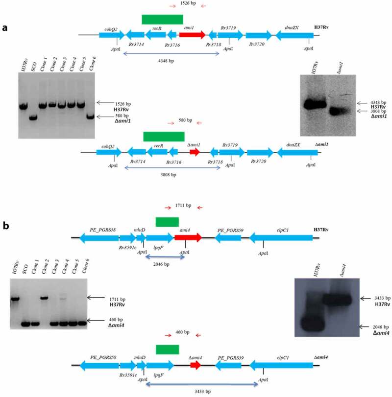
Peptidoglycan (PG), a heteropolysaccharide component of the mycobacterial cell wall can be shed during tuberculosis infection with immunomodulatory consequences. As such, changes in PG structure are expected to have important implications on disease progression and host responses during infection with Mycobacterium tuberculosis. Mycobacterial amidases have important roles in remodeling of PG during cell division and are implicated in susceptibility to antibiotics. However, their role in modulating host immunity remains unknown. We assessed the bacterial burden and host immune responses to M. tuberculosis mutants defective for either one of two PG N-acetylmuramyl-L-alanine amidases, Ami1 and Ami4, in bone marrow-derived macrophages (BMDM) and C57BL/6 mice. In infected BMDM, the single deletion of both genes resulted in increased proinflammatory cytokine responses. In mice, infection with the Δami1 mutant led to differential induction of pro-inflammatory cytokines and chemokines, decreased cellular recruitment and reduced lung pathology during the acute phase of the infection. While increased proinflammatory cytokines production was observed in BMDM infected with the Δami4 mutant, these effects did not prevail in mice. Infection using the Δami1 and Δami4 Mtb mutants showed that these genes are dispensable for intracellular mycobacterial growth in macrophages and mycobacterial burden in mice. These findings suggest that both Ami1 and Ami4 in M. tuberculosis are not essential for mycobacterial growth within the host. In summary, we show that amidases are important for modulating host immunity during Mtb infection in murine macrophages and mice.

Keywords: Ami1; Ami4; Mycobacterium tuberculosis; amidase; host–pathogen interaction; immunopathology; macrophage; mice; mutant.


Conflict of interest statement

No potential conflict of interest was reported by the author(s).

Figures

Figure 1.
Figure 1.
Genotypic analysis of the amidase Mtb strains by PCR and Southern blot analysis. The genomic map of the relevant locus is shown for the (a) H37RvΔami1 strain, (b) H37RvΔami4 strain and the wild-type H37Rv strain. Also shown on the left is the PCR confirmation of the genotype and Southern blotting is shown on the right. For PCR confirmation of ami1 and ami4, chromosomal DNA was used to amplify the ami1 alleles from the wild type and mutant strains using the primers described in Table S1 and indicated as red arrows above. The expected sizes of the amplicons are as follows: ami1/Rv3717:1526 bp and Δami1: 580 bp; ami4/Rv3594, 1711 bp and Δami4, 460 bp. For the Southern blot analysis, chromosomal DNA from the parental and mutant strain was digested with ApaI. The probe used for hybridization is shown as a solid green box and the expected sizes are indicated by the blue arrows. The figures are not drawn to scale
Figure 2.
Figure 2.
Mtb infection of IFN-γ-stimulated macrophages with the Δami1 and Δami4 mutant induced elevated proinflammatory responses with no effect on macrophage cell viability and intracellular Mtb growth. Cell viability of IFN-γ-stimulated (100 U/mL for ~12 h) BMDM infected with the (a) Δami1 mutant, complemented strain (Δami1::ami1) and wild-type H37Rv and (b) CFU counts of BMDM infected with the Δami1 mutant, complemented strain (Δami1::ami1) and wild-type H37Rv. Proinflammatory cytokine production from infected macrophages was measured via ELISA at 4- and 6-days post-infection for BMDMs infected with the (c) Δami1 mutant, complemented strain (Δami1::ami1) and wild-type H37Rv. (d) Cell viability of IFN-γ-stimulated (100 U/mL for ~12 h) BMDM infected with the Δami4 mutant, complemented strain (Δami4::ami4) and wild-type H37Rv was measured via Cell Titer Blue at 0 (4 hours), 2, 4 and 6 days post-infection. (e) CFU counts of BMDM infected with the Δami4 mutant, complemented strain (Δami4::ami4) and wild-type H37Rv. (f) Proinflammatory cytokine production from infected macrophages was measured via ELISA at 4- and 6-days post-infection for BMDMs infected with the Δami4 mutant, complemented strain (Δami4::ami4) and wild-type H37Rv. (*P ≤ 0.05, **P ≤ 0.01, ***P ≤ 0.001, ****P ≤ 0.0001, one-way ANOVA, n = 3). DPI = days post-infection. Data are representative of one (1A, 1D) and two (1B, 1 C, 1E and 1 F) experiments
Figure 3.
Figure 3.
Infection of mice with the Δami1 mutant results in an elevated cytokine/chemokine response at 3-WPI, reduced specific myeloid and lymphoid cell populations in the lung and reduced lung pathology. C57BL/6 mice were infected intranasally with 100 CFU/mouse of the Mtb Δami1 mutant, complemented Δami1::ami1 and WT, H37Rv. (a) Cytokine and chemokine levels from whole lung homogenates of Mtb-infected mice were measured by ELISA at 3-WPI. (b) H&E histology staining of Mtb-infected lungs at 3-WPI. Inflammation was quantified as a measure of alveolar space (Magnification 10x). Percentage of lung alveolar airspaces were quantified from 4 deep cuts of H&E lung sections per mice (30 μm apart). Each data point represents an individual cut. (C) Mice were sacrificed at 3- and 6-WPI to measure mycobacterial burden by CFU enumeration in the lungs. Infected mice were sacrificed at 3-WPI and lungs were collected to measure the frequency and cell numbers of (d) myeloid and (e) lymphoid cell populations. Alveolar macrophages (Alv MΦ) = CD64+SiglecF+CD11c+, recruited interstitial macrophages (Rec MΦ) = CD64+CD11cSiglecF+, CD103 dendritic cells (DC) = MHCII+CD11c+CD103+CD11b, CD11b DC = MHCII+CD11c+CD103CD11b+, neutrophils (Nφ) = LY6G+CD11b+, monocytes (MoM) = CD64+ CD11b+CD11c+, eosinophils (Eos) = CD64SiglecF+CD11b+, B cells = CD19+CD3, CD8+ T cells = CD3+CD4CD8+, CD4+ T cells = CD3+CD4+CD8, naïve T cells = CD62L+CD44+, memory T cells = CD62L+CD44 effector T cells = CD62LCD44+ (*P ≤ 0.05, **P ≤ 0.01, ***P ≤ 0.001, ****P ≤ 0.0001, one-way ANOVA, n = 5–6). Data in panels A to E are representative of two independent experiments
Figure 4.
Figure 4.
Mtb Ami4 is dispensable for the modulation of the host immune response during acute and chronic-phase infection and has no effect on lung pathology in mice. C57BL/6 mice were infected intranasally with 100 CFU/mouse of the Mtb Δami4 mutant, complemented Δami4::ami4 and WT, H37Rv. (a) Cytokine and chemokine levels from whole lung homogenates of Mtb-infected mice were measured by ELISA at 3-WPI. (b) H&E histology staining of Mtb-infected lungs at 3-WPI. Inflammation was quantified as a measure of alveolar space (Magnification 10x). Percentage of lung alveolar airspaces were quantified from 4 deep cuts of H&E lung sections per mice (30 μm apart). Each data point represents an individual cut. (c) Mice were sacrificed at 3- and 6-WPI to measure mycobacterial burden by CFU enumeration in the lungs. Infected mice were sacrificed at 3-WPI and lungs were collected to measure the frequency and cell numbers of (d) myeloid and (e) lymphoid cell populations. Alveolar macrophages (Alv MΦ) = CD64+SiglecF+CD11c+, recruited interstitial macrophages (Rec MΦ) = CD64+CD11cSiglecF+, CD103 dendritic cells (DC) = MHCII+CD11c+CD103+CD11b, CD11b DC = MHCII+CD11c+CD103CD11b+, neutrophils (Nφ) = LY6G+CD11b+, monocytes (MoM) = CD64+ CD11b+CD11c+, eosinophils (Eos) = CD64SiglecF+CD11b+, B cells = CD19+CD3, CD8+ T cells = CD3+CD4CD8+, CD4+ T cells = CD3+CD4+CD8, naïve T cells = CD62L+CD44+, memory T cells = CD62L+CD44 effector T cells = CD62LCD44+ (*P ≤ 0.05, **P ≤ 0.01, ***P ≤ 0.001, ****P ≤ 0.0001, one-way ANOVA, n = 5–6). Data in panels A to E are representative of one experiment

References

    1. World Health Organization . Global tuberculosis report 2019. Geneva: WHO. 2019.
    1. Cohen A, Mathiasen VD, Schön T, et al. The global prevalence of latent tuberculosis: a systematic review and meta-analysis. Eur Respir J. 2019;54:1900655. - PubMed
    1. Barry CE, Crick DC, McNeil MR.. Targeting the formation of the cell wall core of M. tuberculosis. Infect Disord Drug Targets. 2007;7:182–202. - PMC - PubMed
    1. Kaur D, Guerin ME, Skovierova H, et al. Chapter 2: biogenesis of the cell wall and other glycoconjugates of Mycobacterium tuberculosis. Adv Appl Microbiol. 2009;69:23–78. - PMC - PubMed
    1. Heijenoort J. Formation of the glycan chains in the synthesis of bacterial peptidoglycan. Glycobiology. 2001;11:25R–36R. - PubMed

Publication types