

Corneal nerve structure in patients with primary Sjögren's syndrome in China
- PMID: 33980205
- PMCID: PMC8117565
- DOI: 10.1186/s12886-021-01967-7
Corneal nerve structure in patients with primary Sjögren's syndrome in China
Abstract
Background: The aim of this study was to evaluate the in vivo confocal microscopic morphology of corneal subbasal nerves and its relationship with clinical parameters in patients with primary Sjögren's syndrome in China.
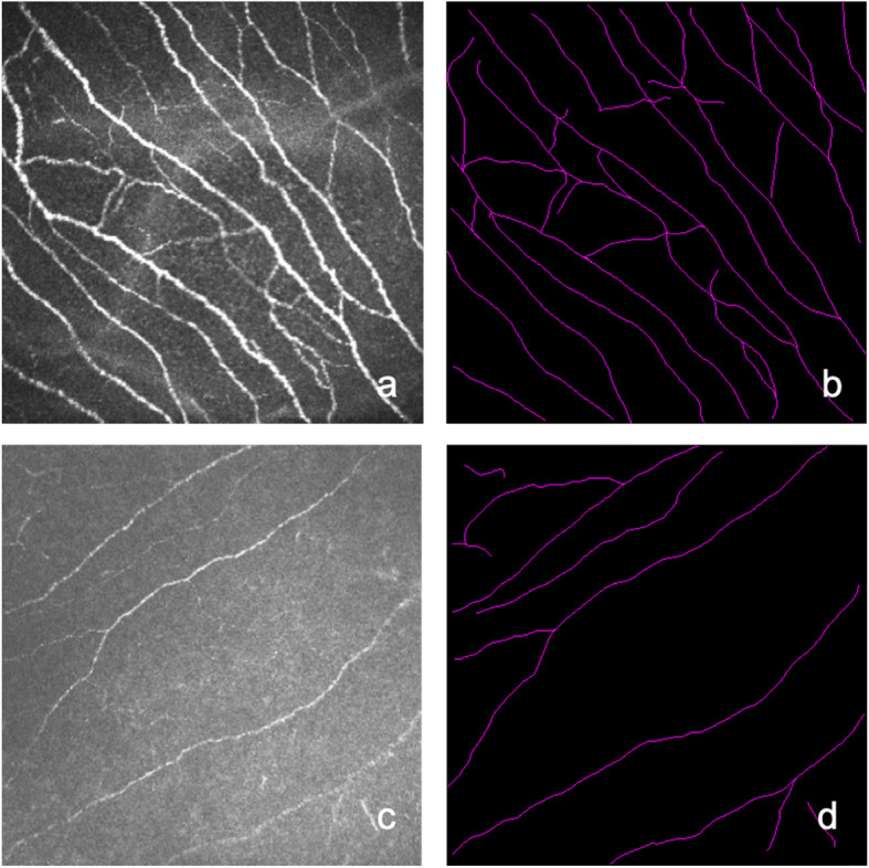
Methods: This was a case control study of 22 dry eye disease (DED) patients with primary Sjögren's syndrome (pSS) and 20 control subjects with non-Sjögren dry eye disease (NSDE). Each patient underwent an evaluation of ocular surface disease using the tear film break-up time (TBUT), noninvasive tear film break-up time (NIKBUT), noninvasive tear meniscus height (NIKTMH), corneal staining (National Eye Institute scale, NEI), Schirmer I test, meibography, and corneal subbasal nerve analysis with in vivo confocal microscopy (IVCM). The right eye of each subject was included in this study.
Results: SS patients showed a shorter TBUT (P = 0.009) and Schirmer I test results (P = 0.028) than the NSDE group. However, there was no significant difference in NIKBUT between the two groups (P = 0.393). The nerve density of subbasal nerves, number of nerves and tortuosity of the SS group were significantly lower than those of the NSDE group (P = 0.001, P < 0.001 and P = 0.039, respectively). In the SS group, the mean nerve length was correlated with age and the Schirmer I test (r = - 0.519, P = 0.013 and r = 0.463, P = 0.035, respectively). Corneal staining was correlated with nerve density and the number of nerves (r = - 0.534, P = 0.013 and r = - 0.487, P = 0.025, respectively).
Conclusions: Sjögren syndrome dry eye (SSDE) patients have more severe clinical dry eye parameters than non-Sjögren dry eye disease (NSDE) patients. Compared with NSDE patients, we found that SSDE patients showed decreased corneal subbasal nerve density and numbers.
Keywords: Corneal nerve; Dry eye disease; In vivo confocal microscopy (IVCM); Sjögren’s syndrome.
Conflict of interest statement
The authors declare that they have no competing interests.
Figures
References
-
- Craig JP, Nelson JD, Azar DT, Belmonte C, Bron AJ, Chauhan SK, de Paiva CS, Gomes JAP, Hammitt KM, Jones L, Nichols JJ, Nichols KK, Novack GD, Stapleton FJ, Willcox MDP, Wolffsohn JS, Sullivan DA. TFOS DEWS II report executive summary. Ocul Surf. 2017;15(4):802–812. doi: 10.1016/j.jtos.2017.08.003. - DOI - PubMed
-
- Solomon A, Dursun D, Liu Z, Xie Y, Macri A, Pflugfelder SC. Pro- and anti-inflammatory forms of interleukin-1 in the tear fluid and conjunctiva of patients with dry-eye disease. Invest Ophthalmol Vis Sci. 2001;42(10):2283–2292. - PubMed
MeSH terms
Grants and funding
LinkOut - more resources
Full Text Sources
Other Literature Sources
Medical
