. 2021 May 12;17(1):190.
doi: 10.1186/s12917-021-02887-w.

Identification of four insertion sites for foreign genes in a pseudorabies virus vector

Affiliations

Identification of four insertion sites for foreign genes in a pseudorabies virus vector

Chuanjian Zhang et al. BMC Vet Res. .

Abstract

Background: Pseudorabies virus (PRV) is a preferred vector for recombinant vaccine construction. Previously, we generated a TK&gE-deleted PRV (PRVΔTK&gE-AH02) based on a virulent PRV AH02LA strain. It was shown to be safe for 1-day-old piglets with maternal PRV antibodies and 4 ~ 5 week-old PRV antibody negative piglets and provide rapid and 100 % protection in weaned pigs against lethal challenge with the PRV variant strain. It suggests that PRVTK&gE-AH02 may be a promising live vaccine vector for construction of recombinant vaccine in pigs. However, insertion site, as a main factor, may affect foreign gene expression.

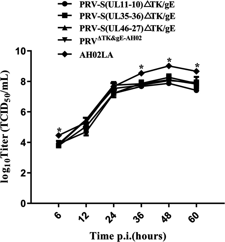
Results: In this study, we constructed four recombinant PRV-S bacterial artificial chromosomes (BACs) carrying the same spike (S) expression cassette of a variant porcine epidemic diarrhea virus strain in different noncoding regions (UL11-10, UL35-36, UL46-27 or US2-1) from AH02LA BAC with TK, gE and gI deletion. The successful expression of S gene (UL11-10, UL35-36 and UL46-27) in recombinant viruses was confirmed by virus rescue, PCR, real-time PCR and indirect immunofluorescence. We observed higher S gene mRNA expression level in swine testicular cells infected with PRV-S(UL11-10)ΔTK/gE and PRV-S(UL35-36)ΔTK/gE compared to that of PRV-S(UL46-27)ΔTK/gE at 6 h post infection (P < 0.05). Moreover, at 12 h post infection, cells infected with PRV-S(UL11-10)ΔTK/gE exhibited higher S gene mRNA expression than those infected with PRV-S(UL35-36)ΔTK/gE (P = 0.097) and PRV-S(UL46-27)ΔTK/gE (P < 0.05). Recovered vectored mutant PRV-S (UL11-10, UL35-36 and UL46-27) exhibited similar growth kinetics to the parental virus (PRVΔTK&gE-AH02).

Conclusions: This study focuses on identification of suitable sites for insertion of foreign genes in PRV genome, which laids a foundation for future development of recombinant PRV vaccines.

Keywords: Bacterial artificial chromosome; Insertion site; Noncoding region; Pseudorabies virus; Spike gene.


Conflict of interest statement

The authors declare that they have no competing interests.

Figures

Fig. 1
Fig. 1
Construction of PRV-S(UL11-10)ΔTK/gE, PRV-S(UL35-36)ΔTK/gE and PRV-S(UL46-27)ΔTK/gE. a The S expression cassette with a kanamycin resistance gene was inserted into the noncoding area (UL11-10, UL35-36, UL46-27 or US2-1) through the first recombination to generate four recombinant BAC clones (BACPRV−S−KAN(UL11−10)ΔTK/gE/gI, BACPRV−S−KAN(UL35–36)ΔTK/gE/gI, BACPRV−S−KAN(UL46−27)ΔTK/gE/gI and BACPRV−S−KAN(US2−1)ΔTK/gE/gI). b The second recombination was performed to delete the kanamycin resistance gene and generate the final recombinants (BACPRV−S(UL11−10)ΔTK/gE/gI, BACPRV−S(UL35–36)ΔTK/gE/gI, BACPRV−S(UL46−27)ΔTK/gE/gI and BACPRV−S(US2−1)ΔTK/gE/gI). c Homologous recombination was performed to recover the intact gI gene and part of gE gene (1299 to 1735 bp of gE open reading frame). d Schematic presentation of the PRV-S(UL11-10)ΔTK/gE, PRV-S(UL35-36)ΔTK/gE and PRV-S(UL46-27)ΔTK/gE were shown
Fig. 2
Fig. 2
RFLP of BACPRVΔTK/gE/gI, BACPRV−S−KAN(UL11−10)ΔTK/gE/gI, BACPRV−S(UL11−10)ΔTK/gE/gI, BACPRV−S−KAN(UL35–36)ΔTK/gE/gI, BACPRV−S(UL35–36)ΔTK/gE/gI, BACPRV−S−KAN(UL46−27)ΔTK/gE/gI, BACPRV−S(UL46−27)ΔTK/gE/gI, BACPRV−S−KAN(US2−1)ΔTK/gE/gI and BACPRV−S(US2−1)ΔTK/gE/gI. DNA of BACPRVΔTK/gE/gI, BACPRV−S−KAN(UL11−10)ΔTK/gE/gI, BACPRV−S(UL11−10)ΔTK/gE/gI, BACPRV−S−KAN(UL35–36)ΔTK/gE/gI, BACPRV−S(UL35–36)ΔTK/gE/gI, BACPRV−S−KAN(UL46−27)ΔTK/gE/gI, BACPRV−S(UL46−27)ΔTK/gE/gI, BACPRV−S−KAN(US2−1)ΔTK/gE/gI and BACPRV−S(US2−1)ΔTK/gE/gI were digested with BamH I. Predicted RFLP pattern with BamH I was performed using PRV ZJ01 strain (GenBank: KM061380.1) as a reference
Fig. 3
Fig. 3
Images of PRV-S(UL11-10)ΔTK/gE, PRV-S(UL11-10)-mini-FΔTK/gE/gI, PRV-S(UL35-36)ΔTK/gE, PRV-S(UL35-36)-mini-FΔTK/gE/gI, PRV-S(UL46-27)ΔTK/gE and PRV-S(UL46-27)-mini-FΔTK/gE/gI under UV excitation and phase contrast are shown. Each panel represents a view of 200 × 200 μm in size
Fig. 4
Fig. 4
PCR identification of S expression cassette and gI/ΔgE. S expression cassette of PRV-S(UL11-10)ΔTK/gE, PRV-S(UL35-36)ΔTK/gE and PRV-S(UL46-27)ΔTK/gE was identified by PCR with primers (S cas check F/S cas check R), and gI/ΔgE was identified by PCR with primers (PRV ΔgE check F/PRV ΔgE check R)
Fig. 5
Fig. 5
Confirmation of S and PRV protein expression by the recombinant PRV using indirect immunofluorescence assay. Pig anti-sera against PEDV or PRV in conjunction with FITC labelled anti-pig secondary antibodies were employed to verify S and PRV protein expression. Each panel represents a view of 200 × 200 μm in size
Fig. 6
Fig. 6
S gene mRNA expression on ST cells infected with PRV-S(UL11-10)ΔTK/gE, PRV-S(UL35-36)ΔTK/gE and PRV-S(UL46-27)ΔTK/gE at a MOI of 10 at 6 and 12 h post infection. Data are expressed in relative arbitrary units, in comparison with the values measured in ST cells infected with PRV-S(UL11-10)ΔTK/gE at 6 h post infection and taken as 1.00. Data were presented as mean ± SEM, and analyzed using one-way ANOVA with a Tukey’s post − hoc test (SPSS Inc., Chicago, IL, USA)
Fig. 7
Fig. 7
Multi-step growth curves of AH02LA, PRVΔTK&gE−AH02, PRV-S (UL11-10)ΔTK/gE, PRV-S(UL35-36)ΔTK/gE and PRV-S(UL46-27)ΔTK/gE. ST cells were infected with AH02LA and the four mutants at a MOI of 0.01. At 6, 12, 24, 36, 48 and 60 h post infection, the culture cells were harvested and then were titrated in ST cells. Asterisks indicate statistical significance between AH02LA and the four mutants (*p < 0.05). Data were presented as mean ± SEM, and analyzed using one-way ANOVA with a Tukey’s post − hoc test (SPSS Inc., Chicago, IL, USA)

Similar articles

Cited by

References

    1. Mettenleiter TC. Aujeszky’s disease (pseudorabies) virus: the virus and molecular pathogenesis–state of the art, June 1999. Vet Res. 2000;31(1):99–115. - PubMed
    1. Dong B, Zarlenga DS, Ren X: An overview of live attenuated recombinant pseudorabies viruses for use as novel vaccines. J Immunol Res. 2014;2014:824630. - PMC - PubMed
    1. Zhang C, Guo L, Jia X, Wang T, Wang J, Sun Z, et al. Construction of a triple gene-deleted Chinese Pseudorabies virus variant and its efficacy study as a vaccine candidate on suckling piglets. Vaccine. 2015;33(21):2432–37. doi: 10.1016/j.vaccine.2015.03.094. - DOI - PubMed
    1. Wang J, Song Z, Ge A, Guo R, Qiao Y, Xu M, et al. Safety and immunogenicity of an attenuated Chinese pseudorabies variant by dual deletion of TK&gE genes. BMC Vet Res. 2018;14(1):287. doi: 10.1186/s12917-018-1536-7. - DOI - PMC - PubMed
    1. Tong W, Zheng H, Li GX, Gao F, Shan TL, Zhou YJ, et al. Recombinant pseudorabies virus expressing E2 of classical swine fever virus (CSFV) protects against both virulent pseudorabies virus and CSFV. Antiviral Res. 2020;173:104652. doi: 10.1016/j.antiviral.2019.104652. - DOI - PubMed

LinkOut - more resources