

Comparison of single-voxel 1H-cardiovascular magnetic resonance spectroscopy techniques for in vivo measurement of myocardial creatine and triglycerides at 3T
- PMID: 33980263
- PMCID: PMC8117273
- DOI: 10.1186/s12968-021-00748-x
Comparison of single-voxel 1H-cardiovascular magnetic resonance spectroscopy techniques for in vivo measurement of myocardial creatine and triglycerides at 3T
Abstract
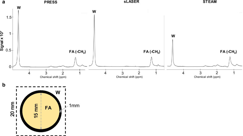
Background: Single-voxel proton cardiovascular magnetic resonance spectroscopy (1H-CMRS) benefits from 3 T to detect metabolic abnormalities with the quantification of intramyocardial fatty acids (FA) and creatine (Cr). Conventional point resolved spectroscopy (PRESS) sequence remains the preferred choice for CMRS, despite its chemical shift displacement error (CSDE) at high field (≥ 3 T). Alternative candidate sequences are the semi-adiabatic Localization by Adiabatic SElective Refocusing (sLASER) recommended for brain and musculoskeletal applications and the localized stimulated echo acquisition mode (STEAM). In this study, we aim to compare these three single-voxel 1H-CMRS techniques: PRESS, sLASER and STEAM for reproducible quantification of myocardial FA and Cr at 3 T. Sequences are compared both using breath-hold (BH) and free-breathing (FB) acquisitions.
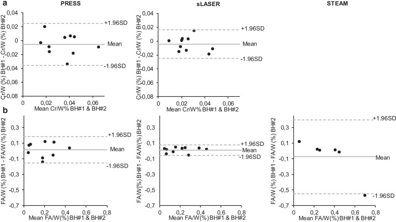
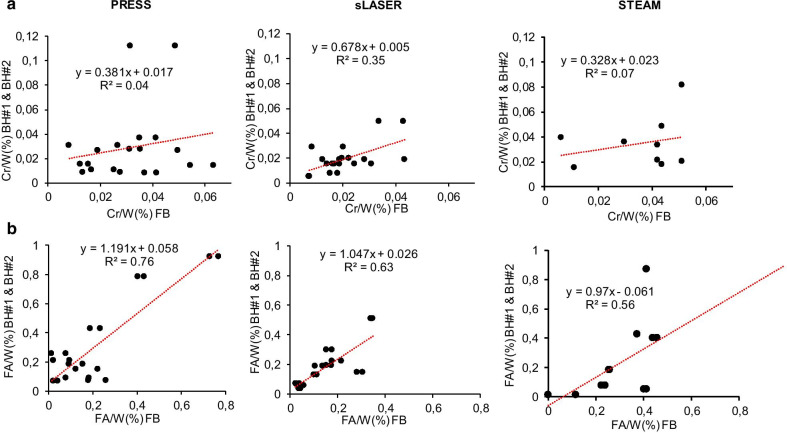
Methods: CMRS accuracy and theoretical CSDE were verified on a purposely-designed fat-water phantom. FA and Cr CMRS data quality and reliability were evaluated in the interventricular septum of 10 healthy subjects, comparing repeated BH and free-breathing with retrospective gating.
Results: Measured FA/W ratio deviated from expected phantom ratio due to CSDE with all sequences. sLASER supplied the lowest bias (10%, vs -28% and 27% for PRESS and STEAM). In vivo, PRESS provided the highest signal-to-noise ratio (SNR) in FB scans (27.5 for Cr and 103.2 for FA). Nevertheless, a linear regression analysis between the two BH showed a better correlation between myocardial Cr content measured with sLASER compared to PRESS (r = 0.46; p = 0.03 vs. r = 0.35; p = 0.07) and similar slopes of regression lines for FA measurements (r = 0.94; p < 0.001 vs. r = 0.87; p < 0.001). STEAM was unable to perform Cr measurement and was the method with the lowest correlation (r = 0.59; p = 0.07) for FA. No difference was found between measurements done either during BH or FB for Cr, FA and triglycerides using PRESS, sLASER and STEAM.
Conclusion: When quantifying myocardial lipids and creatine with CMR proton spectroscopy at 3 T, PRESS provided higher SNR, while sLASER was more reproducible both with single BH and FB scans.
Keywords: 3 T; CMR spectroscopy; Cardiac metabolism; Creatine; Lipids; Proton magnetic resonance spectroscopy; SLASER.
Conflict of interest statement
The authors declare that they have no competing interests. Dr. Robert Weiss served as a
Figures


Similar articles
-
Myocardial triglycerides in cardiac amyloidosis assessed by proton cardiovascular magnetic resonance spectroscopy.J Cardiovasc Magn Reson. 2019 Jan 31;21(1):10. doi: 10.1186/s12968-019-0519-6. J Cardiovasc Magn Reson. 2019. PMID: 30700314 Free PMC article.
-
Repeatability of proton magnetic resonance spectroscopy of the brain at 7 T: effect of scan time on semi-localized by adiabatic selective refocusing and short-echo time stimulated echo acquisition mode scans and their comparison.Quant Imaging Med Surg. 2021 Jan;11(1):9-20. doi: 10.21037/qims-20-517. Quant Imaging Med Surg. 2021. PMID: 33392007 Free PMC article.
-
MASE-sLASER, a short-TE, matched chemical shift displacement error sequence for single-voxel spectroscopy at ultrahigh field.NMR Biomed. 2018 Jul;31(7):e3940. doi: 10.1002/nbm.3940. Epub 2018 Jun 1. NMR Biomed. 2018. PMID: 29856517
-
Measurement sequences for single voxel proton MR spectroscopy.Eur J Radiol. 2008 Aug;67(2):194-201. doi: 10.1016/j.ejrad.2008.03.023. Epub 2008 Jul 2. Eur J Radiol. 2008. PMID: 18599235 Review.
-
Magnetic Resonance Spectroscopy for Cervical Cancer: Review and Potential Prognostic Applications.Cancers (Basel). 2024 Jun 5;16(11):2141. doi: 10.3390/cancers16112141. Cancers (Basel). 2024. PMID: 38893260 Free PMC article. Review.
Cited by
-
Investigating neural dysfunction with abnormal protein deposition in Alzheimer's disease through magnetic resonance spectroscopic imaging, plasma biomarkers, and positron emission tomography.Neuroimage Clin. 2024;41:103560. doi: 10.1016/j.nicl.2023.103560. Epub 2023 Dec 22. Neuroimage Clin. 2024. PMID: 38147791 Free PMC article.
-
sLASER and PRESS perform similarly at revealing metabolite-age correlations at 3 T.Magn Reson Med. 2024 Feb;91(2):431-442. doi: 10.1002/mrm.29895. Epub 2023 Oct 24. Magn Reson Med. 2024. PMID: 37876339 Free PMC article.
-
In Vivo Magnetic Resonance Spectroscopy Methods for Investigating Cardiac Metabolism.Metabolites. 2022 Feb 18;12(2):189. doi: 10.3390/metabo12020189. Metabolites. 2022. PMID: 35208262 Free PMC article. Review.
-
Magnetic resonance imaging of cardiac metabolism in heart failure: how far have we come?Eur Heart J Cardiovasc Imaging. 2022 Sep 10;23(10):1277-1289. doi: 10.1093/ehjci/jeac121. Eur Heart J Cardiovasc Imaging. 2022. PMID: 35788836 Free PMC article.
-
Long-term fasting: Multi-system adaptations in humans (GENESIS) study-A single-arm interventional trial.Front Nutr. 2022 Nov 17;9:951000. doi: 10.3389/fnut.2022.951000. eCollection 2022. Front Nutr. 2022. PMID: 36466423 Free PMC article.
References
-
- Reingold JS, McGavock JM, Kaka S, Tillery T, Victor RG, Szczepaniak LS. Determination of triglyceride in the human myocardium by magnetic resonance spectroscopy: reproducibility and sensitivity of the method. Am J Physiol Endocrinol Metab. 2005;289:E935–939. doi: 10.1152/ajpendo.00095.2005. - DOI - PubMed
Publication types
MeSH terms
Substances
LinkOut - more resources
Full Text Sources
Other Literature Sources
