. 2021 May 12;11(1):84.
doi: 10.1186/s13578-021-00560-1.

The YAP/HIF-1α/miR-182/EGR2 axis is implicated in asthma severity through the control of Th17 cell differentiation

Affiliations

The YAP/HIF-1α/miR-182/EGR2 axis is implicated in asthma severity through the control of Th17 cell differentiation

Jing Zhou et al. Cell Biosci. .

Abstract

Background: Asthma is a heterogeneous chronic inflammatory disease of the airway, involving reversible airflow limitation and airway remodeling. T helper 17 (Th17) cells play an important role in the pathogenesis of allergic asthma. However, there is limited understanding of the signaling pathways controlling Th17 cell differentiation in asthma. The aim of this study was to investigate if the Yes-associated protein (YAP)/hypoxia inducible factor-1α (HIF-1α)/microRNA-182 (miR-182)/early growth response 2 (EGR2) axis is involved in mediating Th17 cell differentiation and disease severity in asthma.

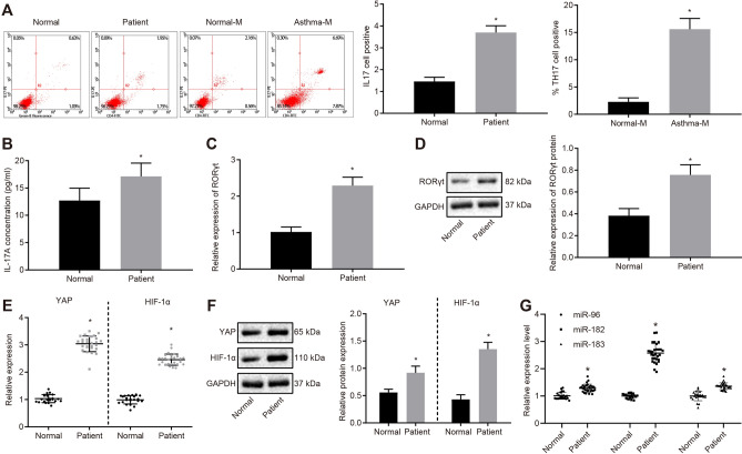
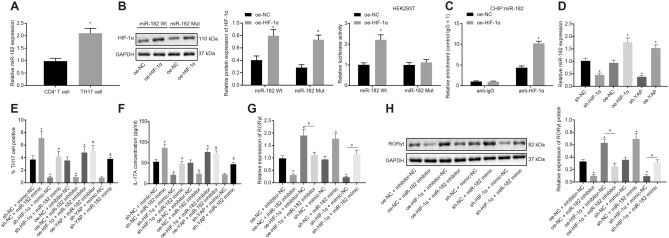
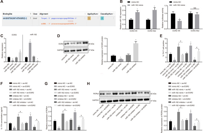
Methods: The study included 29 pediatric patients with asthma, 22 healthy volunteers, ovalbumin-induced murine asthma models, and mouse naive CD4+ T cells. The subpopulation of Th17 cells was examined by flow cytometry. The levels of interleukin-17A were determined by enzyme linked immunosorbent assay. Chromatin immunoprecipitation-quantitative polymerase chain reaction assays and dual-luciferase reporter gene assays were performed to examine interactions between HIF-1α and miR-182, and between miR-182 and EGR2.

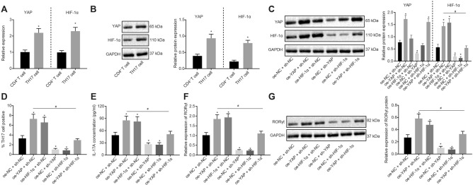
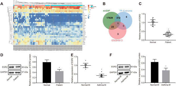
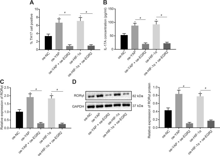
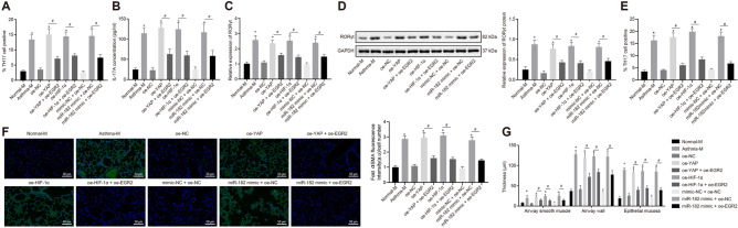
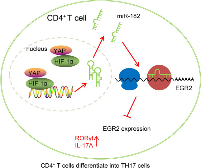
Results: YAP, HIF-1α, and miR-182 were upregulated but EGR2 was downregulated in human and mouse peripheral blood mononuclear cells from the asthma group. Abundant expression of YAP and HIF-1α promoted miR-182 expression and then inhibited EGR2, a target of miR-182, thus enhancing Th17 differentiation and deteriorating asthma and lipid metabolism dysfunction. In addition, in vivo overexpression of EGR2 countered the promoting effect of the YAP/HIF-1α/miR-182 axis on asthma and lipid metabolism dysfunction.

Conclusion: These results indicate that activation of the YAP/HIF-1α/miR-182/EGR2 axis may promote Th17 cell differentiation, exacerbate asthma development, and aggravate lipid metabolism dysfunction, thus suggesting a potential therapeutic target for asthma.

Keywords: Asthma; Differentiation; Dyslipidemia; EGR2; HIF-1α; MiR-182; Th17 cells; YAP.


Conflict of interest statement

The authors declare that they have no conflicts of interest.

Figures

Fig. 1
Fig. 1
Increased YAP, HIF-1α and miR-182 and decreased HDL-C level identified in asthma. a Th17 cell proportion in human PBMCs and mouse spleen cells detected by flow cytometry; b the serum level of IL-17A in asthma patients measured by ELISA; c RORγt mRNA expression in human PBMCs determined by RT-qPCR; d Western blot analysis of RORγt protein in human PBMCs; e mRNA expression of YAP and HIF-1α in human PBMCs determined by RT-qPCR; f Western blot analysis of YAP and HIF-1α proteins in human PBMCs; g miR-183/96/182 expression in human PBMCs determined by RT-qPCR. Comparisons between two groups were conducted by unpaired t test; * p < 0.05, compared with the normal individuals (normal); n = 22 in normal individuals; n = 29 in asthma patients. Each experiment was repeated 3 times independently
Fig. 2
Fig. 2
YAP and HIF-1α promote the differentiation of CD4+ T cells into Th17 cells. a mRNA expression of YAP and HIF-1α in Th17 cells determined by RT-qPCR; b Western blot analysis of YAP and HIF-1α proteins in Th17 cells; c Western blot analysis of YAP and HIF-1α proteins in Th17 cells following different treatments; d Th17 cell proportion after different treatments detected by flow cytometry; e IL-17A serum level in cell supernatant after different treatments by ELISA; f RORγt mRNA expression in cells after different treatments determined by RT-qPCR; g Western blot analysis of RORγt protein in cells after different treatments. Comparisons between two groups were conducted by unpaired t test, and those among multiple groups were conducted by one-way ANOVA with Tukey’s post hoc test; the experiments were repeated 3 times independently; * p < 0.05, compared with the CD4+ T cells, or cells treated with sh-NC, oe-NC, or sh-NC + oe-NC; # p < 0.05, compared with the cells treated with oe-YAP + sh-NC
Fig. 3
Fig. 3
HIF-1α upregulates miR-182 and then promotes Th17 cell differentiation. a miR-182 expression in Th17 cells determined by RT-qPCR; b the HIF-1α protein expression by Western blot analysis and the effect of HIF-1α on the miR-182 promoter activity detected by dual luciferase reporter gene assay; c the binding of HIF-1α to miR-182 promoter detected by ChIP assay; d miR-182 expression after HIF-1α/YAP overexpression or knockdown determined by RT-qPCR; e Th17 cell proportion after different treatments detected by flow cytometry; f the level of IL-17A in cell supernatant after different treatments determined by ELISA; g RORγt mRNA expression in cells after different treatments determined by RT-qPCR; h Western blot analysis of RORγt protein in cells after different treatments. Comparisons between two groups were conducted by unpaired t test, and those between multiple groups were conducted by one-way ANOVA with Tukey’s post hoc test; the experiments were repeated 3 times independently; * p < 0.05, compared with the normal individuals (normal), normal mice (normal-M), CD4+ T cells, cells treated with sh-NC, oe-NC, oe-NC + inhibitor-NC, or sh-NC + mimic-NC; # p < 0.05, compared with the cells treated with oe-HIF-1α + inhibitor-NC or sh-HIF-1α + mimic-NC; & p < 0.05, compared with the cells treated with oe-YAP + inhibitor-NC or sh-YAP + mimic-NC. n = 22 in normal individuals; n = 29 in asthma patients; n = 12 in normal mice; n = 12 in asthma mice
Fig. 4
Fig. 4
EGR2 expression is reduced in asthma. a the heatmap of DEGs related to asthma in respiratory epithelial cells, obtained from the GSE64913 dataset; the right upper histogram represents color gradation; b the Venn diagram showing the DEGs in respiratory epithelial cells from the GSE64913 dataset, downstream genes of miR-182 from the miRDIP database, and human transcription factors from the Cistrome database; c EGR2 mRNA expression in human PBMCs determined by RT-qPCR; d Western blot analysis of EGR protein in human PBMCs; e EGR2 mRNA expression in mouse spleen cells detected by RT-qPCR; f Western blot analysis of EGR2 protein in mouse spleen cells; Comparisons between two groups were conducted by unpaired t test. * p < 0.05, compared with the normal individuals (normal), normal mice (normal-M); n = 22 in normal individuals; n = 29 in asthma patients; n = 12 in normal mice; n = 12 in asthma mice. Each experiment was repeated 3 times independently
Fig. 5
Fig. 5
miR-182 inhibits EGR2 expression to promote Th17 cell differentiation. a the binding site between miR-182 and EGR2 3′UTR in mice predicted using starbase; b miR-182 expression determined by RT-qPCR and the binding of miR-182 to EGR2 confirmed by dual luciferase reporter gene assay; c miR-182 expression and EGR2 mRNA expression determined by RT-qPCR; d Western blot analysis of the EGR2 protein in cells after different treatments; e the Th17 cell proportion after different treatments determined by flow cytometry; f the level of IL-17A after different treatments by ELISA; g RORγt mRNA expression in cells after different treatments determined by RT-qPCR; h Western blot analysis of the RORγt protein in cells after different treatments. Comparisons between two groups were conducted by unpaired t test, and those between multiple groups were conducted by one-way ANOVA with Tukey’s post hoc test; the experiments were repeated 3 times independently; * p < 0.05, compared with cells treated with mimic-NC, inhibitor-NC, oe-NC + mimic-NC, or sh-NC + inhibitor-NC; # p < 0.05, compared with the cells treated with miR-182 mimic + oe-NC or miR-182 inhibitor + sh-NC
Fig. 6
Fig. 6
Overexpression of EGR2 inhibits Th17 cell differentiation induced by the YAP/HIF-1α/miR-182 signaling. a Th17 cell proportion after different treatments determined by flow cytometry; b the level of IL-17A in cell supernatant after different treatments measured by ELISA; c RORγt mRNA expression in cells after different treatments determined by RT-qPCR; d Western blot analysis of RORγt protein in cells after different treatments. Comparisons between multiple groups were conducted by one-way ANOVA with Tukey’s post hoc test; the experiments were repeated 3 times independently; * p < 0.05, compared with the cells treated with oe-NC; # p < 0.05, compared with the cells treated with oe-YAO or oe-HIF-1α
Fig. 7
Fig. 7
EGR2 overexpression alleviates asthma and lipid metabolism dysfunction induced by the YAP/HIF-1α/miR-182 axis in vivo. a the Th17 cell proportion in mouse spleen cells determined by flow cytometry; b the level of IL-17A in mouse serum measured by ELISA; c RORγt mRNA expression in mouse spleen cells determined by RT-qPCR; d Western blot analysis of RORγt protein in mouse spleen cells; e Th17 cell proportion in mouse lung tissues detected by flow cytometry. f Immunofluorescence staining showing the expression of α-SMA in the lung tissues of asthma mice
Fig. 8
Fig. 8
A schematic diagram illustrating the role of the YAP/HIF-1α/miR-182/EGR2 axis in asthma. The overexpression of YAP and HIF-1α promoted the expression of miR-182, thereby inhibiting EGR2 expression, increasing the expression of RORγt and IL-17A, and prompting the differentiation of CD4+T cells into Th17 cells, which ultimately aggravated asthma and lipid metabolism dysfunction

Similar articles

Cited by

References

    1. Dharmage SC, Perret JL, Custovic A. Epidemiology of asthma in children and adults. Front Pediatr. 2019 doi: 10.3389/fped.2019.00246. - DOI - PMC - PubMed
    1. To T, Stanojevic S, Moores G, Gershon AS, Bateman ED, Cruz AA, et al. Global asthma prevalence in adults: findings from the cross-sectional world health survey. BMC Public Health. 2012 doi: 10.1186/1471-2458-12-204. - DOI - PMC - PubMed
    1. Coleman SL, Shaw OM. Progress in the understanding of the pathology of allergic asthma and the potential of fruit proanthocyanidins as modulators of airway inflammation. Food Funct. 2017;8(12):4315–4324. doi: 10.1039/C7FO00789B. - DOI - PubMed
    1. Khan DA. Allergic rhinitis and asthma: epidemiology and common pathophysiology. Allergy Asthma Proc. 2014;35(5):357–361. doi: 10.2500/aap.2014.35.3794. - DOI - PubMed
    1. Castillo JR, Peters SP, Busse WW. Asthma exacerbations: pathogenesis, prevention, and treatment. J Allergy Clin Immunol Pract. 2017;5(4):918–927. doi: 10.1016/j.jaip.2017.05.001. - DOI - PMC - PubMed

LinkOut - more resources