Review
. 2021 Dec;105(12):1623-1631.
doi: 10.1136/bjophthalmol-2021-319228. Epub 2021 May 12.

Structural evaluation in inherited retinal diseases

Affiliations
Review

Structural evaluation in inherited retinal diseases

Malena Daich Varela et al. Br J Ophthalmol. 2021 Dec.

Abstract

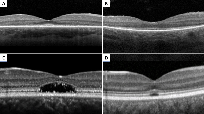
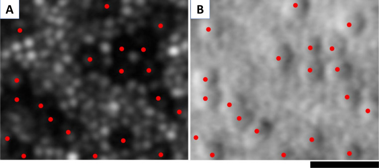
Ophthalmic genetics is a field that has been rapidly evolving over the last decade, mainly due to the flourishing of translational medicine for inherited retinal diseases (IRD). In this review, we will address the different methods by which retinal structure can be objectively and accurately assessed in IRD. We review standard-of-care imaging for these patients: colour fundus photography, fundus autofluorescence imaging and optical coherence tomography (OCT), as well as higher-resolution and/or newer technologies including OCT angiography, adaptive optics imaging, fundus imaging using a range of wavelengths, magnetic resonance imaging, laser speckle flowgraphy and retinal oximetry, illustrating their utility using paradigm genotypes with on-going therapeutic efforts/trials.

Keywords: clinical trial; dystrophy; genetics; imaging; retina.


Conflict of interest statement

Competing interests: The authors alone are responsible for the content and writing of this article. MM consults for MeiraGTx.

Figures

Figure 1
Figure 1
Examples of fundus autofluorescence patters in inherited retinal disease. Hypoautofluorescent defects secondary to: (A) reduced concentration of lipofuscin in RDH5-fundus albipunctatus, (B) retinal pigment epithelium atrophy in choroideraemia and (C) fibrotic tissue in late-stage BEST1 vitelliform macular dystrophy. Hyperautofluorescent defects secondary to: (D) increase in lipofuscin appearing as flecks in ABCA4-retinopathy, (E) drusen in EFEMP1-autosomal dominant drusen and (F) window defect in NMNAT1-Leber congenital amaurosis. (A–E) Short-wavelength and (F) near infrared autofluorescence images.
Figure 2
Figure 2
Optical coherence tomography evaluation of the ellipsoid zone (EZ). Foveal EZ preservation, with peripheral EZ loss, in (A) RPGR-RP and (B) RPE65-LCA. Foveal EZ loss, with peripheral EZ preservation, in (C) ABCA4-MD and (D) CNGB3-ACHM. RP: retinitis pigmentosa; LCA: Leber congenital amaurosis; ACHM, achromatopsia; MD, macular dystrophies.
Figure 3
Figure 3
Cellular imaging with adaptive optics scanning light ophthalmoscopy (AOSLO). AOSLO imaging in CNGA3-associated achromatopsia: (A) confocal image, with red dots marking the ‘dark’ (non-waveguiding) cones. Cones are surrounded by waveguiding rods. (B) Non-confocal (split detection) image over the exact same region with overlying red dots showing the cones marked in (A), which colocalise with cone inner segments, surrounded by rods. Scale bar: 20 µm.

References

    1. Rahman N, Georgiou M, Khan KN, et al. . Macular dystrophies: clinical and imaging features, molecular genetics and therapeutic options. Br J Ophthalmol 2020;104:451–60. 10.1136/bjophthalmol-2019-315086 - DOI - PMC - PubMed
    1. Gill JS, Georgiou M, Kalitzeos A, et al. . Progressive cone and cone-rod dystrophies: clinical features, molecular genetics and prospects for therapy. Br J Ophthalmol 2019;103:711–20. 10.1136/bjophthalmol-2018-313278 - DOI - PMC - PubMed
    1. Kumaran N, Moore AT, Weleber RG, et al. . Leber congenital amaurosis/early-onset severe retinal dystrophy: clinical features, molecular genetics and therapeutic interventions. Br J Ophthalmol 2017;101:1147–54. 10.1136/bjophthalmol-2016-309975 - DOI - PMC - PubMed
    1. Aboshiha J, Dubis AM, Carroll J, et al. . The cone dysfunction syndromes. Br J Ophthalmol 2016;100:115–21. 10.1136/bjophthalmol-2014-306505 - DOI - PMC - PubMed
    1. Hirji N, Aboshiha J, Georgiou M, et al. . Achromatopsia: clinical features, molecular genetics, animal models and therapeutic options. Ophthalmic Genet 2018;39:149–57. 10.1080/13816810.2017.1418389 - DOI - PubMed

Publication types