Systemic vascular air embolus following CT-guided transthoracic needle biopsy: a potentially fatal complication
- PMID: 33980551
- PMCID: PMC8118070
- DOI: 10.1136/bcr-2020-240406
Systemic vascular air embolus following CT-guided transthoracic needle biopsy: a potentially fatal complication
Abstract
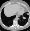
Following an uncomplicated CT-guided transthoracic biopsy, a patient becomes unconscious and subsequently dies despite immediate cardiac resuscitation. The patient felt well during the procedure but started complaining about dizziness and chest pain when he sat up. When he again was put in a supine position, cardiac arrest was noted. A CT scan performed when the symptoms initiated was afterwards rigorously reviewed by the team and revealed air located in the left ventricle, aorta and right coronary artery.We present a rare but potentially lethal complication following CT-guided transthoracic needle biopsy-systemic vascular air embolus. Knowledge and evidence about the complication are sparse because of low incidence and varying presentation. However, immediate initiation of treatment can save a life, and awareness of the complication is therefore crucial.
Keywords: cancer intervention; lung cancer (oncology); radiology (diagnostics).
© BMJ Publishing Group Limited 2021. No commercial re-use. See rights and permissions. Published by BMJ.
Conflict of interest statement
Competing interests: None declared.
Figures


Similar articles
-
Systemic air embolism as a complication of percutaneous computed tomography guided transthoracic lung biopsy.Ann R Coll Surg Engl. 2017 Jul;99(6):e174-e176. doi: 10.1308/rcsann.2017.0091. Ann R Coll Surg Engl. 2017. PMID: 28660818 Free PMC article.
-
Fatal cardiac air embolism after CT-guided percutaneous needle lung biopsy: medical complication or medical malpractice?Forensic Sci Med Pathol. 2024 Mar;20(1):199-204. doi: 10.1007/s12024-023-00639-w. Epub 2023 May 9. Forensic Sci Med Pathol. 2024. PMID: 37160632 Free PMC article.
-
Coronary artery air embolism complicating a CT-guided transthoracic needle biopsy of the lung.Chest. 2002 Mar;121(3):993-6. doi: 10.1378/chest.121.3.993. Chest. 2002. PMID: 11888990
-
Complications of CT-guided transthoracic lung biopsy : A short report on current literature and a case of systemic air embolism.Wien Klin Wochenschr. 2018 Apr;130(7-8):288-292. doi: 10.1007/s00508-018-1317-0. Epub 2018 Jan 23. Wien Klin Wochenschr. 2018. PMID: 29362884 Free PMC article. Review.
-
Cerebral air embolism during percutaneous computed tomography scan-guided liver biopsy.J Cancer Res Ther. 2018;14(7):1650-1654. doi: 10.4103/jcrt.JCRT_1035_17. J Cancer Res Ther. 2018. PMID: 30589054 Review.
Cited by
-
Risk factors associated with air embolism following computed tomography-guided percutaneous lung biopsy: a retrospective case-control study.PeerJ. 2024 Oct 15;12:e18232. doi: 10.7717/peerj.18232. eCollection 2024. PeerJ. 2024. PMID: 39430567 Free PMC article.
References
Publication types
MeSH terms
LinkOut - more resources
Full Text Sources
Other Literature Sources
Research Materials
