. 2021 Jul 12;95(15):e0036121.
doi: 10.1128/JVI.00361-21. Epub 2021 Jul 12.

Inhibition of Antiviral Innate Immunity by Foot-and-Mouth Disease Virus Lpro through Interaction with the N-Terminal Domain of Swine RNase L

Affiliations

Inhibition of Antiviral Innate Immunity by Foot-and-Mouth Disease Virus Lpro through Interaction with the N-Terminal Domain of Swine RNase L

Chao Sui et al. J Virol. .

Abstract

Foot-and-mouth disease virus (FMDV) is the pathogen of foot-and-mouth disease (FMD), which is a highly contagious disease in cloven-hoofed animals. To survive in the host, FMDV has evolved multiple strategies to antagonize host innate immune responses. In this study, we showed that the leader protease (Lpro) of FMDV, a papain-like proteinase, promoted viral replication by evading the antiviral interferon response through counteracting the 2',5'-oligoadenylate synthetase (OAS)/RNase L system. Specifically, we observed that the titers of Lpro deletion virus were significantly lower than those of wild-type FMDV (FMDV-WT) in cultured cells. Our mechanistic studies demonstrated that Lpro interfered with the OAS/RNase L pathway by interacting with the N-terminal domain of swine RNase L (sRNase L). Remarkably, Lpro of FMDV exhibited species-specific binding to RNase L in that the interaction was observed only in swine cells, not human, monkey, or canine cells. Lastly, we presented evidence that by interacting with sRNase L, FMDV Lpro inhibited cellular apoptosis. Taken together, these results demonstrate a novel mechanism that Lpro utilizes to escape the OAS/RNase L-mediated antiviral defense pathway. IMPORTANCE FMDV is a picornavirus that causes a significant disease in agricultural animals. FMDV has developed diverse strategies to escape the host interferon response. Here, we show that Lpro of FMDV antagonizes the OAS/RNase L pathway, an important interferon effector pathway, by interacting with the N-terminal domain of sRNase L. Interestingly, such a virus-host interaction is species-specific because the interaction is detected only in swine cells, not in human, monkey, or canine cells. Furthermore, Lpro inhibits apoptosis through interacting with sRNase L. This study demonstrates a novel mechanism by which FMDV has evolved to inhibit host innate immune responses.

Keywords: FMDV; ISGs; Lpro; RNase L; antagonistic mechanism.; foot-and-mouth disease virus.


Figures
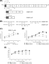
FIG 1
FIG 1
The growth kinetics of FMDV-WT and FMDV-L87. (A) Diagram of the FMDV-WT genome and FMDV-L87 genome. The viral open reading frame is boxed, and the shaded box represents the 87 nucleotides between the two initiation codons of L gene. (B) Sequences of FMDV-WT and FMDV-L87 surrounding the two initiation codons for Lab and Lb. (C) BHK-21 cells were infected with FMDV-WT and FMDV-L87 at an MOI of 1. Culture supernatants were collected at indicated time intervals and the virus titers were analyzed at indicated time points. (D and E) PK-15 and sRNase L KO-PK cells were individually infected with FMDV-WT and FMDV-L87 at an MOI of 1. The culture supernatants were collected at indicated time intervals. One part of the culture supernatants was titrated for TCID50. The other part was used to extract viral RNAs for quantifying viral cDNA copies by a real-time RT-PCR assay. (D) The TCID50s of FMDV-WT and FMDV-L87 on PK-15 and sRNase L KO-PK cells were determined at indicated time intervals. (E) Viral cDNA copies of FMDV-WT and FMDV-L87 on PK-15 and sRNase L KO-PK cells were quantified at indicated time points. Data represent the means ± the standard deviations (error bars) of three experiments. *, P < 0.05.
FIG 2
FIG 2
Lpro prevents RNA degradation. A total of 1 × 106 of PK-15 and sRNase L KO-PK cells were seeded in 12-well plates and grown to 80% confluence. Cells were transfected with 1 μg of pXJ41, pXJ41-L, pXJ41-L C51A, or pXJ41-L87 for 24 h and then stimulated with 2.5 μg/ml poly(I:C) for 7 h or 2.5 μM 2-5A for 5 h. Total RNAs were extracted and analyzed for RNA degradation by 1% agarose gel electrophoresis. (A) RNA degradation in PK-15 cells induced by 2-5A. (B) RNA degradation in PK-15 or sRNase L KO-PK cells induced by poly(I:C). (C) RNA degradation in PK-15 cells treated by poly(I:C). (D) PK-15 and sRNase L KO-PK cells were individually infected with FMDV-WT or FMDV-L87 at an MOI of 1. At 12 h postinfection, total RNAs were extracted and analyzed for RNA degradation. Data are from three independent experiments with similar results.
FIG 3
FIG 3
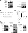
FMDV Lpro interacts with sRNase L. (A) HEK-293T cells were seeded in 6-well plates and grown to 80% confluence. Cells were cotransfected with 1 μg of pXJ41-sRNase L (HA tag) and indicated dose of pXJ41-L (FLAG tag) or pXJ41-L C51A (FLAG tag) for 24 h followed by Western blotting. (B) Schematic representation of LC-sRNase L, LN-L, and LN-L87. (C) HEK-293T cells were seeded in 12-well plates and grown to 80% confluence. One μg of LC-sRNase L and 1 μg of LN-L or LN-L87 were cotransfected into cells. At 24 h posttransfection, cells were lysed and determined for recombination Renilla luciferase reporter activity by luciferase assay system. The data represent the means of three independent experiments, with each sample assayed in triplicate. Error bars indicate the standard deviations of three experiments. *, P < 0.05. (D) HEK-293T cells were seeded in 60-mm-diameter dishes and grown to 80% confluence. Cells were cotransfected with 3 μg of pXJ41-sRNase L (HA tag) and 3 μg of pXJ41-L (FLAG tag) or pXJ41-L87 (FLAG tag) for 24 h. Cell lysates were subjected to coimmunoprecipitation using indicated antibodies (IP: FLAG or IP: HA). Immunocomplexes were analyzed by Western blotting using indicated antibodies (WB: HA or WB: FLAG). Whole-cell lysis (WCL) was also subjected to Western blotting using anti-HA, anti-FLAG, or anti-β-actin antibody. (E) Three μg of pXJ41-sRNase L (HA tag) and 3 μg of pXJ41-L C51A (FLAG tag) were transfected into HEK293-293T cells for 24 h. Cell lysates were subjected to coimmunoprecipitation using anti-FLAG antibody. Immunocomplexes were analyzed by Western blotting using anti-HA antibody. Whole-cell lysis (WCL) was also subjected to Western blotting using anti-HA, anti-FLAG, or anti-β-actin antibody. Data are representative of three independent experiments with similar results.
FIG 4
FIG 4
FMDV Lpro interacts with the N-terminal domain of sRNase L. (A) Schematic representation of human, swine, and chimeric RNase L structure. (B) HEK-293T cells were seeded in 60-mm-diameter dishes and grown to 80% confluence. Cells were cotransfected with 3 μg of pXJ41-L (FLAG tag) and 3 μg of pXJ41-sRNase L (HA tag), pXJ41-hRNase L (HA tag), pXJ41-Nh-Cs (HA tag), or pXJ41-Ns-Ch (HA tag) for 24 h. Cell lysates were subjected to coimmunoprecipitation using anti-FLAG antibody. Immunocomplexes were analyzed by Western blotting using anti-HA antibody. Whole-cell lysis (WCL) was also subjected to Western blotting using anti-HA, anti-FLAG, or anti-β-actin antibody. Data are representative of three independent experiments with similar results.
FIG 5
FIG 5
FMDV Lpro inhibits activation of RNase L in a species-specific manner. (A) A total of 1 × 106 of monkey embryonic kidney epithelial cells (MARC-145), Madin-Darby canine kidney cells (MDCK), porcine kidney cells (PK-15), and human cervical cancer cells (HeLa) were seeded in 12-well plates and grown to 80% confluence. Cells were transfected with 1 μg of pXJ41 or pXJ41-L and subsequently transfected with 2-5A for 5 h to activate RNase L. RNA degradation was assessed by agarose gel electrophoresis. (B) Expression of Lpro in MARC-145, MDCK, PK-15, and HeLa cells was detected by Western blotting using anti-FLAG antibody.
FIG 6
FIG 6
sRNase L dimerization is inhibited by FMDV Lpro. (A) Three μg of pXJ41-sRNase L (HA tag) and 3 μg of pXJ41-L (FLAG tag) or pXJ41-L87 (FLAG tag) were cotransfected into HEK-293T cells for 18 h. After being treated with 2-5A at a concentration of 5 μM for 5 h, cell lysates were prepared and analyzed by Western blotting. pXJ41-L87 (FLAG tag) transfected into HEK-293T cells without 2-5A treatment was used as a negative control. The upper panel shows sRNase L monomers and dimers using native-PAGE and Western blotting. The lower panel shows expression of Lpro or L87 by SDS-PAGE and Western blotting. (B) Three μg of pXJ41-sRNase L (HA tag) and 3 μg of pXJ41-L C51A (FLAG tag) were transfected into HEK-293T cells for 18 h. After treated with 5 μM 2-5A for 5 h, sRNase L dimerization was analyzed by native-PAGE and Western blotting. Expression of L C51A was analyzed by SDS-PAGE and Western blotting. β-actin served as a loading control. The data presented here are results representing data from three Western blotting experiments.
FIG 7
FIG 7
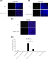
Apoptosis in FMDV-WT- and FMDV-L87-infected cells. (A to C) The nicked DNA in apoptotic cells induced by FMDV-WT (A), FMDV-L87 (B), or mock (C) was stained in green color by TUNEL assay and the nuclei were stained in blue color by DAPI. (D) The percentage of TUNEL positive cells. The number of cells representing apoptosis was determined by counting 100 cells in each random microscopic field. Each experiment was conducted in triplicate and repeated three times. Error bars indicate the standard deviations of three experiments. *, P < 0.05.
FIG 8
FIG 8
The working model for the negative regulation of OAS/RNase L signaling pathway by FMDV Lpro. FMDV Lpro inhibits the OAS/RNase L pathway through interaction with the N-terminal domain of sRNase L, which occurs downstream of the OAS catalytic activity.

Similar articles

Cited by

References

    1. Fry EE, Stuart DI, Rowlands DJ. 2005. The structure of foot-and-mouth disease virus. Curr Top Microbiol Immunol 288:71–101. 10.1007/3-540-27109-0_4. - DOI - PubMed
    1. Grubman MJ, Baxt B. 2004. Foot-and-mouth disease. Clin Microbiol Rev 17:465–493. 10.1128/CMR.17.2.465-493.2004. - DOI - PMC - PubMed
    1. Conda-Sheridan M, Lee SS, Preslar AT, Stupp SI. 2014. Esterase-activated release of naproxen from supramolecular nanofibres. Chem Commun (Camb) 50:13757–13760. 10.1039/c4cc06340f. - DOI - PMC - PubMed
    1. Strebel K, Beck E. 1986. A second protease of foot-and-mouth disease virus. J Virol 58:893–899. 10.1128/JVI.58.3.893-899.1986. - DOI - PMC - PubMed
    1. Steinberger J, Skern T. 2014. The leader proteinase of foot-and-mouth disease virus: structure-function relationships in a proteolytic virulence factor. Biol Chem 395:1179–1185. 10.1515/hsz-2014-0156. - DOI - PMC - PubMed

Publication types

MeSH terms

Substances

LinkOut - more resources