. 2021 Jul 12;95(15):e0075121.
doi: 10.1128/JVI.00751-21. Epub 2021 Jul 12.

The 5-HT3 Receptor Affects Rotavirus-Induced Motility

Affiliations

The 5-HT3 Receptor Affects Rotavirus-Induced Motility

Marie Hagbom et al. J Virol. .

Abstract

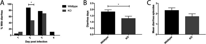
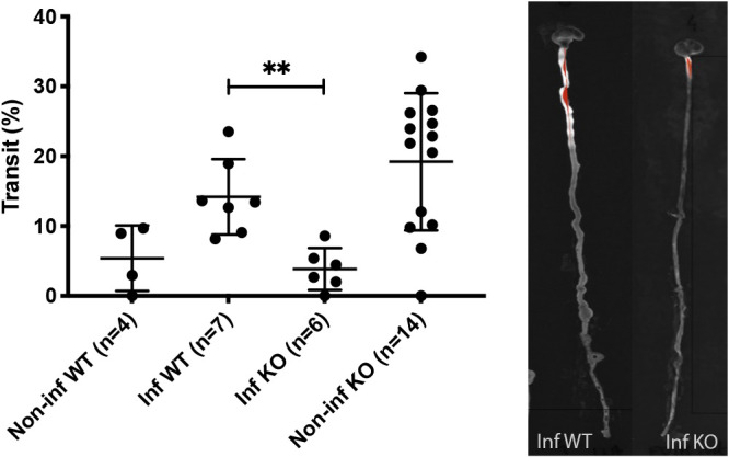
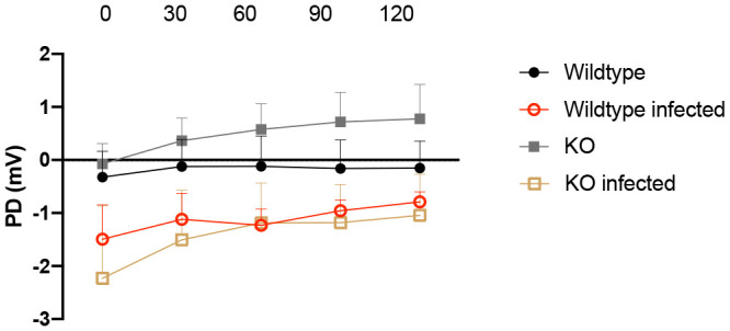
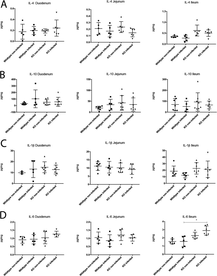
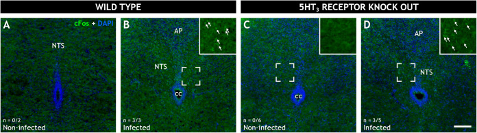
Rotavirus infection is highly prevalent in children, and the most severe effects are diarrhea and vomiting. It is well accepted that the enteric nervous system (ENS) is activated and plays an important role, but knowledge of how rotavirus activates nerves within ENS and to the vomiting center is lacking. Serotonin is released during rotavirus infection, and antagonists to the serotonin receptor subtype 3 (5-HT3 receptor) can attenuate rotavirus-induced diarrhea. In this study, we used a 5-HT3 receptor knockout (KO) mouse model to investigate the role of this receptor in rotavirus-induced diarrhea, motility, electrolyte secretion, inflammatory response, and vomiting reflex. The number of diarrhea days (P = 0.03) and the number of mice with diarrhea were lower in infected 5-HT3 receptor KO than wild-type pups. In vivo investigation of fluorescein isothiocyanate (FITC)-dextran transit time showed that intestinal motility was lower in the infected 5-HT3 receptor KO compared to wild-type mice (P = 0.0023). Ex vivo Ussing chamber measurements of potential difference across the intestinal epithelia showed no significant difference in electrolyte secretion between the two groups. Immediate early gene cFos expression level showed no difference in activation of the vomiting center in the brain. Cytokine analysis of the intestine indicated a low effect of inflammatory response in rotavirus-infected mice lacking the 5-HT3 receptor. Our findings indicate that the 5-HT3 receptor is involved in rotavirus-induced diarrhea via its effect on intestinal motility and that the vagus nerve signaling to the vomiting center occurs also in the absence of the 5-HT3 receptor. IMPORTANCE The mechanisms underlying rotavirus-induced diarrhea and vomiting are not yet fully understood. To better understand rotavirus pathophysiology, characterization of nerve signaling within the ENS and through vagal efferent nerves to the brain, which have been shown to be of great importance to the disease, is necessary. Serotonin (5-HT), a mediator of both diarrhea and vomiting, has been shown to be released from enterochromaffin cells in response to rotavirus infection and the rotavirus enterotoxin NSP4. Here, we investigated the role of the serotonin receptor 5-HT3, which is known to be involved in the nerve signals that regulate gut motility, intestinal secretion, and signal transduction through the vagus nerve to the brain. We show that the 5-HT3 receptor is involved in rotavirus-induced diarrhea by promoting intestinal motility. The findings shed light on new treatment possibilities for rotavirus diarrhea.

Keywords: 5-HT3 receptor; diarrhea; disease mechanisms; disease symptoms; motility; rotavirus; serotonin; vomiting.


Figures

FIG 1
FIG 1
Severity of diarrhea in rotavirus-infected wild-type and 5-HT3 receptor KO mice. (A) Percentage of KO pups and wild-type pups with diarrhea on day 2 (*, P = 0.012), which is the time point when the most severe diarrhea occurs in infant mice. (B) Number of mean diarrhea days in KO and wild-type mice (*, P = 0.03). (C) The mean number of diarrhea episodes in wild-type and KO pups show that there were fewer diarrhea episodes in the KO pups (P = 0.07). Wild-type mice, n = 18; KO mice, n = 27. Data are expressed as mean with SEM. (A) Fisher’s exact test; (B and C) Mann-Whitney test.
FIG 2
FIG 2
Immunofluorescence staining showing rotavirus infection in the small intestine of wild-type and 5-HT3 receptor KO mice. The images depict rotavirus infection in the duodenum (A and D), jejunum (B and E), and ileum (C and F) of wild-type (A to C) and 5-HT3 receptor KO (D to F) pups at 16 hpi. The intestine samples were stained with a primary antibody against viral protein 6 (an in-house guinea pig antibody) followed by goat anti-guinea-pig Alexa594 (red, virus). Nuclear staining with DAPI (blue). (G) Rotavirus fluorescence quantification was performed on a surface area with minimum of seven intestinal villi from each segment of wild-type and 5-HT3 receptor KO mice. The graph presents the percentage of rotavirus fluorescence correlated with the total surface area. ns, no significance; n = 7 villi.
FIG 3
FIG 3
Rotavirus-induced intestinal motility in wild-type and 5-HT3 receptor KO mice. The percentage transit of a 4-kDa FITC-dextran probe through the intestinal tract (from the pylorus to the rectum) (left) was visualized by a UV spectrophotometer as a fluorescence-dependent white color (right). The infected wild-type mice showed higher percentage of transit than infected 5-HT3 receptor KO mice. This indicate that increased motility due to rotavirus infection is partly dependent of the presence of the 5-HT3 receptor. Data are expressed as median with interquartile range. **, P = 0.0023; Mann-Whitney test.
FIG 4
FIG 4
Role of the 5-HT3 receptor in intestinal secretion induced by rotavirus infection. Potential difference (PD) was measured in jejunal segments (4 mice per group) from wild-type and 5-HT3 receptor KO pups at 48 hpi. PD was measured every 30 min over 2 h in Ussing chambers. There was no significant difference between infected wild-type and 5-HT3 receptor KO mice. This suggests that the 5-HT3 receptor does not play a major role in rotavirus-induced electrolyte secretion. mV, millivolt. Data are expressed as mean with SEM. Two-way ANOVA test.
FIG 5
FIG 5
Role of the 5-HT3 receptor in modulating the immune response during rotavirus infection. The levels of pro- and anti-inflammatory cytokines were measured in segments of the small intestine. No difference was detected in the anti-inflammatory response of IL-4 (A) and IL-10 (B) nor the proinflammatory cytokines IL-1β (C) or TNF-α (below the detection limits). The levels of IL-6 (D), which can act as both a pro- and anti-inflammatory cytokine, were higher in the infected and noninfected 5-HT3 receptor KO mice, but infected KO mice had higher IL-6 levels than the infected wild-type pups (P = 0.0006). This difference was also observed between noninfected 5-HT3 receptor KO mice and wild-type mice. This indicates that the 5HT3 receptor may not be of importance for inflammatory response during rotavirus infection. n = 5 to 8 mice per group. Data are expressed as mean with SD. One-way ANOVA.
FIG 6
FIG 6
Role of the 5-HT3 receptor in rotavirus-induced cFos expression in the nucleus of the solitary tract of wild-type and 5HT3R receptor KO mice. Representative wide-field low-power fluorescence micrographs of coronal mouse brain sections from 5- to 7-day-old noninfected (A and C) and infected (B and D) wild-type (A and B) and 5HT3 receptor KO (C and D) pups. The sections were stained for the immediate early gene cFos (green) and 4′,6-diamidino-2-phenylindole (DAPI; blue). The inserts (B, C, and D) present magnified views of the marked regions. cFos immunoreactive cell nuclei (arrows) can be observed in the nucleus of the solitary tract (NTS) of the infected mice (B and D) but not the noninfected mice (A and C). The micrograph indicates that there was no difference in cFos expression in the vomiting center of the brains of infected wild-type and 5-HT3 receptor KO-infected mice. This means that the receptor was not involved in the neural vomiting reflex mechanism. The number of animals (n) with cFos activity in the NTS is denoted in the bottom left corner. AP, area postrema; cc, central canal; NTS, nucleus of the solitary tract. Scale bar in panel D, 100 μm for panels A to D.

References

    1. Tate JE, Burton AH, Boschi-Pinto C, Parashar UD, World Health Organization-Coordinated Global Rotavirus Surveillance Network. 2016. Global, regional, and national estimates of rotavirus mortality in children <5 years of age, 2000–2013. Clin Infect Dis 62(Suppl 2):S96–S105. 10.1093/cid/civ1013. - DOI - PMC - PubMed
    1. Troeger C, Khalil IA, Rao PC, Cao S, Blacker BF, Ahmed T, Armah G, Bines JE, Brewer TG, Colombara DV, Kang G, Kirkpatrick BD, Kirkwood CD, Mwenda JM, Parashar UD, Petri WA, Jr., Riddle MS, Steele AD, Thompson RL, Walson JL, Sanders JW, Mokdad AH, Murray CJL, Hay SI, Reiner RC, Jr.. 2018. Rotavirus vaccination and the global burden of rotavirus diarrhea among children younger than 5 years. JAMA Pediatr 172:958–965. 10.1001/jamapediatrics.2018.1960. - DOI - PMC - PubMed
    1. Lundgren O, Peregrin AT, Persson K, Kordasti S, Uhnoo I, Svensson L. 2000. Role of the enteric nervous system in the fluid and electrolyte secretion of rotavirus diarrhea. Science 287:491–495. 10.1126/science.287.5452.491. - DOI - PubMed
    1. Hagbom M, Istrate C, Engblom D, Karlsson T, Rodriguez-Diaz J, Buesa J, Taylor JA, Loitto VM, Magnusson KE, Ahlman H, Lundgren O, Svensson L. 2011. Rotavirus stimulates release of serotonin (5-HT) from human enterochromaffin cells and activates brain structures involved in nausea and vomiting. PLoS Pathog 7:e1002115. 10.1371/journal.ppat.1002115. - DOI - PMC - PubMed
    1. Cloez-Tayarani I, Changeux JP. 2007. Nicotine and serotonin in immune regulation and inflammatory processes: a perspective. J Leukoc Biol 81:599–606. 10.1189/jlb.0906544. - DOI - PubMed

Publication types

MeSH terms

Substances