Review
. 2021 May 12;34(3):e00228-20.
doi: 10.1128/CMR.00228-20. Print 2021 Jun 16.

Tools and Techniques for Severe Acute Respiratory Syndrome Coronavirus 2 (SARS-CoV-2)/COVID-19 Detection

Affiliations
Review

Tools and Techniques for Severe Acute Respiratory Syndrome Coronavirus 2 (SARS-CoV-2)/COVID-19 Detection

Seyed Hamid Safiabadi Tali et al. Clin Microbiol Rev. .

Abstract

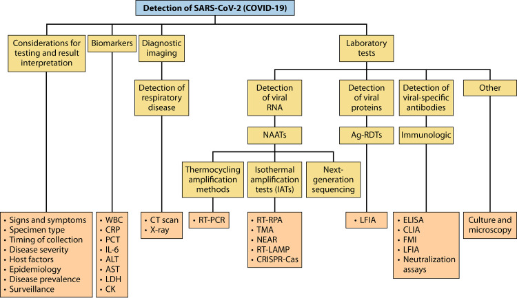
The coronavirus disease 2019 (COVID-19) pandemic, caused by severe acute respiratory disease coronavirus 2 (SARS-CoV-2), has led to millions of confirmed cases and deaths worldwide. Efficient diagnostic tools are in high demand, as rapid and large-scale testing plays a pivotal role in patient management and decelerating disease spread. This paper reviews current technologies used to detect SARS-CoV-2 in clinical laboratories as well as advances made for molecular, antigen-based, and immunological point-of-care testing, including recent developments in sensor and biosensor devices. The importance of the timing and type of specimen collection is discussed, along with factors such as disease prevalence, setting, and methods. Details of the mechanisms of action of the various methodologies are presented, along with their application span and known performance characteristics. Diagnostic imaging techniques and biomarkers are also covered, with an emphasis on their use for assessing COVID-19 or monitoring disease severity or complications. While the SARS-CoV-2 literature is rapidly evolving, this review highlights topics of interest that have occurred during the pandemic and the lessons learned throughout. Exploring a broad armamentarium of techniques for detecting SARS-CoV-2 will ensure continued diagnostic support for clinicians, public health, and infection prevention and control for this pandemic and provide advice for future pandemic preparedness.

Keywords: 2019-nCoV; COVID-19; NAAT; PCR; SARS-CoV-2; antigen; biomarkers; coronavirus; next-generation sequencing; serology.


Figures

FIG 1
FIG 1
Timeline of COVID-19 spread and the global response to it (10, 13, 23–30). Of note, while SARS-CoV-2 was initially thought to have emerged from China in December 2020, there are data to suggest that it may have circulated more broadly earlier than initially recorded in other countries, and further studies are under way to investigate this possibility in other areas (571–574).
FIG 2
FIG 2
Physical and genome structure of SARS-CoV-2. (A) Diagram of the SARS-CoV-2 virion. (B) Genome organization and proteins with known or unknown functions.
FIG 3
FIG 3
Real-time RT-PCR analysis. (A) Typical steps required for the detection of SARS-CoV-2 with real-time RT-PCR. (B) Principle of real-time RT-PCR. (1) During reverse transcription, reverse transcriptase (RT) creates a cDNA from the viral RNA template, with the aid of the reverse primer (or random oligonucleotides). The RNase H activity of the RT digests the initial RNA template. (2) The DNA polymerase activity of RT (or of the Taq polymerase) completes the second DNA strand guided by the forward primer and cDNA. (3) The newly formed double-stranded DNA (dsDNA) is used as a template for the PCR portion of the assay. At the annealing stage, the reverse primer binds to the sense strand of dsDNA in a sequence-specific manner, and the forward primer and a dually labeled probe bind to the antisense strand of the DNA. In this stage, the fluorophore (F) present on the probe is masked by the quencher (Q). During the extension step, the DNA polymerase extends the forward primer and, in the process, hydrolyzes the probe, resulting in the release of the fluorophore. Next, following excitation, fluorescence emission can be captured by the real-time thermocycler. With each round of PCR amplification, the dsDNA amplicon is multiplied by a 2-fold factor, with a proportional increase in the overall fluorescence signal. After 30 to 40 cycles of amplification, the RT-PCR is complete. The PCR cycle at which the fluorescence signal crosses the threshold for positivity is called the threshold cycle (CT), and CT values are inversely proportional to the quantity of the target present in the reaction mixture.
FIG 4
FIG 4
Mechanism of RT-RPA. The RT-RPA reactions typically occur at between 37°C and 42°C in the following steps. (1) The reaction is initiated by the binding of a recombinase (e.g., T4 UvsX) and a loading factor (e.g., T4 UvsY) to each of the forward and reverse primers. (2) These recombinase/loading factor/oligonucleotide complexes search for homologous sequences in dsDNA, formed in the RT reaction from viral RNA (not depicted). (3) Once sequence homology is found, the recombinase complex invades the duplex DNA, forming a structure called a D-loop in an ATP-dependent reaction, where there is the unwinding of dsDNA and binding of the primer to its complementary sequence. Access to the primer-binding sequence is possible due to the stabilization of the opposite strand by SSBs (e.g., T4 gp32). Subsequently, the recombinase and loading factor disassemble and are released to initiate other rounds of target recognition. (4) Following the binding of the forward and reverse primers, these primers are extended at their 3′ ends using a strand displacement DNA polymerase (e.g., Bsu), and during the elongation process, there is a further separation of the two strands. (5) Eventually, SSBs are displaced, and the replication of both strands is complete.
FIG 5
FIG 5
Principle of TMA. (1) The reactions use a reverse primer that is complementary to the sequence of the RNA template, but the reverse primer also contains an overhang with a promoter sequence for T7 RNA polymerase at its 5′ end. (2) Reverse transcription is conducted by the RT; the newly transcribed cDNA includes both the target sequence and the T7 promoter. (3) The RNA template is digested by the RNase H activity of the RT. (4) dsDNA is produced by the DNA polymerase activity of the RT. (5) The produced dsDNA is used as the template for transcription mediated by the T7 RNA polymerase. RNA is thereby amplified severalfold and, through the activity of the same enzyme(s), can serve as the template for a new TMA reaction. As the cycle progress, exponential amplification ensues. Detection of the amplified RNA is usually accomplished using sequence-specific molecular beacons (“torch”) or hybridization probes targeting the single-stranded RNA (ssRNA).
FIG 6
FIG 6
Principle of NEAR technology. The NEAR amplification reactions occur at 60°C and can be broken down into two milestones: NEAR amplification duplex formation and product formation. (1) The target recognition region (B′) of the reverse primer (R) binds to the complementary sequence (B) of the target DNA sense strand and is fully extended by the strand displacement DNA polymerase. (2) A second R primer binds to the template DNA and, during extension, displaces the elongated product of the first R primer extension. (3) The recognition region (A) of the forward primer (F) binds to its complementary sequence (A′) in the R extension product, and F is extended to create a double-stranded nicking enzyme recognition site (N). (4) The nicking enzyme recognizes N and cleaves a single strand of DNA in a sequence-specific manner at the cut site (X). (5) This releases a fragment of the R extension product. The remaining fragment serves as a primer and is extended at its 3′ end. (6) This extension completes the double-stranded complex, termed the NEAR amplification duplex, which is the starting point for product formation. (7) Nicking enzymes bind to the nicking enzyme recognition sites on both ends of the NEAR amplification duplex and cleave at X. (8) The resulting single-strand nicks create two complexes, each consisting of a single-stranded target region flanked by a nicking enzyme recognition region. (9) Repeated nicking, polymerization, and strand displacement activities result in the amplification of the AB and A′B′ target products. Cleaved complexes are regenerated, while the AB and A′B′ products can anneal to R and F primers, respectively. In turn, the bidirectional extension of the primer and product each creates duplexes that lead to the generation of the opposite product upon cleavage. Product amplification continues until reagents or enzymes are depleted.
FIG 7
FIG 7
Amplification of nucleic acids using RT-LAMP. Overall, there are four core primers that mediate all the processes in a LAMP reaction by recognizing six distinct regions of the target DNA through several steps. (1) After the conversion of the template RNA into dsDNA via reverse transcription (not shown), the LAMP reaction starts from strand invasion by the forward inner primer (FIP), which hybridizes through its F2 region to the F2c region of the target DNA. This initiates complementary-strand synthesis using a strand displacement DNA polymerase. (2) The forward outer primer (FOP) (also termed the F3 primer) then hybridizes to the F3c region of the target DNA and, during extension, displaces the newly elongated strand from the FIP. (3) Given that the FIP also contains an F1c sequence, the strand displacement triggered by the DNA polymerase and the FOP leads to the formation of a self-annealing loop in the 5′ end of the FIP-linked strand (regions F1 and F1c). (4) This single-stranded DNA with a stem-loop at its 5′ end then serves as a template for the backward inner primer (BIP), which hybridizes to the B2c region of the template DNA through its B2 sequence. (5) During elongation, the complementary strand opens the 5′-end stem-loop. Next, the backward outer primer (BOP) (also termed the B3 primer) hybridizes to the B3c region of the target DNA, and its elongation displaces the BIP-linked complementary strand. (6) The displacement of the BIP-linked strand results in self-hybridization on both the 5′ and 3′ ends, leading to two stem-loops and the formation of a dumbbell-shaped DNA. (7 to 9) The amplification of the dumbbell structure with the FIP leads to a concatemer and the formation of a second dumbbell structure that can be amplified with the BIP. Amplification can occur from the 3′ end of each dumbbell structure or with the annealing of primers such as the FIP and BIP. Additional loop primers (i.e., loop F [LF] and loop B [LB] primers) can also be used to increase the speed and sensitivity (41, 350, 351). Visualization of LAMP amplification is typically done by using pH-sensitive colorimetric or intercalating fluorescent dyes.
FIG 8
FIG 8
Principle of CRISPR-Cas technology for viral RNA detection. First, the viral RNA is subjected to reverse transcription and amplification, e.g., in an RT-RPA reaction at 37°C to 42°C, to generate dsDNA. The dsDNA can be targeted by guide RNAs (gRNAs) directly in a CRISPR-Cas12 detection system, whereas RNA detection using the CRISPR-Cas13 system requires an additional T7 transcription step. When Cas12 or Cas13 is activated by the recognition of gRNA, there will be cleavage of the target as well as nonspecific cleavage of dually labeled oligonucleotide probes. The probes are ssDNA or ssRNA for the CRISPR-Cas12 or CRISPR-Cas13 systems, respectively. The readout for either method can be colorimetric by the incorporation of fluorescein amidite (FAM)/fluorescein isothiocyanate (FITC)-biotin probes and the use of lateral flow dipsticks, or fluorometric readouts can be used by the incorporation of dually labeled fluorophore (F)-quencher (Q) probes. Upon collateral cleavage, the unquenched fluorophore can be excited with blue light, and the resulting emission of fluorescence can be visualized or captured with a fluorometer.
FIG 9
FIG 9
Sequencing techniques for identification of SARS-CoV-2. (A) Sanger sequencing. First, SARS-CoV-2 RNA is often amplified by RT-PCR (not depicted). Sanger sequencing reactions can be undertaken to analyze either of the DNA strands, but only one strand per reaction can be assessed. The extension of the primer annealing to the template DNA occurs in the presence of DNA polymerase, buffer, cofactors, deoxynucleotide triphosphates (dNTPs), and fluorescently labeled dideoxynucleotide triphosphates (ddNTPs). The binding of the ddNTPs to the oligonucleotide strands will cease the extension, resulting in various DNA structures with different lengths. Next, the extended DNAs undergo capillary gel electrophoresis in which the shorter DNA strands move faster, resulting in the detection of the fluorescently labeled nucleotides in the order of the size of the DNA strands. Finally, as DNA fragments are resolved and nucleotide-specific fluorescence signals are captured by a detector, a chromatogram is assembled to reveal the sequence of the template. (B) Next-generation sequencing (NGS) by synthesis. First, a library of millions of DNA fragments is created from the template (or enhanced by multiplex RT-PCR for SARS-CoV-2). Adapters are bound to the two ends of each DNA fragment. The adapters consist of a universal primer-binding site and a unique sequence (i.e., barcode) that can be hybridized to a specific sequence on the support (e.g., flow cell). Following hybridization, with complementary sequences of the adapters, bridge amplification is used to amplify each DNA fragment at a defined physical position. In the sequencing and detection steps, fluorescently labeled nucleotides are bound to the forward strands in the presence of a primer and a polymerase, which results in the generation of fluorescent light that is detected by an analyzer in real time. Many other NGS technologies are also available. (C) For example, NGS by nanopore technology is presented. After creating a library of DNA fragments by multiplex RT-PCR and barcoding, the library is loaded onto a membrane containing nanopores. The nanopores are proteins that open the DNA double strand, and as each nucleotide is passed through the membrane, it causes a specific change in the ionic current that can then be translated into the nucleotide sequence of the templates.
FIG 10
FIG 10
Antigen testing for the detection of SARS-CoV-2. (A) Principle of a lateral flow immunochromatographic assay (LFIA). The design of the LFIA for antigen detection is a qualitative immunological reaction confined to a small portable device (e.g., cassette or dipstick) that can be performed in the laboratory or a POC setting. Briefly, antigens in specimens (e.g., nasal swabs, nasopharyngeal swabs, and saliva) are placed in a well with a sample pad, and the fluid containing the antigen flows through the device via capillary action. The bottom of the well where the specimen is inoculated contains a sample pad, which is in contact with the conjugate pad used as a support for SARS-CoV-2-specific monoclonal antibodies (mAbs) that are labeled with colloidal gold nanoparticles (AuNPs) or other tags. If present, SARS-CoV-2 antigen (usually S or N protein) forms a complex with the mAbs bound to the AuNPs, and the entire complex migrates via capillary action until it is captured by other SARS-CoV-2 antigen-specific mAbs immobilized on the nitrocellulose membrane (i.e., the test line). As antigen-antibody complexes are trapped at this location, they form a line that can be visualized by the naked eye or with the aid of a detector. Also, mAbs-AuNPs, whether conjugated with antigens or not, continue to migrate until captured by an isotype-specific antibody directed against the fragment crystallizable (Fc) portion of the mAb at the control line. This ensures proper liquid flow through the device and test validity. (B) Point-of-care detection of SARS-CoV-2 antigen using a FET-based sensor. Upon binding of spike (S) proteins to the anti-S monoclonal antibodies immobilized on the graphene sheet via the PBASE linker, a change in the voltage-ampere diagram reveals the presence of the virus. (Panel B is adapted from reference with permission of the American Chemical Society.)
FIG 11
FIG 11
Common serological immunoassays for the detection of SARS-CoV-2-specific antibodies. (A) Common designs of ELISA methods, including indirect, modified indirect, and double-antigen sandwich assays. (B) Magnetic bead-based CLIA.
FIG 12
FIG 12
Serological lateral flow immunoassays for the detection of SARS-CoV-2-specific antibodies. (A) Schematic of a lateral flow immunoassay device for the simultaneous detection of IgM and IgG antibodies. Upon the addition of the sample, the liquid moves toward the preimmobilized reagents through capillary action and reacts with them. If IgM/IgG antibodies are present in the sample, they bind and form a complex with the recombinant SARS-CoV-2 antigen conjugated with colloidal gold nanoparticles (AuNPs). The complex is then captured in one of the test lines by anti-human IgM/IgG antibodies, resulting in a pink color due to the accumulation of the AuNPs. There is also an AuNP-rabbit IgG conjugate that will be captured in the control line, indicating proper liquid flow through the device. The results will be observable in ∼15 min. (B) Variations of results of the lateral flow assay device in a cassette format. (This figure was inspired by the work in reference .)
FIG 13
FIG 13
Methods used for SARS-CoV-2 detection or identification of COVID-19. Of note, cell culture and microscopy are not used for clinical diagnosis but are used for research purposes. Abbreviations: WBC, white blood cell; CRP, C-reactive protein; PCT, procalcitonin; IL-6, interleukin 6; ALT, alanine aminotransferase; AST, aspartate aminotransferase; LDH, lactate dehydrogenase; CK, creatine kinase; NAAT, nucleic acid amplification test; RT-PCR, reverse transcription-PCR; TMA, transcription-mediated amplification; RT-LAMP, reverse transcription–loop-mediated isothermal amplification; RT-RPA, reverse transcription-recombinase polymerase amplification; CRISPR-Cas, clustered regularly interspaced short palindromic repeat (CRISPR)-CRISPR associated (Cas); NEAR, nicking enzyme-assisted reaction; Ag-RDT, antigen rapid diagnostic test; LFIA, lateral flow immunoassay; ELISA, enzyme-linked immunosorbent assay; CLIA, chemiluminescence immunoassay; FMI, fluorescent microparticle immunoassay; CT scan, computed tomography scan.

References

    1. LeBlanc JJ, Li Y, Bastien N, Forward KR, Davidson RJ, Hatchette TF. 2009. Switching gears for an influenza pandemic: validation of a duplex reverse transcriptase PCR assay for simultaneous detection and confirmatory identification of pandemic (H1N1) 2009 influenza virus. J Clin Microbiol 47:3805–3813. 10.1128/JCM.01344-09. - DOI - PMC - PubMed
    1. Lu R, Zhao X, Li J, Niu P, Yang B, Wu H, Wang W, Song H, Huang B, Zhu N, Bi Y, Ma X, Zhan F, Wang L, Hu T, Zhou H, Hu Z, Zhou W, Zhao L, Chen J, Meng Y, Wang J, Lin Y, Yuan J, Xie Z, Ma J, Liu WJ, Wang D, Xu W, Holmes EC, Gao GF, Wu G, Chen W, Shi W, Tan W. 2020. Genomic characterisation and epidemiology of 2019 novel coronavirus: implications for virus origins and receptor binding. Lancet 395:565–574. 10.1016/S0140-6736(20)30251-8. - DOI - PMC - PubMed
    1. Harding H, Broom A, Broom J. 2020. Aerosol-generating procedures and infective risk to healthcare workers from SARS-CoV-2: the limits of the evidence. J Hosp Infect 105:717–725. 10.1016/j.jhin.2020.05.037. - DOI - PMC - PubMed
    1. Tang S, Mao Y, Jones RM, Tan Q, Ji JS, Li N, Shen J, Lv Y, Pan L, Ding P, Wang X, Wang Y, MacIntyre CR, Shi X. 2020. Aerosol transmission of SARS-CoV-2? Evidence, prevention and control. Environ Int 144:106039. 10.1016/j.envint.2020.106039. - DOI - PMC - PubMed
    1. Jarvis MC. 2020. Aerosol transmission of SARS-CoV-2: physical principles and implications. Front Public Health 8:590041. 10.3389/fpubh.2020.590041. - DOI - PMC - PubMed

Publication types

MeSH terms