. 2021 Dec 20;27(6):355-365.
doi: 10.5761/atcs.oa.20-00398. Epub 2021 May 12.

Effect of CTLA4-Ig on Obliterative Bronchiolitis in a Mouse Intrapulmonary Tracheal Transplantation Model

Affiliations

Effect of CTLA4-Ig on Obliterative Bronchiolitis in a Mouse Intrapulmonary Tracheal Transplantation Model

Yamato Suzuki et al. Ann Thorac Cardiovasc Surg. .

Abstract

Objectives: One of the serious problems after lung transplantation is chronic lung allograft dysfunction (CLAD). Most CLAD patients pathologically characterized by obliterative bronchiolitis (OB). Cytotoxic T-lymphocyte-associated antigen 4 (CTLA4)-Ig is a combination protein of the Fc fragment of human IgG1 linked to the extracellular domain of CTLA4. The aim of the study was to examine the effect of CTLA4-Ig therapy on OB using a mouse intrapulmonary tracheal transplantation (IPTT) model.

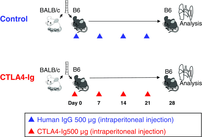
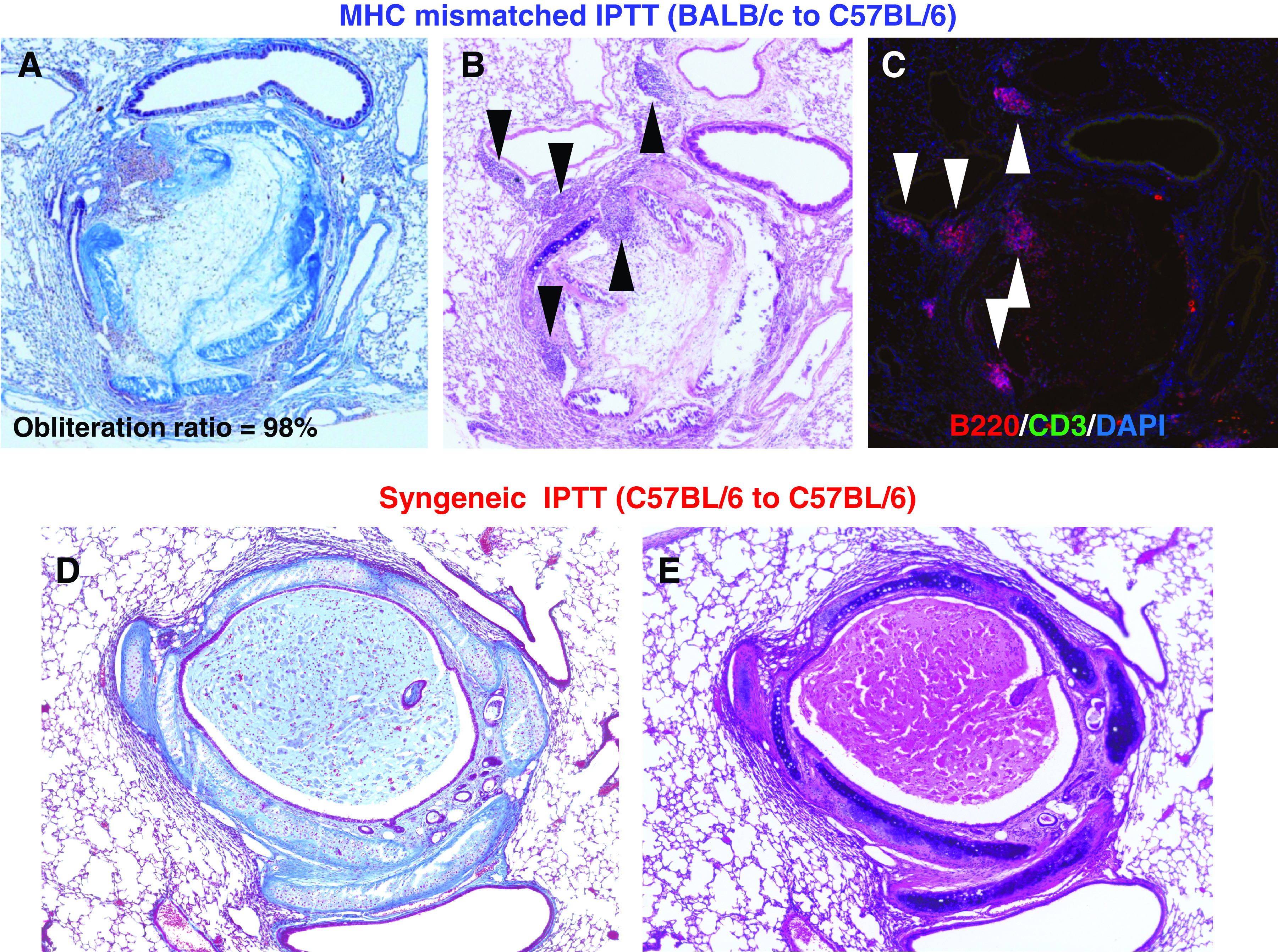
Methods: IPTT was performed between BALB/c (donor) and C57BL/6 (recipient) mice. Abatacept, which is a commercially available form of CTLA4-Ig, was intraperitoneally injected in recipient mice immediately after surgery, on days 7, 14, and 21. The mice in the control group received human IgG.

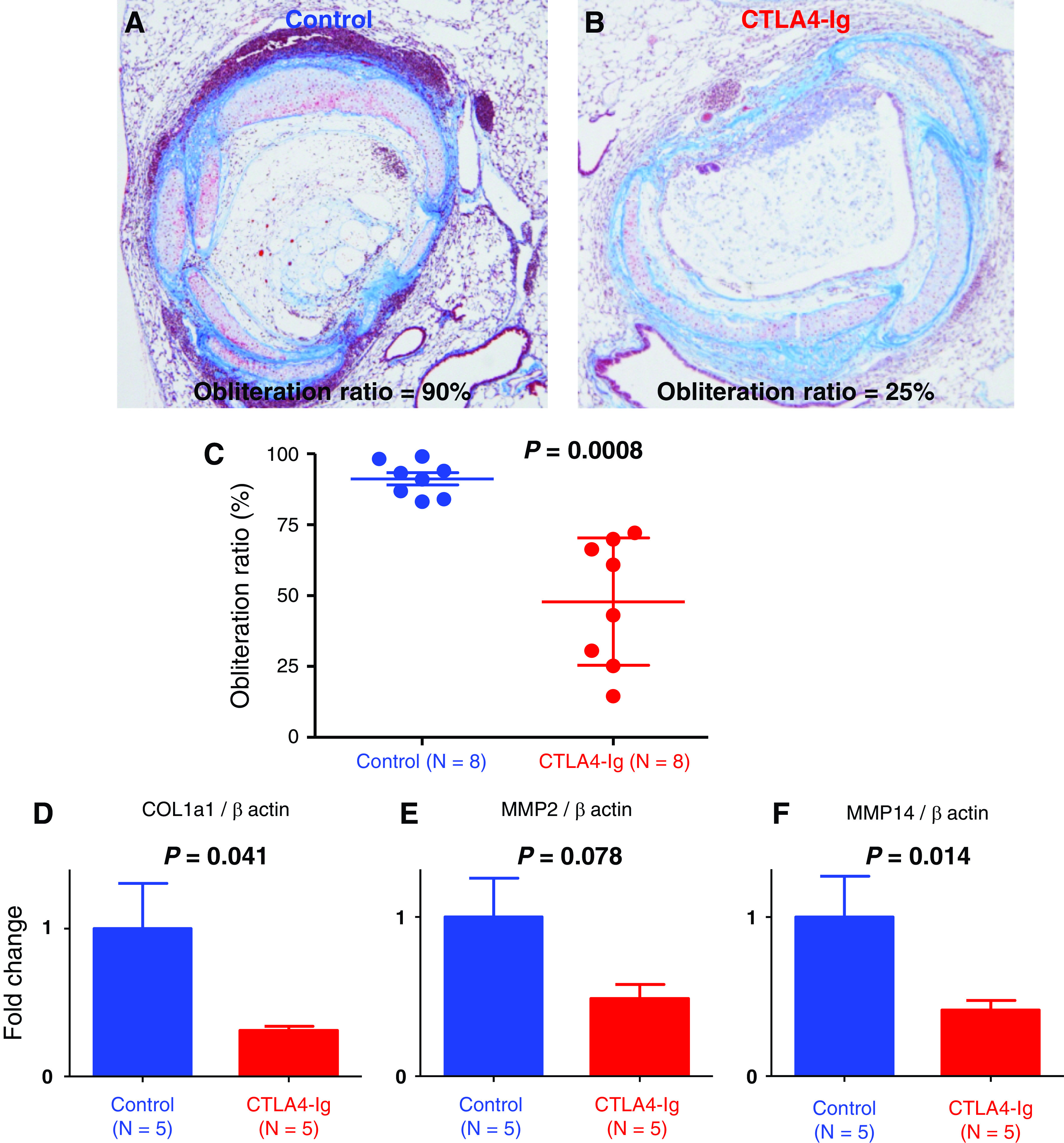
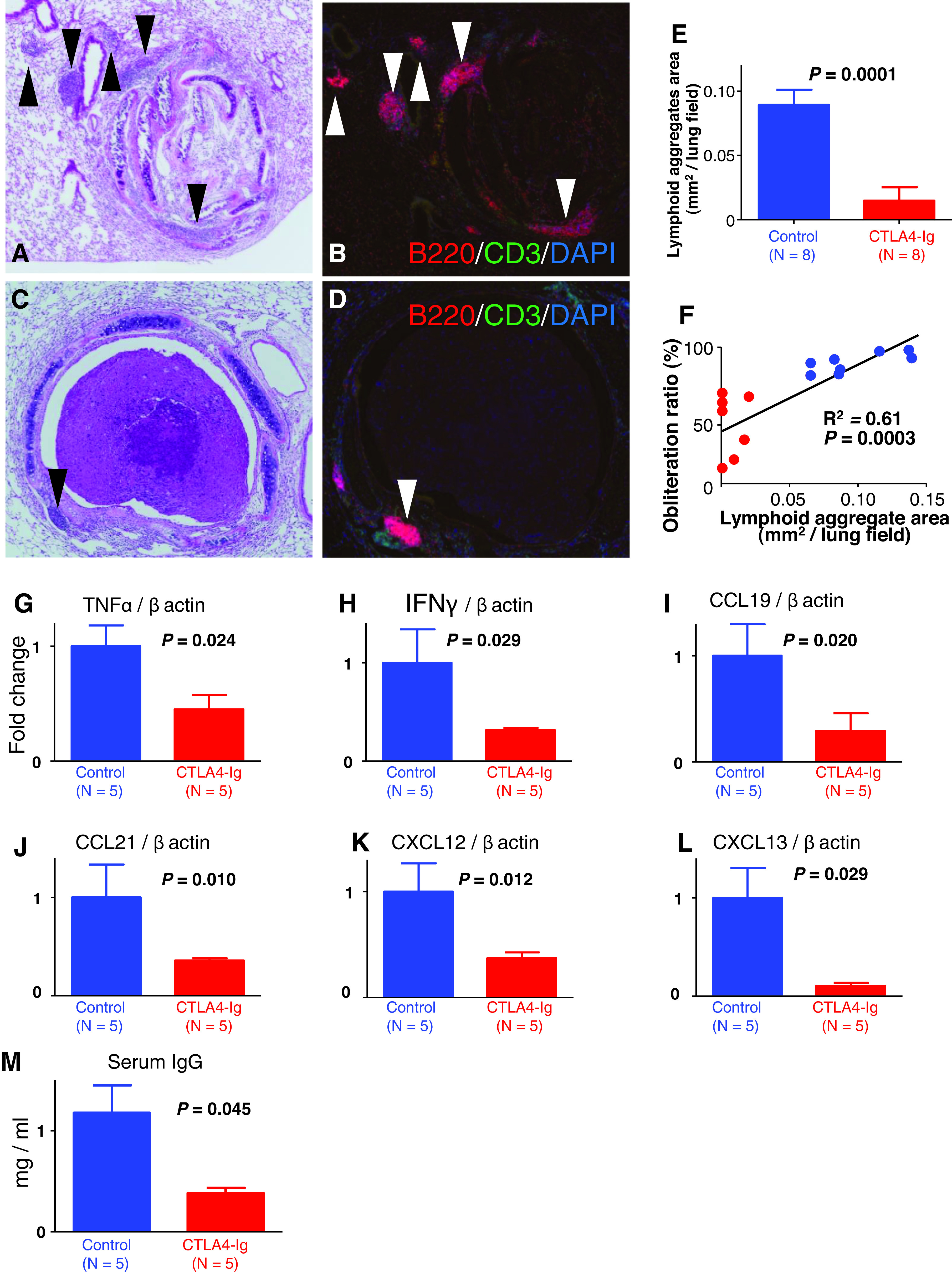
Results: We performed semi-quantitative analysis of graft luminal obliteration at post-transplant day 28. We calculated the obliteration ratio of the lumen of the transplanted trachea in each case. The obliteration ratio was significantly lower in the CTLA4-Ig group than that in the control group (91.2 ± 2.1% vs. 47.8 ± 7.9%, p = 0.0008). Immunofluorescent staining revealed significantly decreased lymphoid neogenesis in the lung.

Conclusions: CTLA4-Ig therapy attenuated tracheal obliteration with fibrous tissue in the mouse IPTT model. The attenuation of fibrous obliteration was correlated with the inhibition of lymphoid neogenesis.

Keywords: chronic lung allograft dysfunction; cytotoxic T-lymphocyte-associated antigen 4; lung transplantation; obliterative bronchiolitis.


Figures

Fig. 1
Fig. 1. Study design. C57BL/6 recipients were intraperitoneally injected with 500 μg of abatacept, which is a commercially available form of CTLA4-Ig, diluted in 200 μl PBS immediately after surgery, on days 7, 14, and 21. The recipients in the control group received human IgG (isotype control) in the same manner. On post-transplant day 28, the mice in both control and CTLA4-Ig groups were sacrificed and lungs were extracted. CTLA4: cytotoxic T-lymphocyte- associated antigen 4; PBS: phosphate-buffered saline
Fig. 2
Fig. 2. Fibrous obliteration and lymphoid neogenesis after MHC mismatched IPTT on day 28 after IPTT. (A and B) Pathological findings from a case of MHC mismatched IPTT, BALB/c to C57BL/6. Masson’s trichrome and hematoxylin–eosin staining, magnification 100. The transplanted trachea shows typical luminal obliteration with collagen and fibrous tissue. The destruction of the tracheal structure (tracheal ring) is also observed. Lymphoid aggregates in the recipient lung are seen adjacent to the transplanted trachea. (C) Immunofluorescent staining from a case of MHC mismatched IPTT. Immunofluorescent staining, magnification 100. Areas of CD3 positive T cells and B220 positive B cells in the lymphoid aggregates are observed. (D and E) Pathological findings from a case of syngeneic IPTT (C57BL/6 to C57BL/6). Masson’s trichrome and hematoxylin–eosin staining, magnification 100. There is no fibrous obliteration in the tracheal lumen, and the structure of the transplanted trachea is well preserved. There is no lymphoid aggregate in the recipient lung. IPTT: intrapulmonary tracheal transplantation; MHC: major histocompatibility complex
Fig. 3
Fig. 3. Effect of CTLA4-Ig treatment on fibrous obliteration in allografts. (A and B) Representative histology of a case from the control group (mice treated with human IgG) and from the CTLA4-Ig group (mice treated with CTLA4-Ig) on post-transplant day 28. Masson’s trichrome staining, magnification 100. A case from the control group shows severe fibrous obliteration of the transplanted trachea and the obliteration ration is 90%. A case from the CTLA4-Ig group 3B shows mild obliteration and the obliteration ration is 25%. (C) Obliteration ratio of the transplanted trachea on post-transplant day 28. The obliteration ratio was significantly lower in the CTLA4-Ig group than that in the control group (91.2 ± 2.1% vs. 47.8 ± 7.9%, p <0.001; control group, n = 8; CTLA4-Ig group, n = 8). (D) Analysis of mRNA expression of COL1a1 gene in lung grafts on post-transplant day 28 (control group, n = 5; CTLA4-Ig group, n = 5). The expression of COL1a1 in the CTLA4-Ig group was significantly decreased compared with that in the control group (p = 0.041). (E and F) Analysis of mRNA expression of MMP-2 and MMP-14 gene in lung grafts on post-transplant day 28 (control group, n = 5; CTLA4-Ig group, n = 5). The expression of MMP-2 tended to decrease in the CTLA4-Ig group (p = 0.078). The expression of MMP-14 in the CTLA4-Ig group was significantly decreased compared with that in the control group (p = 0.014). COL1a1: collagen type I alpha 1; CTLA4: cytotoxic T-lymphocyte- associated antigen 4; MMP-2: matrix metalloproteinase-2
Fig. 4
Fig. 4. Effect of CTLA4-Ig treatment on lymphoid neogenesis, mRNA expression of cytokines and chemokines in the lung and plasma IgG. (A and B) Representative histology of a case from the CTLA4-Ig group (mice treated with CTLA4-Ig) on post-transplant day 28. Hematoxylin–eosin staining and immunofluorescent staining for B220/CD3/DAPI, magnification 100. Black and white arrow heads indicate lymphoid aggregates. (C and D) Representative histology of a case from the control group (mice treated with human IgG) on post-transplant day 28. Hematoxylin–eosin staining and immunofluorescent staining for B220/CD3/DAPI, magnification 100. Each arrow head indicates (black and white) lymphoid aggregates on post-transplant day 28. (E) Total area of the lymphoid aggregates in each group on post-transplant day 28 (control group, n = 8; CTLA4-Ig group, n = 8). The total area of the lymphoid aggregates in the CTLA4-Ig group was significantly decreased compared to the control group (0.096 ± 0.010 mm2 vs. 0.0066 ± 0.0028 mm2, p = 0.001). (F) Correlation between the total lymphoid aggregates area and the obliteration on post-transplant day 28 (control group, red dots, n = 8; CTLA4-Ig group, blue dots, n = 8). There was a significant positive correlation between them (R² = 0.61, p = 0.001). (G and H) Analysis of mRNA expressions of TNF-α and in the lung on post-transplant day 28 (control group, n = 5; CTLA4-Ig group, n = 5). The expression of TNF-α was significantly lower in the CTLA4-Ig group than that in the control group (p = 0.024). The expression of IFN-γ in the CTLA4-Ig group (p = 0.29). (I–L) Analysis of mRNA expressions of CCL19, CCL21, CXCL 12 and CXCL 13 in the lung on post-transplant day 28 (control group, n = 5; CTLA4-Ig group, n = 5). The expression of CCL19 in the CTLA4-Ig group was significantly lower than that in the control group (p = 0.020). The expression of CCL21 showed a trend toward a decrease with the CTLA4-Ig treatment (p = 0.10). The expression of CXCL12 in the CTLA4-Ig group was significantly lower than that in the control group (p = 0.012). The expression of CXCL13 in the CTLA4-Ig group was significantly lower than in the control group (p = 0.042). (M) Analysis of plasma IgG level on post-transplant day 28 (control group, n = 5; CTLA4-Ig group, n = 5). Plasma IgG was significantly lower in the CTLA4-Ig group than that in the control group (p = 0.042). CCL19: C-C motif chemokine ligand 19; CTLA4: cytotoxic T-lymphocyte-associated antigen 4; CXCL 12: C-X-C motif chemokine ligand 12; IFN-γ: interferon-gamma; TNF-α: tumor necrosis factor-alpha

Similar articles

Cited by

References

    1. Chambers DC, Cherikh WS, Goldfarb SB, et al. . The International Thoracic Organ Transplant Registry of the International Society for Heart and Lung Transplantation: thirty-fifth adult lung and heart-lung transplant report—2018. Focus theme: Multiorgan Transplantation. J Heart Lung Transplant 2018; 37: 1169– 83. - PubMed
    1. Verleden GM, Glanville AR, Lease ED, et al. . Chronic lung allograft dysfunction: definition, diagnostic criteria, and approaches to treatment—A consensus report from the Pulmonary Council of the ISHLT. J Heart Lung Transplant 2019; 38: 493– 503. - PubMed
    1. Matsumura Y, Zuo XJ, Prehn J, et al. . Soluble CTLA4Ig modifies parameters of acute inflammation in rat lung allograft rejection without altering lymphocytic infiltration or transcription of key cytokines. Transplantation 1995; 59: 551– 8. - PubMed
    1. Vincenti F, Rostaing L, Grinyo J, et al. . Belatacept and long-term outcomes in kidney transplantation. N Engl J Med 2016; 374: 333– 43. - PubMed
    1. Iasella CJ, Winstead RJ, Moore CA, et al. . Memory T cells in transplantation: Old challenges define new directions. Transplantation 2018; 104: 2024– 34. - PMC - PubMed