. 2021 May 12;12(5):474.
doi: 10.1038/s41419-021-03749-x.

Fas/FasL mediates NF-κBp65/PUMA-modulated hepatocytes apoptosis via autophagy to drive liver fibrosis

Affiliations

Fas/FasL mediates NF-κBp65/PUMA-modulated hepatocytes apoptosis via autophagy to drive liver fibrosis

Siwei Tan et al. Cell Death Dis. .

Abstract

Fas/Fas ligand (FasL)-mediated cell apoptosis involves a variety of physiological and pathological processes including chronic hepatic diseases, and hepatocytes apoptosis contributes to the development of liver fibrosis following various causes. However, the mechanism of the Fas/FasL signaling and hepatocytes apoptosis in liver fibrogenesis remains unclear. The Fas/FasL signaling and hepatocytes apoptosis in liver samples from both human sections and mouse models were investigated. NF-κBp65 wild-type mice (p65f/f), hepatocytes specific NF-κBp65 deletion mice (p65Δhepa), p53-upregulated modulator of apoptosis (PUMA) wild-type (PUMA-WT) and PUMA knockout (PUMA-KO) littermate models, and primary hepatic stellate cells (HSCs) were also used. The mechanism underlying Fas/FasL-regulated hepatocytes apoptosis to drive HSCs activation in fibrosis was further analyzed. We found Fas/FasL promoted PUMA-mediated hepatocytes apoptosis via regulating autophagy signaling and NF-κBp65 phosphorylation, while inhibition of autophagy or PUMA deficiency attenuated Fas/FasL-modulated hepatocytes apoptosis and liver fibrosis. Furthermore, NF-κBp65 in hepatocytes repressed PUMA-mediated hepatocytes apoptosis via regulating the Bcl-2 family, while NF-κBp65 deficiency in hepatocytes promoted PUMA-mediated hepatocytes apoptosis and enhanced apoptosis-linked inflammatory response, which contributed to the activation of HSCs and liver fibrogenesis. These results suggest that Fas/FasL contributes to NF-κBp65/PUMA-modulated hepatocytes apoptosis via autophagy to enhance liver fibrogenesis, and this network could be a potential therapeutic target for liver fibrosis.


Conflict of interest statement

The authors declare no competing interests.

Figures
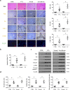
Fig. 1
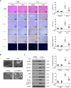
Fig. 1. Fas/FasL-mediated apoptosis involved in liver fibrogenesis.
a H&E staining, Fas, FasL, collagen-I (COL-I), α-SMA immunohistochemical staining (brown), and TUNEL staining (green) in the related liver tissues were presented. FasL, Fas, and COL-I area from the histological staining were also determined. Values are presented as mean ± SEM. *P < 0.05, n = 6 per group. CTRL, the control mice (olive oil-treated mice); CCl4, 20% carbon tetrachloride-induced mouse fibrosis; Normal, healthy volunteers; LF, human liver fibrosis. b The apoptotic index from TUNEL staining was presented. n = 6 in each group, values are presented as mean ± SEM. *P < 0.05. c Western blotting represented that the levels of Fas, FasL, α-SMA, COL-I, COL-IV, and cleaved caspase-3 were enhanced in liver fibrotic sections. β-actin was used as the loading control. d The ratio of densitometry units of the normalized FasL/β-actin, Fas/β-actin, and cleaved caspase-3/β-actin was also presented (n = 6 per group), values are presented as mean ± SEM. *P < 0.05.
Fig. 2
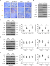
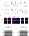
Fig. 2. Autophagy participated in Fas/FasL-mediated hepatic apoptosis in liver fibrosis.
a H&E staining, Fas, BECN1, COL-I, α-SMA staining (brown), and TUNEL staining (green) were presented in the indicated sections from olive oil- (as CTRL) or CCl4-treated mice, with or without 3-MA administration. α-SMA area and COL-I area from the histological staining, and the apoptotic index (TUNEL staining) were also determined. *P < 0.05 versus CTRL mice, #P < 0.05 versus CCl4-treated mice without 3-MA treatment, n = 6 per group. b Ultrastructural features in the hepatocytes of liver tissues from human tissues and mouse models were presented (red arrows indicating autophagosomes). c Western blotting depicted that inhibition of autophagy by 3-MA downregulated the expressions of BECN1, α-SMA, COL-I, COL-IV, PUMA, and cleaved caspase-3, without affecting the state of Fas and FasL, in CCl4-treated mice. The ratio of densitometry units of the normalized α-SMA/β-actin and COL-I/β-actin was presented (n = 6 per group), values are presented as mean ± SEM. *P < 0.05 versus CTRL mice, #P < 0.05 versus CCl4-treated mice without 3-MA treatment.
Fig. 3
Fig. 3. PUMA responded to Fas/FasL/autophagy-mediated hepatocytes apoptosis during liver fibrosis.
a Sirius red staining (red), α-SMA (brown), PUMA staining (brown), and TUNEL staining (green) were presented in the indicated sections from CTRL and CCl4-treated mice (n = 6 per group). b Double IF staining by utilizing HSCs marker α-SMA (red) and hepatocytes marker transferrin (green) were presented (upper panel). Double staining of transferrin (red) and TUNEL (green), α-SMA (red), and TUNEL (green) indicated that the apoptotic cells mainly located in hepatocytes in liver fibrotic mice (middle panel). Co-staining of BECN1 (green) and PUMA (red), Fas (green), and PUMA (red) further revealed that PUMA involved in Fas and autophagy-regulated signaling (lower panel). Nuclei (blue) were counterstained with DAPI (4′6-diamidino-2-phenylindole dihydrochloride). c PUMA immunohistochemistry staining (brown) and TUNEL staining (green) in the indicated humans liver sections were presented. d PUMA and α-SMA levels were determined by western blotting.
Fig. 4
Fig. 4. Targeted deletion of PUMA ameliorated hepatocytes apoptosis and liver fibrosis.
a Sirius red staining (red) and cleaved caspase-3 staining (brown) presented that targeted deletion of PUMA ameliorated hepatic apoptosis and collagen deposition. Western blotting presented that targeted deletion of PUMA ameliorated the levels of collagen-IV (COL-IV), collagen-I (COL-I), and α-SMA in CCl4-treated mice (n = 6 per group). b Immunohistochemistry staining and western blotting revealed that PUMA deficiency did not affect the status of Fas and BECN1 in CCl4-treated mice. c Double immunofluorescence staining (transferrin (red) and TUNEL (green), α-SMA (red) and TUNEL (green)) and the analysis of the apoptotic index of hepatocytes or HSCs indicated that targeted deletion of PUMA mainly ameliorated hepatocytes apoptosis during liver fibrosis, n = 6 per group. Nuclei (blue) were counterstained with DAPI. *P < 0.05. NS, no significance. d α-SMA (red) staining in the primary HSCs dissociated from the indicated PUMA-WT and PUMA-KO mice was represented, nuclei (blue) were counterstained with DAPI. e The indicated proteins from primary HSCs analyzed by western blotting. The ratio of densitometry units of the normalized α-SMA/β-actin was also determined, n = 6 per group, values are presented as mean ± SEM. *P < 0.05 versus primary HSCs from CTRL mice, #P < 0.05 versus primary HSCs from CCl4-treated PUMA-WT mice.
Fig. 5
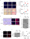
Fig. 5. Fas/FasL repressed the activation of NF-κBp65 in hepatocytes in liver fibrogenesis.
a Immunohistochemical staining (brown) of Fas, FasL, and p-p65 (the phosphorylation of NF-κBp65) in the indicated liver sections was presented (n = 6 per group). b Expressions of Fas, FasL, p65, p-p65, and COL-I proteins from the related humans liver sections were detected by western blotting. c Western blotting was adopted to analyze the levels of Fas, FasL, p65, p-p65, and COL-I in the liver tissues of mouse models. The ratio of densitometry units of the normalized Fas/β-actin and p-p65/β-actin from (b) and (c) was also determined, n = 6 per group, values are presented as mean ± SEM. *P < 0.05. CTRL, the control mice (olive oil-treated mice); CCl4, 20% carbon tetrachloride-induced mouse fibrosis; Normal, healthy volunteers; LF, human liver fibrosis. d Western blotting depicted that knockdown of Fas upregulated the level of p-p65 in the primary hepatocytes isolated from CCl4-treated mice. The ratio of densitometry units of the normalized p65/β-actin and p-p65/β-actin was also presented, n = 6 per group, values are presented as mean ± SEM. *P < 0.05 versus primary hepatocytes from CTRL group, #P < 0.05 versus primary hepatocytes from CCl4-treated group without siFas treatment. e FasL treatment enhanced the expression of Fas and repressed the phosphorylation of NF-κBp65 (p-p65) in the primary hepatocytes, while knockdown of Fas by siRNA upregulated the level of p-p65 in FasL-treated group. The ratio of densitometry units of the normalized p65/β-actin and p-p65/β-actin was also presented, n = 6 per group, values are presented as mean ± SEM. *P < 0.05 versus primary hepatocytes from PBS group, #P < 0.05 versus primary hepatocytes from FasL-treated group without siFas treatment.
Fig. 6
Fig. 6. NF-κBp65 inhibited PUMA-mediated hepatocytes apoptosis and attenuated liver fibrosis.
a TUENL staining (red) and sirius red staining at the indicated time points were adopted to showed that NF-κBp65 deficiency in hepatocytes promoted apoptosis and collagen deposition during fibrogenesis. The apoptotic index and sirius red area were also analyzed. *P < 0.05. p65Δhepa: hepatocytes specific NF-κBp65 deletion; p65f/f: NF-κBp65 wild-type. b Immunohistochemical staining revealed NF-κBp65 deficiency in hepatocytes induced PUMA expression and cell apoptosis (brown). Double staining of TUNEL (green) and PUMA (red) indicated that PUMA expression and TUNEL signaling were located in similar cells, and co-staining of TUNEL (green) and transferrin (red) was also presented. Cell nuclei (blue) were counterstained by DAPI. c Western blotting represented hepatocytes specific NF-κBp65 deletion promoted the upregulation of PUMA and downregulated the levels of anti-apoptotic proteins Bcl-2, Bcl-xL, and Mcl-1 in primary hepatocytes isolated from CCl4-treated mice, and the phosphorylation of NF-κBp65 (p-p65) was repressed in primary hepatocytes isolated from CCl4-treated p65f/f mice. d Primary HSCs dissociated from the indicated mice were stained by α-SMA (red). Nuclei (blue) were counterstained with DAPI. e The indicated proteins from primary isolated HSCs in the mouse models were detected by western blotting. The ratio of densitometry units of the normalized α-SMA/β-actin was also determined. *P < 0.05 versus primary HSCs from CTRL mice, #P < 0.05 versus HSCs from CCl4-treated p65f/f mice. n = 6 per group.
Fig. 7
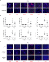
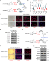
Fig. 7. NF-κBp65/PUMA-regulated hepatocytes apoptosis enhanced hepatic inflammatory response.
a The indicated inflammatory cytokines in the mouse livers were analyzed via immunofluorescence staining (red), the related cytokines were significantly repressed in CCl4-treated PUMA-KO mice, nuclei (blue) were counterstained with DAPI. b Inflammatory factors mRNA expression in the livers from either CCl4-treated PUMA-WT or PUMA-KO mice were analyzed by quantitative reverse-transcription PCR. *P < 0.05 versus CTRL mice, #P < 0.05 versus CCl4-treated PUMA-WT mice. The expression of β-actin in each tissue was quantified as the internal control. n = 6 per group. c Expressions of indicated inflammatory cytokines in the livers were analyzed via immunofluorescence staining (red), the related cytokines were repressed in CCl4-treated p65Δhepa mice following PUMA antisense oligonucleotides (AS), nuclei (blue) were counterstained with DAPI.
Fig. 8
Fig. 8. NF-κBp65/PUMA-regulated hepatocytes apoptosis-linked inflammatory response to promote HSCs activation and liver fibrosis.
a The area of the indicated inflammatory cytokines from immunofluorescence staining of Fig. 7c was analyzed. *P < 0.05 versus p65f/f mice, #P < 0.05 versus p65Δhepa mice with vehicle administration. b The administration of TGF-β1 inhibitor pirfenidone, TNF-α inhibitor pentoxifylline or monocytes inactivator clodronate-loaded liposomes, attenuated HSCs activation (α-SMA staining of primary HSCs, red) and liver fibrosis (α-SMA staining, red) in p65Δhepa/PUMA-WT mice. Nuclei (blue) were counterstained with DAPI, n = 6 per group. IF, immunofluorescence staining. c, d Western blotting presented that pirfenidone, pentoxifylline, or clodronate-loaded liposomes attenuated liver fibrosis (liver section) and the activation of HSCs (primary HSCs section) in CCl4-induced p65Δhepa/PUMA-WT mouse model, respectively. n = 6 per group.
Fig. 9
Fig. 9. NF-κBp65/PUMA-driven liver inflammation-induced HSCs activation and liver fibrosis.
a Schematic diagram of the serum test in vitro. b The genes expressions of the indicated inflammatory cytokines from CCl4-treated PUMA-WT or PUMA-KO mice were detected by quantitative reverse-transcription PCR. The expression of β-actin in each tissue was quantified as the internal control. n = 6 per group. *P < 0.05. c Representative images of the growth with activation of primary isolated HSCs following the treatment of the distinct serum extracted from CCl4-treated PUMA-WT or PUMA-KO mice. Nuclei (blue) were counterstained with DAPI. d Expressions of the related proteins of primary HSCs after serum treatment were detected by western blotting, revealing that PUMA-WT serum enhanced the activation of HSCs without affecting cell apoptosis. n = 6 per group. e Schematic diagram of the serum test in vivo. f Sirius red staining (red) and α-SMA staining (red) were examined in the liver tissues of PUMA-KO mice following the treatment of the serum extracted either from CCl4-treated PUMA-WT or PUMA-KO mice. The primary isolated HSCs were also analyzed by α-SMA staining (red). Nuclei (blue) were counterstained with DAPI. g The indicated proteins of the primary isolated HSCs from PUMA-KO mice after serum treatment were detected by western blotting, n = 6 per group.

Similar articles

Cited by

References

    1. Lee YA, Wallace MC, Friedman SL. Pathobiology of liver fibrosis: a translational success story. Gut. 2015;64:830–841. doi: 10.1136/gutjnl-2014-306842. - DOI - PMC - PubMed
    1. Yin C, Evason KJ, Asahina K, Stainier DY. Hepatic stellate cells in liver development, regeneration, and cancer. J. Clin. Invest. 2013;123:1902–1910. doi: 10.1172/JCI66369. - DOI - PMC - PubMed
    1. Pradere JP, et al. Hepatic macrophages but not dendritic cells contribute to liver fibrosis by promoting the survival of activated hepatic stellate cells in mice. Hepatology. 2013;58:1461–1473. doi: 10.1002/hep.26429. - DOI - PMC - PubMed
    1. Suh YG, et al. CD11b(+) Gr1(+) bone marrow cells ameliorate liver fibrosis by producing interleukin-10 in mice. Hepatology. 2012;56:1902–1912. doi: 10.1002/hep.25817. - DOI - PMC - PubMed
    1. Kornek M, Popov Y, Libermann TA, Afdhal NH, Schuppan D. Human T cell microparticles circulate in blood of hepatitis patients and induce fibrolytic activation of hepatic stellate cells. Hepatology. 2011;53:230–242. doi: 10.1002/hep.23999. - DOI - PMC - PubMed

Publication types