. 2021 May 12;12(5):476.
doi: 10.1038/s41419-021-03761-1.

CBP/p300 HAT maintains the gene network critical for β cell identity and functional maturity

Affiliations

CBP/p300 HAT maintains the gene network critical for β cell identity and functional maturity

Linlin Zhang et al. Cell Death Dis. .

Abstract

Loss of β cell identity and functional immaturity are thought to be involved in β cell failure in type 2 diabetes. CREB-binding protein (CBP) and its paralogue p300 act as multifunctional transcriptional co-activators and histone acetyltransferases (HAT) with extensive biological functions. However, whether the regulatory role of CBP/p300 in islet β cell function depends on the HAT activity remains uncertain. In this current study, A-485, a selective inhibitor of CBP/p300 HAT activity, greatly impaired glucose-stimulated insulin secretion from rat islets in vitro and in vivo. RNA-sequencing analysis showed a comprehensive downregulation of β cell and α cell identity genes in A-485-treated islets, without upregulation of dedifferentiation markers and derepression of disallowed genes. A-485 treatment decreased the expressions of genes involved in glucose sensing, not in glycolysis, tricarboxylic acid cycle, and oxidative phosphorylation. In the islets of prediabetic db/db mice, CBP/p300 displayed a significant decrease with key genes for β cell function. The deacetylation of histone H3K27 as well as the transcription factors Hnf1α and Foxo1 was involved in CBP/p300 HAT inactivation-repressed expressions of β cell identity and functional genes. These findings highlight the dominant role of CBP/p300 HAT in the maintenance of β cell identity by governing transcription network.


Conflict of interest statement

The authors declare no competing interests.

Figures
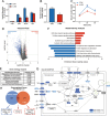
Fig. 1
Fig. 1. Effects of A-485 on insulin secretion and transcriptome profile of rat islets.
A Isolated rat islets were pretreated with 3 μM A-485 at 5.6 mM glucose for 16 h, followed by stimulation with 3.3, 8.3 and 16.7 mM glucose or 35 mM KCl for 1 h, and insulin secretion was measured. B Random serum insulin levels of control and A-485-treated C57BL/6 mice (n = 5). C Serum insulin levels under glucose loading in control and A-485-treated C57BL/6 mice (n = 5). D Volcano plots of differentially expressed genes. E Top 5 upregulated (red) and downregulated (blue) pathways in KEGG analysis. F GO analysis of downregulated genes. G Visualization of downregulated genes involved in insulin secretion pathway. Dark blue represents fold change ≥2.0 and light blue represents fold change ≥1.5. Gray-labeled genes show no significant change. H Overlap analysis of downregulated genes between CBP/p300 HAT-inhibited rat islets and pancreatic endocrine progenitors-specific CBPHet; p300KO mice islets (fold change ≥2.0, p-value <0.05). Data are given as mean ± SD for three separate experiments. *p < 0.05, **p < 0.01 vs control (CON) group.
Fig. 2
Fig. 2. CBP/p300 HAT inhibition leads to reduced expressions of β and α identity genes.
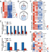
A Heatmap of β cell identity genes. B Venn diagrams of upregulated (red) and downregulated (blue) genes enriched in β and α cell by A-485 treatment. C Heatmap of α cell identity genes, islet hormone genes, and progenitor markers. D Heatmap of disallowed genes and related methylases and demethylases. EF Isolated rat islets were treated with 3 μM A-485 for 16 h and mRNA levels of β and α cell identity genes were detected by RT-qPCR. Data are given as mean ± SD for three separate experiments. ***p < 0.001 vs control group.
Fig. 3
Fig. 3. Impaired β cell identity and functional maturity in CBP/p300 HAT-inhibited β cell.
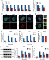
A mRNA expressions of β and α cell identity genes in isolated islets from control and A-485-treated mice. BC Representative pancreatic sections co-immunostained for insulin (Ins, green), Pdx1 or Ucn3 (red) and DAPI (blue) from control and A-485-treated mice (scale bars, 20 μm). D mRNA expressions of Glut2 and Gck in isolated islets from control and A-485-treated mice. E Representative pancreatic sections co-immunostained for insulin (Ins, green), Glut2 (red), and DAPI (blue) from control and A-485-treated mice (scale bars, 20 μm). FG mRNA expressions of Glut2 and Gck at various concentrations of glucose in isolated rat islets treated with A-485 for 16 h. H mtDNA expression in control and A-485-treated INS-1 cells for 16 h. I Protein expressions of mitochondrial MTCO1, ATP5A1 and SDHA in control and A-485-treated INS-1 cells for 16 h. JK mRNA levels of Ins1 and Ins2 at various concentrations of glucose in isolated rat islets treated with A-485 for 16 h. Data are given as mean ± SD for three separate experiments. *p < 0.05, **p < 0.01, ***p < 0.001 vs control group.
Fig. 4
Fig. 4. CBP/p300 HAT mediates expression of Hadh responsible for basal insulin secretion.
A mRNA and protein levels of Hadh and Foxa2 in isolated rat islets treated with A-485 for 16 h. B Representative pancreatic sections co-immunostained for insulin (Ins, green), Hadh (red), and DAPI (blue) from control and A-485-treated C57BL/6 mice (scale bars, 20 μm). C Blood glucose levels of 4-week db/m and db/db mice were measured after 16 h fasting. D Fasting serum insulin levels of 4-week db/m and db/db mice. E Serum insulin levels after glucose loading in 4-week db/m and db/db mice (n = 5). F Isolated islets from 4-week-old db/m and db/db mice were stimulated with 5.6 mM glucose for 1 h to measure insulin secretion. GJ Representative pancreatic sections co-immunostained for insulin (Ins, green), Hadh/MafA/Glut2/CBP/p300 (red), and DAPI (blue) from 4-week-old db/m and db/db mice (scale bars, 20 μm). Data are given as mean ± SD. * p < 0.05, **p < 0.01, ***p < 0.001 vs control group, #p < 0.001 vs 0 min.
Fig. 5
Fig. 5. Role of H3K27Ac in CBP/p300-mediated islet identity gene expressions.
A Representative pancreatic sections co-immunostained for insulin (Ins, green), H3K27Ac (red), and DAPI (blue) from control and A-485-treated mice (scale bars, 20 μm). BC Genomic distribution of H3K27Ac peaks in control and A-485-treated rat islets. D Genomic distribution of downregulated H3K27Ac peaks between two groups (fold change ≥2.0, p value <0.001). E Overlap analysis of annotated genes with hypo-H3K27Ac and downregulated genes identified in RNA-seq. F KEGG pathway analysis of overlapping genes. G Visualization of H3K27ac levels in adjacent area of transcriptional regions. Chromosomal span and gene structure are shown, and the significantly downregulated peaks are highlighted with the gray dotted box.
Fig. 6
Fig. 6. Transcription factors Hnf1α and Foxo1 are regulated by CBP/p300 HAT activity.
A Top 10 of upstream transcriptional regulators predicted by IPA analysis based on the downregulated gene set identified in RNA-seq. BC mRNA and protein levels of Hnf1α in isolated rat islets treated with A-485 for 16 h. D Co-immunoprecipitation using Hnf1α antibody in A-485 treated INS-1 cells for 6 h. EF mRNA and protein levels of Foxo1 in A-485-treated rat islets for 16 h. GH Foxo1 levels in INS-1 cells treated with 3 μM A-485 and 10 μg/ml cycloheximide (CHX) for the indicated time. Signal intensity was quantified by Image J software for statistical comparison. IJ Foxo1 levels in INS-1 cells treated with 3 μM A-485 and 10 μM MG132 for 6 h. Signal intensity was quantified by Image J software for statistical comparison. Data are given as mean ± SD. *p < 0.05, **p < 0.01 vs control group.
Fig. 7
Fig. 7. The schematic illustration summarizes CBP/p300 HAT-governed transcription network in islet β cell.
The HAT activity of CBP/p300 is required for maintaining the expressions of β cell identity genes and functional genes partially via acetylating histone H3K27 as well as transcription factors Hnf1a and Foxo1. Inhibition of CBP/p300 HAT by A-485 leads to the deacetylation of H3K27, which restricts chromatin accessibility. In addition, inactivation of CBP/p300 HAT also decreases the acetylation levels of Hnf1α and Foxo1, repressing the transcription activation of the target genes by preventing the interaction of Hnf1α and p300 as well as promoting Foxo1 protein degradation.

Similar articles

Cited by

References

    1. Talchai C, Xuan S, Lin HV, Sussel L, Accili D. Pancreatic β cell dedifferentiation as a mechanism of diabetic β cell failure. Cell. 2012;150:1223–1234. doi: 10.1016/j.cell.2012.07.029. - DOI - PMC - PubMed
    1. Swisa A, Glaser B, Dor Y. Metabolic stress and compromised identity of pancreatic beta cells. Front. Genet. 2017;8:21. doi: 10.3389/fgene.2017.00021. - DOI - PMC - PubMed
    1. Gu C, et al. Pancreatic beta cells require NeuroD to achieve and maintain functional maturity. Cell Metab. 2010;11:298–310. doi: 10.1016/j.cmet.2010.03.006. - DOI - PMC - PubMed
    1. Taylor BL, Liu FF, Sander M. Nkx6.1 is essential for maintaining the functional state of pancreatic beta cells. Cell Rep. 2013;4:1262–1275. doi: 10.1016/j.celrep.2013.08.010. - DOI - PMC - PubMed
    1. Gao T, et al. Pdx1 maintains β cell identity and function by repressing an α cell program. Cell Metab. 2014;19:259–271. doi: 10.1016/j.cmet.2013.12.002. - DOI - PMC - PubMed

Publication types

Substances