. 2021 May 13;12(5):479.
doi: 10.1038/s41419-021-03763-z.

RNA m6A reader YTHDF2 facilitates lung adenocarcinoma cell proliferation and metastasis by targeting the AXIN1/Wnt/β-catenin signaling

Affiliations

RNA m6A reader YTHDF2 facilitates lung adenocarcinoma cell proliferation and metastasis by targeting the AXIN1/Wnt/β-catenin signaling

Yin Li et al. Cell Death Dis. .

Abstract

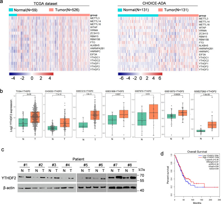
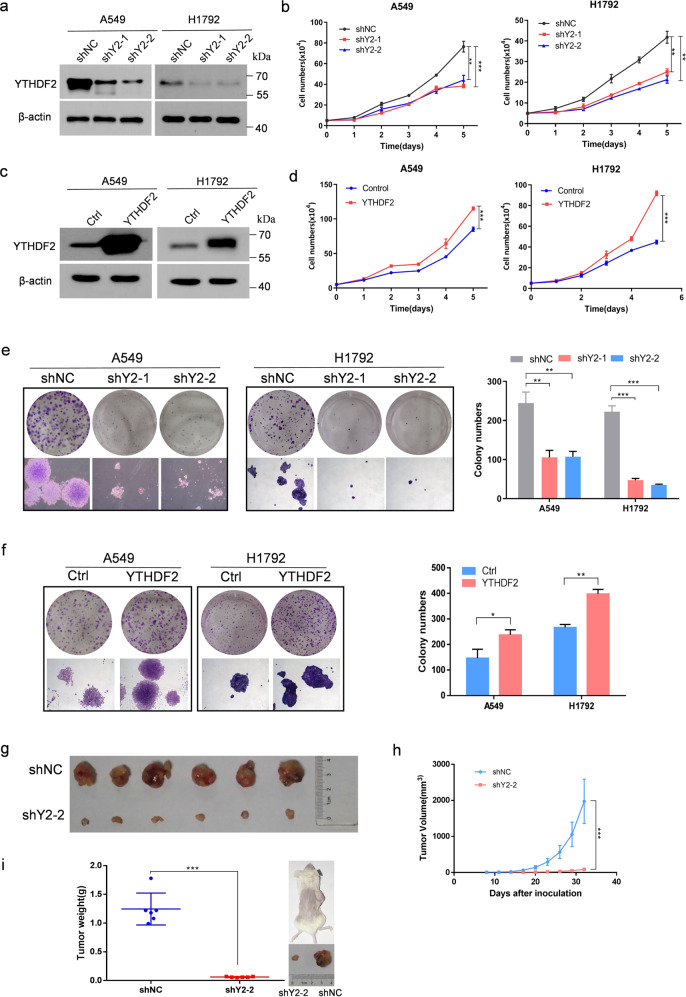
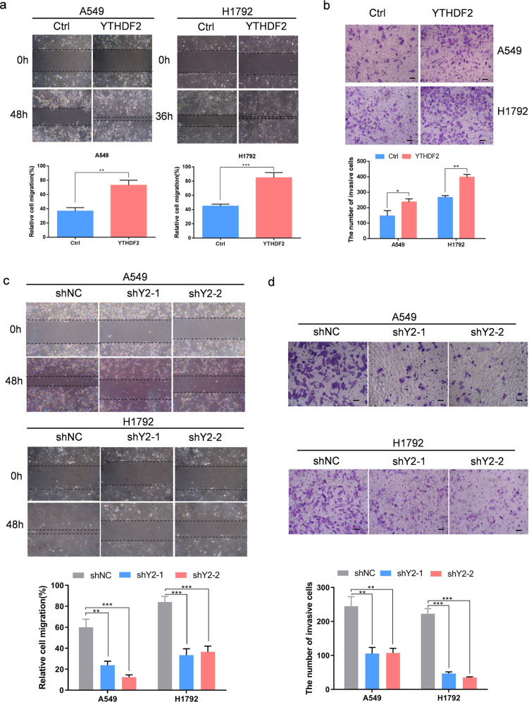
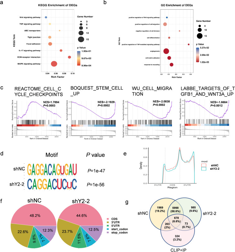
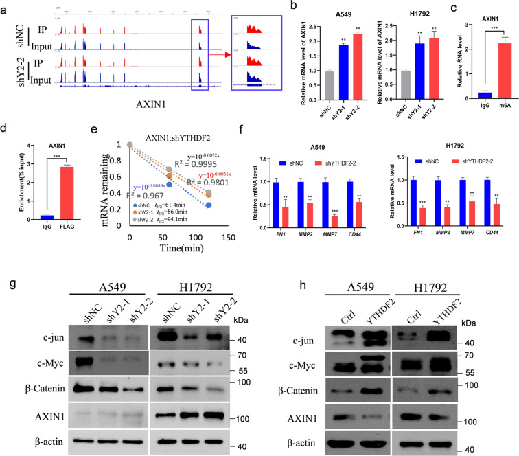
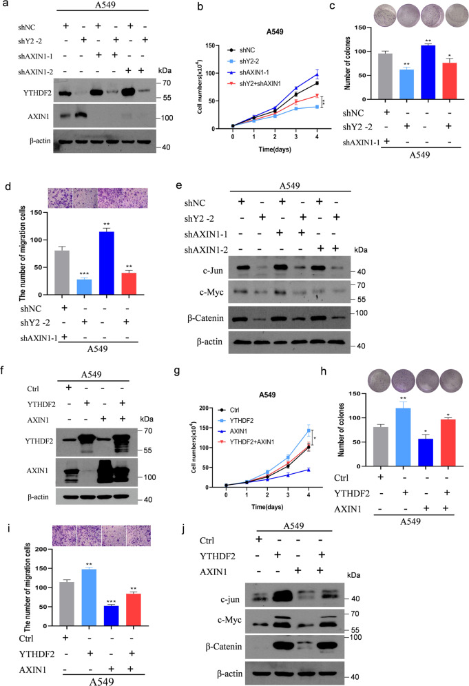
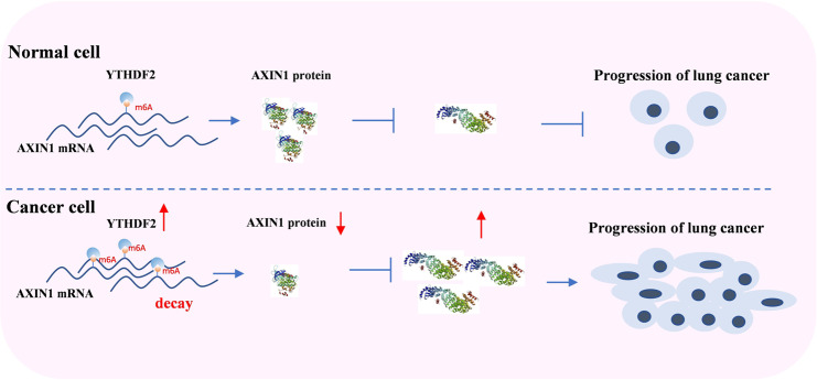
Lung adenocarcinoma (LUAD) remains a leading cause of cancer-related deaths worldwide. YTHDF2 is a reader of N6-methyladenosine (m6A) on RNA and plays a critical role in the initiation and propagation of myeloid leukemia; however, whether YTHDF2 controls the development of LUAD remains to be explored. Here, we found that YTHDF2 was significantly upregulated in LUAD compared with paracancerous normal tissues, and YTHDF2 knockdown drastically inhibited, while its overexpression promoted, cell growth, colony formation and migration of LUAD cells in vitro. In addition, YTHDF2 knockdown significantly inhibited tumorigenesis in a murine tumor xenograft model. Through the integrative analysis of RNA-seq, m6A-seq, CLIP-seq, and RIP-seq datasets, we identified a set of potential direct targets of YTHDF2 in LUAD, among which we confirmed AXIN1, which encodes a negative regulator of the Wnt/β-catenin signaling, as a direct target of YTHDF2. YTHDF2 promoted AXIN1 mRNA decay and subsequently activated the Wnt/β-catenin signaling. Knockout of AXIN1 sufficiently rescued the inhibitory effect of YTHDF2 depletion on lung cancer cell proliferation, colony-formation, and migration. These results revealed YTHDF2 to be a contributor of LUAD development acting through the upregulation of the AXIN1/Wnt/β-catenin signaling, which can be a potential therapeutic target for LUAD.


Conflict of interest statement

The authors declare that they have no conflict of interest.

Figures

Fig. 1
Fig. 1. YTHDF2 was highly expressed in LUAD.
a Heat map profiling the expression of m6A WERs in TCGA (left) and CHOICE (right) databases of LUAD. b Relative RNA levels of YTHDF2 in LUAD tissue and normal lung tissue in TCGA, CHOICE, and GEO datasets. c Immunoblotting assay of YTHDF2 expression in eight paired LUAD primary tumor samples. β-actin was used as a loading control. d) Kaplan–Meier analysis of LUAD cancer in TCGA for the correlations between YTHDF2 expression and overall survival.
Fig. 2
Fig. 2. YTHDF2 regulated LUAD cell proliferation and viability in vitro and vivo.
a Western blot analysis of YTHDF2 expression in A549 and H1792 cells infected with two independent shRNAs targeting YTHDF2 or a control shRNA. b Trypan blue live cell count assays were performed to determine cell growth after YTHDF2 knockdown in A549 and H1792 cells. c Western blot analysis for YTHDF2 expression in A549 and H1792 cells that were lentivirally infected with YTHDF2 or a control vector. d Trypan blue live cell count assays were performed to determine cell growth after YTHDF2 was over-expressed in A549 and H1792 cells. e Colony formation assays of A549 and H1792 cells described in a. f Colony formation assays of A549 and H1792 cells described in c. A549 cells with empty vector or YTHDF2 knockdown vectors were subcutaneously injected into nude mice. h Tumor size was measured every 3 days, and growth curves were plotted. Tumors were dissected from nude mice of each group and photographed at 32 days after transplantation, and the size and weight of tumors were measured (g, i). All data and error bars are presented as the mean ± SDs. *P < 0.05, **P < 0.01, and ***P < 0.001 compared with control cells.
Fig. 3
Fig. 3. YTHDF2 regulated the metastatic capacity of LUAD cells.
a, b Ectopic overexpression of YTHDF2 significantly promoted A549 and H1792 cell migration and invasive capabilities as examined by wound healing (a) and Transwell invasion (b) assays. Data are shown as means ± S.D.; *P < 0.05, **P < 0.01, and ***P < 0.001 compared with control cells. c, d Silencing of YTHDF2 by shRNA effectively inhibited A549 and H1792 cell migration and invasive capabilities. Data are shown as means ± S.D.; **P < 0.01 and ***P < 0.001 compared with control cells.
Fig. 4
Fig. 4. Identification of YTHDF2 targets via MeRIP-seq and RNA-seq in LUAD.
a, b KEGG and GO enrichment analysis of differentially expressed genes (DEGs) identified by RNA-seq. c GSEA plots showing that the pathways of DEGs altered by YTHDF2 knockdown were involved in LUAD. d m6A motif detected by the HOMER motif analysis with m6A-seq data in A549 cells with or without YTHDF2 knockdown. e Metagene profiles of m6A enrichment across the mRNA transcriptome in LUAD. f Graphs of m6A peak distribution showing the proportion of total m6A peaks in the indicated regions in control and YTHDF2-knockdown cells. g Venn diagram illustrating the overlapped targets of YTHDF2 in control and YTHDF2-knockdown cells identified by m6A-seq analysis and the targets identified by eCLIP and RIP.
Fig. 5
Fig. 5. AXIN1/Wnt/β-catenin signaling was targeted by YTHDF2.
a Distribution of m6A peaks across AXIN1 transcripts in control and YTHDF2-deficient cells. b qPCR analysis of AXIN1 mRNA expression in A549 and H1792 with or without YTHDF2 knockdown. Samples were normalized to β-actin mRNA. c Enrichment of m6A modification in AXIN1 as detected by a gene-specific m6A qPCR assay. d RIP–qPCR showing the association of AXIN1 with FLAG-tagged YTHDF2 in A549 cells. e Increased AXIN1 mRNA half-life by silencing YTHDF2 in A549 cells. Values were the mean ± S.D. of n = 3 independent experiments. f Relative mRNA levels of AXIN1/Wnt/β-catenin downstream targets identified by qPCR analysis in A549 and H1792 cells. Data represented means ± S.D. for three independent experiments. Immunoblotting to measure c-jun, c-Myc, β-catenin, and AXIN1 protein levels in transformed A549 and H1792 control cells and cells with YTHDF2 knockdown (g) or YTHDF2 overexpression (h).
Fig. 6
Fig. 6. AXIN1 was critical to YTHDF2-promoted Wnt/β-catenin signaling.
a Immunoblotting of lysates from A549 cells transfected with shNC, shYTHDF2#2, shYTHDF2#2, and/or shAXIN1-1 or shAXIN1-2. Expression levels of YTHDF2 and AXIN1 were measured. β-actin was used as a loading control. b Live cell count, c colony formation, d migration, and e immunoblotting of AXIN1/Wnt/β-catenin downstream targets in cells described in a. f Immunoblotting of lysates from A549 cells transfected with shCtrl, YTHDF2, AXIN1, YTHDF2, and/AXIN1 vector. Expression levels of YTHDF2 and AXIN1 were measured. β-actin was used as a loading control. g Live cell count, h colony formation, i migration, and j immunoblotting of AXIN1/Wnt/β-catenin downstream targets in cells described in f.
Fig. 7
Fig. 7. YTHDF2 high expressed promoted progression and metastasis of lung cancer cells via AXIN1/Wnt/β-catenin signalling.
YTHDF2 facilitated the decay of AXIN1 m6A mRNA and results in the dereased AXIN1protein level.The decreased AXIN1 promote the activity of Wnt/β-catenin signalling and enhance the proliferation and migration of lung cancer.

Similar articles

Cited by

References

    1. Torre LA, et al. Global cancer statistics, 2012. Cancer J. Clin. 2015;65:87–108. doi: 10.3322/caac.21262. - DOI - PubMed
    1. Ettinger DS, et al. NCCN guidelines insights: non-small cell lung cancer, version 1.2020. J. Natl Compr. Cancer Netw. 2019;17:1464–1472. doi: 10.6004/jnccn.2019.0059. - DOI - PubMed
    1. Miller KD, et al. Cancer treatment and survivorship statistics, 2019. Cancer J. Clin. 2019;69:363–385. doi: 10.3322/caac.21565. - DOI - PubMed
    1. Siegel RL, Miller KD, Jemal A. Cancer statistics, 2019. Cancer J. Clin. 2019;69:7–34. doi: 10.3322/caac.21551. - DOI - PubMed
    1. Koch A, et al. Analysis of DNA methylation in cancer: location revisited. Nat. Rev. Clin. Oncol. 2018;15:459–466. doi: 10.1038/s41571-018-0004-4. - DOI - PubMed

Publication types