. 2021 May 12;11(1):10146.
doi: 10.1038/s41598-021-89334-w.

Symmetrically dimethylated histone H3R2 promotes global transcription during minor zygotic genome activation in mouse pronuclei

Affiliations

Symmetrically dimethylated histone H3R2 promotes global transcription during minor zygotic genome activation in mouse pronuclei

Kohtaro Morita et al. Sci Rep. .

Abstract

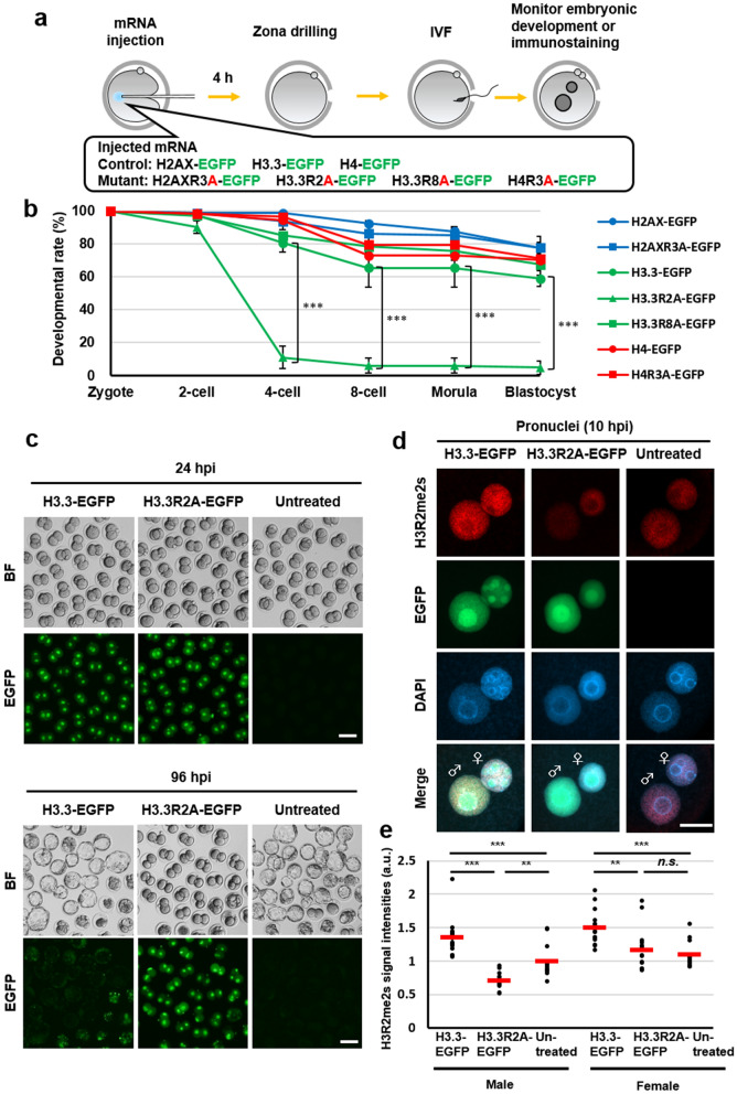
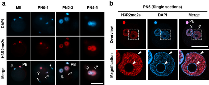
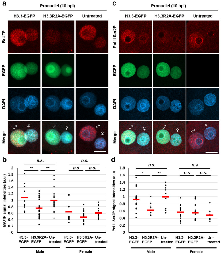
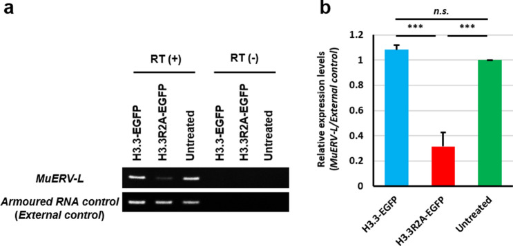
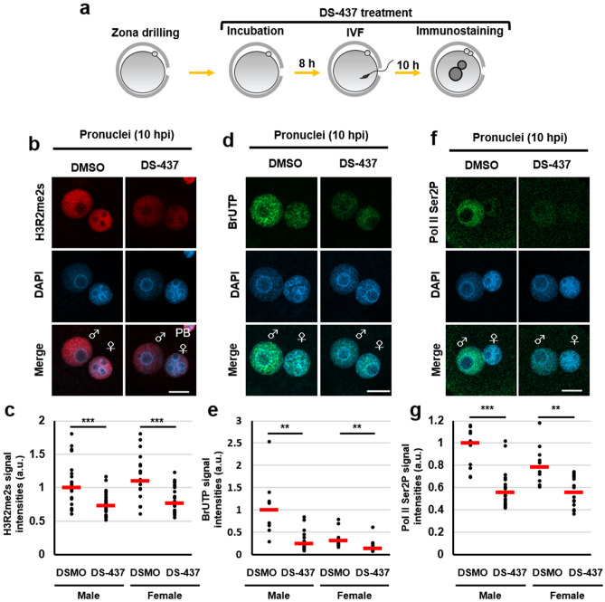
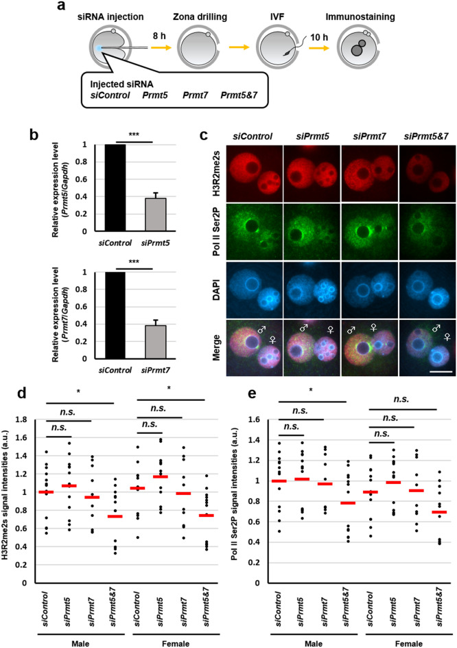
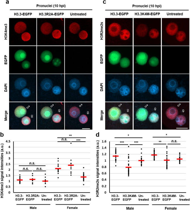
Paternal genome reprogramming, such as protamine-histone exchange and global DNA demethylation, is crucial for the development of fertilised embryos. Previously, our study showed that one of histone arginine methylation, asymmetrically dimethylated histone H3R17 (H3R17me2a), is necessary for epigenetic reprogramming in the mouse paternal genome. However, roles of histone arginine methylation in reprogramming after fertilisation are still poorly understood. Here, we report that H3R2me2s promotes global transcription at the 1-cell stage, referred to as minor zygotic genome activation (ZGA). The inhibition of H3R2me2s by expressing a histone H3.3 mutant H3.3R2A prevented embryonic development from the 2-cell to 4-cell stages and significantly reduced global RNA synthesis and RNA polymerase II (Pol II) activity. Consistent with this result, the expression levels of MuERV-L as minor ZGA transcripts were decreased by forced expression of H3.3R2A. Furthermore, treatment with an inhibitor and co-injection of siRNA to PRMT5 and PRMT7 also resulted in the attenuation of transcriptional activities with reduction of H3R2me2s in the pronuclei of zygotes. Interestingly, impairment of H3K4 methylation by expression of H3.3K4M resulted in a decrease of H3R2me2s in male pronuclei. Our findings suggest that H3R2me2s together with H3K4 methylation is involved in global transcription during minor ZGA in mice.


Conflict of interest statement

The authors declare no competing interests.

Figures

Figure 1
Figure 1
Identification of histone arginine residues for early embryonic development. (a) Schematic diagram of the experimental procedures. (b) Development of H2AX-EGFP—(blue bar with round box), H2AXR3A-EGFP—(blue bar with square box), H3.3-EGFP—(green bar with round box), H3.3R2A-EGFP—(green bar with triangular box), H3.3R8A-EGFP—(green bar with square box), H4-EGFP—(red bar with round box), and H4R3A-EGFP—(red bar with square box) expressed embryos. A chi-squared test was performed for analysing embryonic development through to the blastocyst stage. The asterisk indicates statistical significance (***p < 0.001). (c) Representative images of H3.3-EGFP-expressed, H3.3R2A-EGFP-expressed, and untreated embryos at 24 hpi and 96 hpi. Scale bar = 100 µm. (d) Immunofluorescent images of H3R2me2s (red) and DAPI (blue) in H3.3-EGFP-expressed, H3.3R2A-EGFP-expressed, and untreated zygotes. EGFP (green) shows successful injection. Key: ♂, male pronuclei; ♀, female pronuclei. Scale bar = 20 µm. (e) Quantification of H3R2me2s signal intensities in the pronuclei of H3.3-EGFP-expressed, H3.3R2A-EGFP-expressed, and untreated zygotes. Each dot plot represents a single zygote. Red bars indicate the mean values. The mean value of male pronuclei in untreated zygotes was set as 1. One-way ANOVA and the Tukey–Kramer test were performed to evaluate statistical significance. *Significantly different from the control (**p < 0.01, ***p < 0.001); n.s.: Not significantly different from the control. The number of zygotes was 15 for the H3.3-EGFP group, 15 for the H3.3R2A-EGFP group, and 13 for the untreated group. The pronucleus used for quantification are presented in Fig. S5.
Figure 2
Figure 2
Localisation of H3R2me2s in pronuclei of zygotes. (a) Immunostaining for localisation of H3R2me2s in MII oocytes and PN1–5 stage zygotes. The representative images of zygotes stained with DAPI (blue) and anti-H3R2me2s antibody (red). (b) An image example showing the male pronucleus of a zygote at PN5. Male pronuclei corresponding to dashed squares are shown magnified below. Shown are representative images of zygotes stained with DAPI (blue) and anti-H3R2me2s antibody (red). Key: ♂, male pronuclei; ♀, female pronuclei; PB, polar body; arrowhead, sperm. Scale bar = 50 µm.
Figure 3
Figure 3
Reduction of transcriptional activities by expression of H3.3R2A mutant in zygotes. (a), (c) Immunofluorescent images of BrUTP (red), Pol II Ser2P (red), and DAPI (blue) in H3.3-EGFP-expressed, H3.3R2A-EGFP-expressed, and untreated zygotes. EGFP (green) shows successful injection. Key: ♂, male pronuclei; ♀, female pronuclei. Scale bar = 20 µm. (b), (d) Quantification of the BrUTP and Pol II Ser2P signal intensities in pronuclei of H3.3-EGFP-expressed, H3.3R2A-EGFP-expressed, and untreated zygotes. Red bars indicate the mean values. The mean value of male pronuclei in untreated zygotes was set as 1. One-way ANOVA and the Tukey–Kramer test were performed to evaluate statistical significance. *Significantly different from the control (*p < 0.05, **p < 0.01); n.s.: Not significantly different from the control. The number of zygotes in (b) was 10 for the H3.3-EGFP expressed group, 24 for the H3.3R2A-EGFP expressed group, and 21 for the untreated group. The number of zygotes in (d) was 15 for the H3.3-EGFP expressed group, 13 for the H3.3R2A-EGFP expressed group, and 10 for the untreated group. The pronucleus used for quantification are presented in Fig. S7a, b.
Figure 4
Figure 4
Downregulation of MuERV-L via expression of H3.3R2A mutant in early 2-cell at 24 hpi. (a) Representative images of RT-PCR of MuERV-L and Armoured RNA control (External control). (b) Quantification of the PCR bands. The mean value of male pronuclei in untreated zygotes was set as 1. Mean ± S.E. from three independent experiments. One-way ANOVA and the Tukey–Kramer test were performed to evaluate statistical significance. *Significantly different from the control (***p < 0.001); n.s.: Not significantly different from the control. The full-length gels used for quantification are presented in Fig. S8.
Figure 5
Figure 5
Reduction of transcriptional activities by DS-437-treatment in zygotes. (a) Schematic diagram of the experimental procedures. (b), (d), (f) Immunofluorescent images of H3R2me2s (red), BrUTP (green), Pol II Ser2P (green), and DAPI (blue) in DMSO- and DS-437-treated zygotes. DMSO treatment was used as a control. (c), (e), (g) Quantification of the H3R2me2s, BrUTP, and Pol II Ser2P signal intensities in pronuclei of DMSO- and DS-437-treated zygotes. Red bars indicate the median values. The median value of male pronuclei in untreated zygotes was set as 1. Mann–Whitney U test were performed to evaluate statistical significance. The number of zygotes in (c) was 14 for the DMSO-treated group and 28 for the DS-437-treated group. The number of zygotes in (e) was 9 for the DMSO-treated group and 12 for the DS-437-treated group. The number of zygotes in (g) was 10 for the DMSO-treated group and 16 for the DS-437-treated group. The pronucleus used for quantification are presented in Fig. S9.
Figure 6
Figure 6
Knockdown of Prmt5 and Prmt7 mRNA revealed reduction in global transcription activities during minor ZGA. (a) Schematic diagram of the experimental procedures. (b) Knockdown of Prmt5 and Prmt7 expression by an antisense RNA injection was confirmed by quantitative RT-PCR analysis. The relative ratios were obtained by dividing the expression level of the Prmt5 and Prmt7 with the expression level of the Gapdh. Student’s t-test were performed for gel images. *Significantly different from the control (***p < 0.001). Bars represent the standard error of the mean. Full-length gels are presented in Fig. S10a. (c) Immunofluorescent images of H3R2me2s (red), Pol II Ser2P (green), and DAPI (blue) in Prmt5 and Prmt7 knockdown zygotes. siControl RNA was used as a control. (d), (e) Quantification of the H3R2me2s and Pol II Ser2P signal intensities in pronuclei of Prmt5 and Prmt7 knockdown zygotes. Dunnet test were performed to evaluate statistical significance. The number of zygotes in (d), (e) was 13 for the siControl group, 13 for the siPrmt5 group, 10 for the siPrmt7 group, and 13 for the siPrmt5&7 group. The pronucleus used for quantification are presented in Fig. S10b.
Figure 7
Figure 7
Expression of H3.3K4M mutant impaired H3R2me2s accumulation. (a), (c) Immunofluorescent images of H3K4me3 (red) or H3R2me2s (red) and DAPI (blue) in H3.3-EGFP-expressed, H3.3R2A-EGFP-expressed, H3.3K4M-EGFP-expressed, and untreated zygotes. EGFP (green) shows successful injection. Key: ♂, male pronuclei; ♀, female pronuclei. Scale bar = 20 µm. (b), (d) Quantification of the H3K4me3 and H3R2me2s signal intensities in pronuclei. Red bars indicate the mean values. The mean value of male pronuclei in untreated zygotes was set as 1. One-way ANOVA and the Tukey–Kramer test were performed to evaluate statistical significance. *Significantly different from the control (**p < 0.01, ***p < 0.001); n.s.: Not significantly different from the control. The number of zygotes in (b) was 15 for the H3.3-EGFP expressed group, 10 for the H3.3R2A-EGFP expressed group, and 13 for the untreated group. The pronucleus used for quantification are presented in Fig. S11a. The number of zygotes in (d) was 22 for the H3.3-EGFP expressed group, 29 for the H3.3K4M-EGFP-expressed group, and 23 for the untreated group. The pronucleus used for quantification are presented in Fig. S13.

Similar articles

Cited by

References

    1. McLay DW, Clarke HJ. Remodelling the paternal chromatin at fertilization in mammals. Reproduction. 2003;125:625–633. doi: 10.1530/rep.0.1250625. - DOI - PMC - PubMed
    1. Tosti E, Ménézo Y. Gamete activation: basic knowledge and clinical applications. Hum. Reprod. Update. 2016;22:420–439. doi: 10.1093/humupd/dmw014. - DOI - PMC - PubMed
    1. Hemberger M, Dean W, Reik W. Epigenetic dynamics of stem cells and cell lineage commitment: digging Waddington's canal. Nat. Rev. Mol. Cell. Biol. 2009;10:526–537. doi: 10.1038/nrm2727. - DOI - PubMed
    1. Guo F, et al. Active and passive demethylation of male and female pronuclear DNA in the mammalian zygote. Cell Stem Cell. 2014;15:447–459. doi: 10.1016/j.stem.2014.08.003. - DOI - PubMed
    1. Peat JR, et al. Genome-wide bisulfite sequencing in zygotes identifies demethylation targets and maps the contribution of TET3 oxidation. Cell Rep. 2014;9:1990–2000. doi: 10.1016/j.celrep.2014.11.034. - DOI - PMC - PubMed

Publication types