. 2021 May 12;4(1):569.
doi: 10.1038/s42003-021-02070-9.

Biased action of the CXCR4-targeting drug plerixafor is essential for its superior hematopoietic stem cell mobilization

Affiliations

Biased action of the CXCR4-targeting drug plerixafor is essential for its superior hematopoietic stem cell mobilization

Astrid S Jørgensen et al. Commun Biol. .

Abstract

Following the FDA-approval of the hematopoietic stem cell (HSC) mobilizer plerixafor, orally available and potent CXCR4 antagonists were pursued. One such proposition was AMD11070, which was orally active and had superior antagonism in vitro; however, it did not appear as effective for HSC mobilization in vivo. Here we show that while AMD11070 acts as a full antagonist, plerixafor acts biased by stimulating β-arrestin recruitment while fully antagonizing G protein. Consequently, while AMD11070 prevents the constitutive receptor internalization, plerixafor allows it and thereby decreases receptor expression. These findings are confirmed by the successful transfer of both ligands' binding sites and action to the related CXCR3 receptor. In vivo, plerixafor exhibits superior HSC mobilization associated with a dramatic reversal of the CXCL12 gradient across the bone marrow endothelium, which is not seen for AMD11070. We propose that the biased action of plerixafor is central for its superior therapeutic effect in HSC mobilization.


Conflict of interest statement

The authors declare no competing interest.

Figures

Fig. 1
Fig. 1. Clinical trials involving plerixafor, and comparison of plerixafor vs AMD11070.
a Overview of the types of clinical trials where plerixafor have been tested. The numbers in brackets show the number of trials within the different therapeutic areas. In scientific literature, plerixafor is also known as AMD3100. The drug is used under the trade name Mozobil. This image was created by Anna Sofie Husted using Adobe illustrator and photoshop under the licence of University of Copenhagen. b Chemical structures of plerixafor and AMD11070. The values below show their binding affinity for CXCR4 measured as their ability to inhibit CXCL12 binding in competitive binding, their ability to inhibit viral replication in MT-4 cells infected with the HIV-1 strain, and their ability to block migration of the A375 melanoma cell line towards CXCL12 (10 nM).
Fig. 2
Fig. 2. Internalization of CXCR4 is differentially affected by CXCL12, plerixafor, and AMD11070.
a Antibody feeding experiment with FLAG-tagged CXCR4 and US28 in HEK293 cells. The cells were either immediately fixed (t = 0) or incubated at 37 °C for 30 min to induce internalization (t = 30) and then fixed. CXCL12 was added at 1 μM. Cell surface receptors are labeled with Alexa Fluor 488-conjugated secondary antibody (green, left column), while internalized receptors were labeled with Alexa Flour 568-conjugated secondary antibody (red, second column from left). Nomarski images are included to illustrate the cell outline (right column). Scale bar, 5 μm. Images show representative cells from three independent experiments. b Acute time-course internalization ELISA. HEK293 cells were transiently transfected with FLAG-tagged CXCR4 (white circle) or US28 (black square). For each receptor, the value at a given time point is corrected for background, normalized to the value at t = 0, and presented in percent with mean ± SEM shown. The experiment was performed at least four times in quadruples. c SNAP-tag CXCR4 constitutive internalization. Internalization was determined after preincubation with Tag-lite SNAP-Lumi4-Tb (donor) for 1 h at 4 °C (white circle) or 37 °C (black circle) and shown with mean ± SEM from at least four independent experiments performed in triplicates. d SNAP-tag CXCR4 agonist CXCL12 induced internalization in HEK293A cells. Cells were stimulated with increasing concentration of CXCL12; 0 nM (white circle), 0.1 nM (orange circle), 1 nM (yellow circle), 0.01 µM (blue circle), 0.1 µM (green circle) or 1 µM (black circle). Internalization was determined upon CXCL12 addition after 1-hour preincubation with Tag-lite SNAP-lumi4-Tb at 37 °C. Data are shown with mean ± SEM of triplicates from at least three independent experiments. e SNAP-tagged CXCR4 receptor internalization after stimulation with CXCL12 (black circle), plerixafor (red square), and AMD11070 (white square). The data were normalized to maximal internalization levels by CXCL12 for CXCR4 and presented with mean ± SEM of triplicates from at least six independent experiments.
Fig. 3
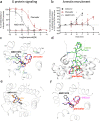
Fig. 3. Plerixafor and AMD11070 show distinct pharmacological profiles at CXCR4 and display different CXR4 binding modes.
a Agonism and antagonism of CXCR4 induced G protein activity. Measured by inositol triphosphate (IP3) accumulation in HEK293 cells transiently transfected with CXCR4 and the chimeric G protein Gqi4myr in the absence (square) or presence (circle) of 5 nM CXCL12 (corresponding to 80% activity). Cells were stimulated with increasing concentration of plerixafor (red) or AMD11070 (white). The data were normalized to signaling levels in response to 5 nM CXCL12 and presented with %mean ± SEM of duplicates from at least three independent experiments. b β-arrestin recruitment to CXCR4 by plerixafor (red square) and AMD11070 (white square). CXCL12 (black circle) was used as a control. β-arrestin2 recruitment was measured in C2C12 cells stably expressing Prolink (PK)-tagged CXCR4 and Enzyme Acceptor (EA)-tagged β-arrestin2. The data were normalized to the maximal recruitment levels by CXCL12 and presented with %mean ± SEM of duplicates from four independent experiments. c Overlay of the proposed binding modes for plerixafor (red) and AMD11070 (black) in the full-length CXCR4 model (extracellular view). CXCR4 transmembrane helices are shown and annotated in gray. Key binding residues are annotated on a light blue background and shown as sticks in green. Interactions: ionic = dotted magenta; H-bonds = dotted yellow; cation-pi = dotted green. d Overlay of the proposed binding mode for plerixafor (red) and the experimental binding mode for the peptide CXCR4 antagonist CVX15 (green; PDB 3OE0) (side view). e Overlay of the proposed binding mode for AMD11070 (black) and the experimental binding mode for the small-molecule CXCR4 antagonist IT1t (orange; PDB 3ODU) (viewed as in Fig. 3c). f The suggested path of the distal CXCL12 N-terminus (magenta) overlaid on plerixafor (red) and AMD11070 (black) (viewed as in Fig. 3c).
Fig. 4
Fig. 4. Transfer of plerixafor to CXCR3 (also) reveals partial agonism.
a Binding pocket of WT CXCR3 (left) and K300A-S304E mutant (right) shown from the extracellular side. Residues involved in plerixafor and AMD11070 binding in CXCR4 (Fig. 3) are shown as sticks. K300 and S304 in WT are mutated to A300 and E304, respectively. The wild-type CXCR3 model was retrieved from the GPCR-HGmod database. Residues are colored according to side chains; red = acidic residue, blue = basic residue, yellow = aromatic residue, green = polar residue. b Antagonistic effect of plerixafor and AMD11070 on CXCL11-induced CXCR3 G protein activity. Measured by inositol triphosphate (IP3) accumulation in HEK293 cells transiently transfected with WT CXCR3 or CXCR3[K300A-S304E] and the chimeric G protein Gqi4myr and stimulated with 10 nM CXCL11. Data of plerixafor previously published in ref. . Data are shown with %mean ± SEM of duplicates from 3 independent experiments, CXCR3 wt (black circle) and CXCR3 [K300A-S304E] stimulated with plerixafor (red square) or AMD11070 (white square). c Agonistic effect of plerixafor and AMD11070 on CXCR3 G protein activity compared to CXCL10 and CXCL11. Measured by inositol triphosphate (IP3) accumulation in HEK293 cells transiently transfected with CXCR3 WT or CXCR3[K300A-S304E] and the chimeric G protein Gqi4myr. The data were normalized to the maximal recruitment levels by CXCL11 on the two receptors and presented with %mean ± SEM of duplicates from at least three indepependent experiments. d Agonistic effect of plerixafor and AM11070 in β-arrestin2 recruitment upon CXCR3 activation compared to CXCL10 and CXCL11. Measured by the BRET-based arrestin recruitment assay in CHO cells transiently transfected with WT CXCR3 or CXCR3[K300A-S304E], Rluc8-Arrestin3, and mem-linker-citrine-SH3 constructs. The data were normalized to the maximal recruitment levels by CXCL11 on the two receptors and presented with %mean ± SEM of duplicates from five independent experiments. In c and d data are presented as CXCL11 (gray square), CXCL10 (black square), plerixafor (red square), and AMD11070 (white square).
Fig. 5
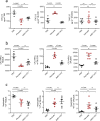
Fig. 5. In vivo effect of plerixafor and AMD11070 on HPC and neutrophil mobilization from the BM and redistribution.
a CXCL12 ELISA on BM and spleen supernatant and blood serum. Values display the concentration of CXCL12 as pg ml−1(n = 4–5). b Counting of CFU-HPCs (colony forming units, hematopoietic progenitor cells) after 12 days of culture following cell harvesting from bone marrow (BM), blood, and spleen, values displayed as cells ml−1 (n = 5). Photos of cultures can be found in Supplementary Fig. 3. c FACS analysis of the total number of neutrophils in the femur, blood, and spleen 1 h after i.p. injection of PBS, plerixafor, or AMD11070 (n = 5). The gating strategy for flow cytometry can be found in Supplementary Fig. 3. Color codes throughout the figure are PBS (black circles), Plerixafor (red squares), AMD11070 (white squares). Mean values and error bars representing SEM are shown. Statistical significances were analyzed by one-way ANOVA, exact P-values are shown for each comparison, ns = P ≥ 0.05.

Similar articles

Cited by

References

    1. Hauser AS, Attwood MM, Rask-Andersen M, Schiöth HB, Gloriam DE. Trends in GPCR drug discovery: new agents, targets and indications. Nat. Rev. Drug Discov. 2017;16:829–842. doi: 10.1038/nrd.2017.178. - DOI - PMC - PubMed
    1. Marchese A, et al. The E3 ubiquitin ligase AIP4 mediates ubiquitination and sorting of the G protein-coupled receptor CXCR4. Dev. Cell. 2003;5:709–722. doi: 10.1016/S1534-5807(03)00321-6. - DOI - PubMed
    1. Gurevich VV, Gurevich EV. GPCR signaling regulation: the role of GRKs and arrestins. Front. Pharmacol. 2019;10:125. doi: 10.3389/fphar.2019.00125. - DOI - PMC - PubMed
    1. Kenakin, T. The Application of Signaling Bias to New Therapeutic Drug Therapy for Seven Transmembrane (G Protein-coupled) Receptors: Quantifying Bias in Biased Signaling in Physiology, Pharmacology and Therapeutics (ed. Arey, J. B.) 81–102 (Elsevier, 2014).
    1. Murphy PM, et al. International union of pharmacology. XXII. Nomencl. Chemokine Recept. Pharmacol. Rev. 2000;52:145–176. - PubMed

Publication types

MeSH terms

LinkOut - more resources