. 2021 Jun;40(23):4050-4059.
doi: 10.1038/s41388-021-01821-6. Epub 2021 May 12.

Cell surface GRP78 and Dermcidin cooperate to regulate breast cancer cell migration through Wnt signaling

Affiliations

Cell surface GRP78 and Dermcidin cooperate to regulate breast cancer cell migration through Wnt signaling

Tyson W Lager et al. Oncogene. 2021 Jun.

Abstract

The heat shock protein GRP78 typically resides in the endoplasmic reticulum in normal tissues, but it has been shown to be expressed on the cell surface of several cancer cells, and some stem cells, where it can act as a signaling molecule by not-yet-fully defined mechanisms. Although cell surface GRP78 (sGRP78) has emerged as an attractive chemotherapeutic target, understanding how sGRP78 is functioning in cancer has been complicated by the fact that sGRP78 can function in a cell-context dependent manner, with a diverse array of reported binding partners, to regulate a variety of cellular responses. We had previously shown that sGRP78 was important in regulating pluripotent stem cell (PSC) functions, and hypothesized that embryonic-like mechanisms of GRP78 were critical to regulating aggressive breast cancer cell functions. Here, using proteomics we identify Dermcidin (DCD) as a novel sGRP78 binding partner common to both PSCs and breast cancer cells. We show that GRP78 and DCD cooperate to regulate stem cell and cancer cell migration that is dependent on the cell surface functions of these proteins. Finally, we identify Wnt/β-catenin signaling, a critical pathway in stem cell and cancer cell biology, as an important downstream intermediate in regulating this migration phenotype.


Conflict of interest statement

DISCLOSURE OF CONFLICTS OF INTEREST

The authors declare no competing interests.

Figures
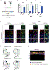
Figure 1.
Figure 1.. DCD and GRP78 colocalize on the surface of iPSCs and breast cancer cells.
(A) Dermcidin was an identified potential binding partner from a mass spectrometry proteomic screen profiling common surface GRP78 binding partners between iPSCs and cancer cells. (B) MDA-MB-231-GRP78ind cells were grown in the absence or presence of Doxycycline for 3 days, then fixed and stained with MemBrite™ (blue) (MB), and antibodies to DCD (green) or GRP78 (red) to examine colocalization on the surface of breast cancer cells using confocal microscopy. Images were taken at 60x magnification. Insets of images are shown to the right of each panel. (C) To assess colocalization of GRP78 and DCD at the cell surface of MDA-MB-231-GRP78ind cells under basal conditions and following GRP78 overexpression, Manders’ Colocalization Coefficient (MCC) values were calculated for GRP78/DCD, GRP78/MB, and DCD/MB colocalization. Bar charts represent results from three independent experiments. Graphs depict whole fields of view (top; each value represents 1 field; results from 6 fields shown) or from individual cells from the field images (bottom; each value represents 1 cell; 40 cells shown, obtained from the 6 fields). (D) MDA-MB-231-GRP78ind cells that had been treated with doxycycline for 3 days and stained with MemBrite™ (MB), anti-DCD or anti-GRP78 antibodies as described were used in confocal microscopy. Z-stack images were taken using a 100x objective and 2.5x optical zoom. 3-D rendering analysis of the X-Z plane to visualize cell surface colocalization was performed using Imaris Software. Examples of colocalization between DCD and GRP78 (purple), and cell surface colocalization of DCD and GRP78 (GRP78/DCD/MB; gold) are shown. (E-G) Relative DCD gene expression was examined between (E) IMR90 and FiPS4F5, (F) MCF7 Non-TICs (CD24+/CD44+) and TICs (CD24/CD44+), as well as (G) MCF7 sGRP78 and sGRP78+ cell populations using real-time PCR. Statistical significance was calculated using a student’s t-test. ** = p<0.01, **** = p<0.0001.
Figure 2.
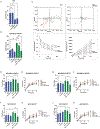
Figure 2.. Cell surface GRP78 and DCD are both important for GRP78-dependent migration.
(A) HUViPS4F1 cells were seeded into the upper compartment of a Transwell chamber and treated with goat IgG, anti-GRP78 or anti-DCD (1μg/mL). Lower compartments were supplemented with media and inserts were incubated for 24h. Inserts were fixed, cells in the upper chamber were removed and remaining migrating cells were fluorescently stained and counted. Bars represent mean ± SD (n=3). Statistical significance was calculated using one-way ANOVA. * = p<0.05 (B) MDA-MB-231-GRP78ind cells +/− doxycycline were seeded into the upper compartment of a Transwell chamber in serum free DMEM media only (untreated, UT), or serum free DMEM media treated with goat IgG, or anti-DCD (1μg/mL). Lower compartments were supplemented with DMEM media + 20% FBS and inserts were incubated for 24 hours. Inserts were fixed, cells in the upper chamber were removed, and remaining migrating cells were fluorescently counted. Bars represent mean ± SD (n=3). Statistical significance was calculated using one-way ANOVA. ** = p<0.01; *** = p<0.001 (C) MDA-MB-231-GRP78ind cells were pre-treated for three days with doxycycline. Cells were imaged for RFP expression, in the absence or presence of anti-DCD (1μg/mL), every 10 minutes for 4.5 hours using live cell time-lapse confocal microscopy. Random cells were manually tracked and cell trajectories from the origin, and mean square displacement and directionality ratio were analyzed. (D-K) MDA-MB-231-GRP78ind or MCF7-GRP78ind cells were pre-treated with doxycycline for 24 hours and then seeded to confluency. A vertical and horizontal wound was generated in each well. Cells were then grown in serum free media only +/− doxycycline or treated with goat IgG, anti-GRP78 (D-G), or anti-DCD (H-K) (1μg/mL). Images were taken every 24 hours for 72 hours. The percentage of wound closure at 72 hours is shown in D, F, H, and J. Bars represent mean ± SD (n=3). The percentage of wound closure for all time points is shown in E, G, I, and K. Line graphs represent wound closure rates at all experimental time points. Statistical analysis was performed using one-way ANOVA, * = p<0.05; ** = p<0.01, *** = p<0.001, **** = p<0.0001.
Figure 3.
Figure 3.. Cell surface GRP78 and DCD are functionally linked to regulate cancer cell migration.
(A) MDA-MB-231-GRP78ind or MDA-MB-231-GRP78ind + DCD-FLAG cells were pre-treated with doxycycline for 24 hours and then seeded to confluency. A vertical and horizontal wound was generated and cells were then grown in serum free media only +/− doxycycline or treated daily with goat IgG, anti-GRP78, or anti-DCD (1μg/mL). The percentage of wound closure at 72 hours is shown in A. Bars represent mean ± SD (n=3). (B) Line graphs displaying the percentage of wound closure across all experimental time points. Statistical analysis was performed using one-way ANOVA. ** = p<0.01, *** = p<0.001, **** = p<0.0001.
Figure 4.
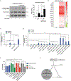
Figure 4.. GRP78 and DCD regulate breast cancer cell migration through Wnt/β-catenin signaling.
(A) MDA-MB-231-GRP78ind and MDA-MB-231-GRP78ind + DCD-FLAG cells were serum deprived for 24 hours (untreated, UT), or treated with 10μM CHIR99021 for 30 minutes, and then lysed. Cell lysates were separated using SDS-PAGE and probed for active β-catenin, total β-catenin, and β-actin protein levels using immunoblot analysis. Quantification of active β-catenin levels compared to β-actin levels is shown on the right. Statistical significance was calculated using a student’s t-test. * = p<0.05. (B) Relative amounts of Wnt target genes were analyzed between MDA-MB-231-GRP78ind and MDA-MB-231-GRP78ind + DCD-FLAG cells using real-time PCR. (C) MDA-MB-231-GRP78ind and MDA-MB-231-GRP78ind + DCD-FLAG cells were seeded in a 96-well plate and transfected with TOPFlash/FOPFlash plasmids. β-catenin transcriptional activity was assessed after 24-hour treatment with 10μM CHIR99021 by measuring luminescence. TOPFlash values are presented as fold change from untreated cells. Bars represent mean ± SD (n=3). * Indicates significance, p<0.05, **** = p<0.0001 analyzed with Welch’s ANOVA. (D) MDA-MB-231-GRP78ind and MDA-MB-231-GRP78ind + DCD-FLAG cells were seeded in a 96-well plate and transfected with TOPFlash/FOPFlash plasmids. Cells were pretreated for 3 hours with IgG control, anti-GRP78, or anti-DCD inhibitory antibodies (1μg/mL). β-catenin transcriptional activity was assessed after 24-hour treatment with 10μM CHIR99021 by measuring luminescence. TOPFlash values are presented as fold change from untreated cells. Bars represent mean ± SD. * indicates significance, p<0.05, ** = p<0.01, **** = p<0.0001 analyzed using an unpaired two-tailed t-test. (E) MDA-MB-231-GRP78ind or MDA-MB-231-GRP78ind + DCDFLAG cells were pre-treated with doxycycline for 24 hours and then grown to confluency. A vertical and horizontal wound was generated and cells were then grown in serum free DMEM media only +/− doxycycline or treated daily with DMSO, or 10μM XAV-939. The percentage of wound closing compared to the initial wound was calculated for each time point. Displayed is the percent gap closure rate at 72 hours. Bars represent mean ± SD (n=3). ** indicates significance compared to untreated cells, p<0.01, *** = p<0.001, **** = p<0.0001 using a Kruskal-Wallis test. (F) Schematic of proposed model. GRP78 and DCD interact at the cell surface – either directly, or indirectly through unknown cofactor(s) – to regulate cell migration by activating the canonical Wnt signaling pathway.

References

    1. Chaffer CL, Weinberg RA: A perspective on cancer cell metastasis. Science 2011, 331:1559–1564. - PubMed
    1. Ben-Porath I, Thomson MW, Carey VJ, Ge R, Bell GW, Regev A, et al.: An embryonic stem cell-like gene expression signature in poorly differentiated aggressive human tumors. Nat Genet 2008, 40:499–507. - PMC - PubMed
    1. Holm F, Hellqvist E, Mason CN, Ali SA, Delos-Santos N, Barrett CL, et al.: Reversion to an embryonic alternative splicing program enhances leukemia stem cell self-renewal. Proc Natl Acad Sci U S A 2015, 112:15444–15449. - PMC - PubMed
    1. Mizuno H, Spike BT, Wahl GM, Levine AJ: Inactivation of p53 in breast cancers correlates with stem cell transcriptional signatures. Proc Natl Acad Sci U S A 2010, 107:22745–22750. - PMC - PubMed
    1. Malta TM, Sokolov A, Gentles AJ, Burzykowski T, Poisson L, Weinstein JN, et al.: Machine Learning Identifies Stemness Features Associated with Oncogenic Dedifferentiation. Cell 2018, 173:338–354 e315. - PMC - PubMed

Publication types