

Nodular fasciitis of the anterior chest wall mimicking myxofibrosarcoma: A case report and literature review
- PMID: 33981377
- PMCID: PMC8085780
- DOI: 10.1016/j.radcr.2021.03.039
Nodular fasciitis of the anterior chest wall mimicking myxofibrosarcoma: A case report and literature review
Abstract
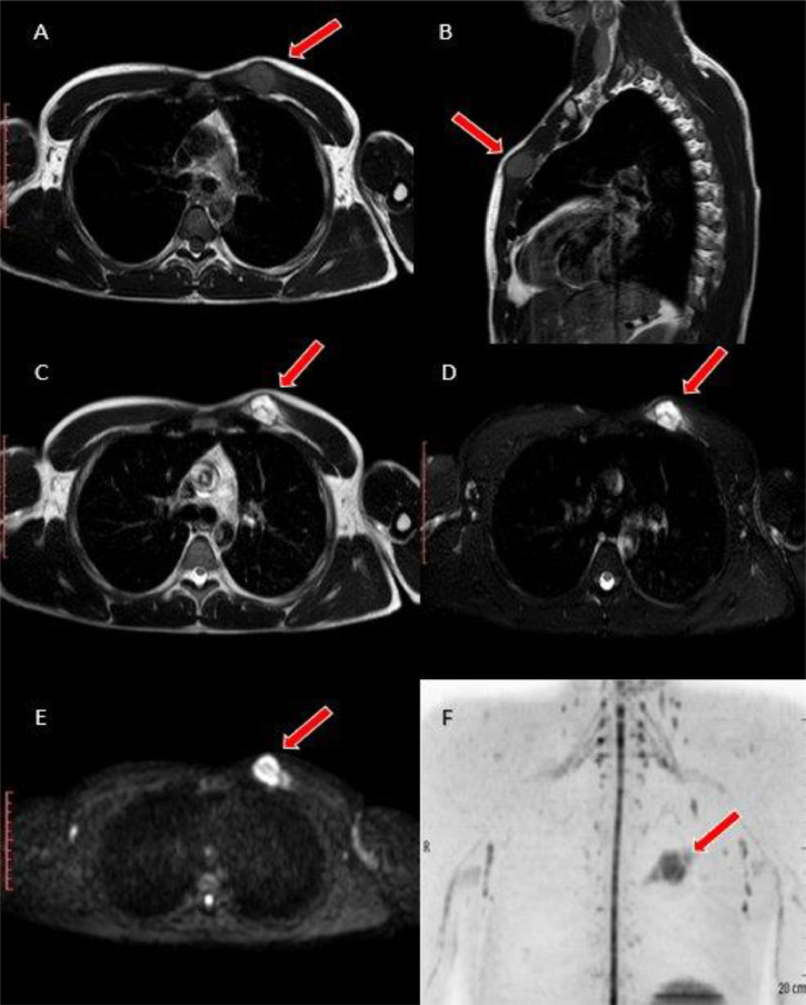
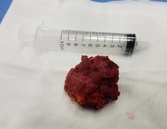
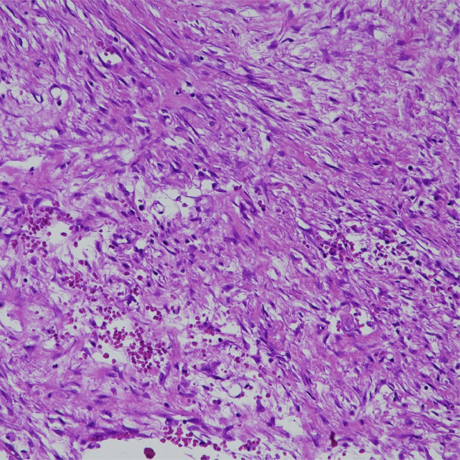
Nodular fasciitis is a benign tumor of soft tissues originating from the proliferation of fibroblasts and myofibroblasts, generally developing between the subcutaneous tissue and the underlying muscular layer. Nodular fasciitis predominantly localizes in the upper extremities, trunk, head and neck. Biomolecular and immunohistochemical analyses result essential to demonstrate the benign origin of the process, also confirmed by very low recurrence rate after complete excision, which represents the gold standard for treatment. We report the case of a 36 years-old man who developed a nodular protuberance clinically evident in the upper-left side of the thorax. We further, highlight the main characteristics of this rare neoplasm trough a thorough review of the literature.
Keywords: Mesenchymal tissue; Nodular fasciitis; Soft tissue mass; Ultrasound Magnetic resonance imaging.
© 2021 The Authors. Published by Elsevier Inc. on behalf of University of Washington.
Figures

References
-
- Stout AP. Pseudosarcomatous fascitis in children. Cancer. 1961;14:1216–1222. - PubMed
-
- Lu L, Lao IW, Liu X, Yu L, Wang J. Nodular fasciitis: a retrospective study of 272 cases from China with clinicopathologic and radiologic correlation. Ann Diagn Pathol. 2015;19(3):180–185. - PubMed
-
- Wagner LM, Gelfand MJ, Laor T, Ryckman FC, Al-Ghawi H, Bove KE. A welcome surprise: nodular fasciitis presenting as soft tissue sarcoma. J Pediatr Hematol Oncol. 2011;33(4):316–319. - PubMed
-
- Plaza JA, Mayerson J, Wakely PE. Nodular fasciitis of the hand: a potential diagnostic pitfall in fine-needle aspiration cytopathology. Am J Clin Pathol. 2005;123(3):388–393. - PubMed
-
- Shimizu S, Hashimoto H, Enjoji M. Nodular fasciitis: an analysis of 250 patients. Pathology (Phila) 1984;16(2):161–166. - PubMed
Publication types
LinkOut - more resources
Full Text Sources
Other Literature Sources
