. 2021 Apr 26:11:639768.
doi: 10.3389/fcimb.2021.639768. eCollection 2021.

Salmonella Typhimurium Triggers Extracellular Traps Release in Murine Macrophages

Affiliations

Salmonella Typhimurium Triggers Extracellular Traps Release in Murine Macrophages

Amy Mónaco et al. Front Cell Infect Microbiol. .

Abstract

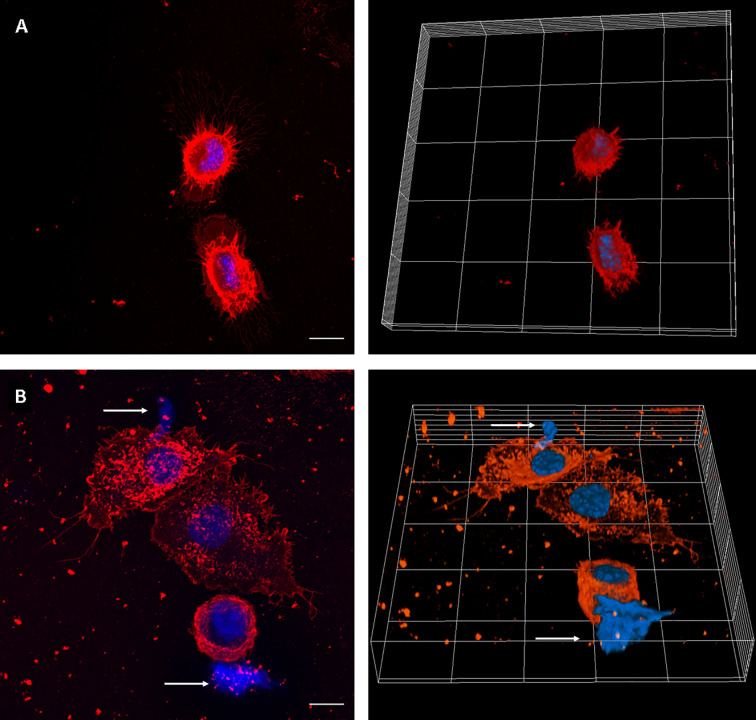
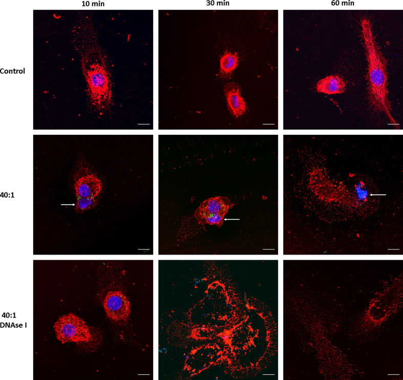
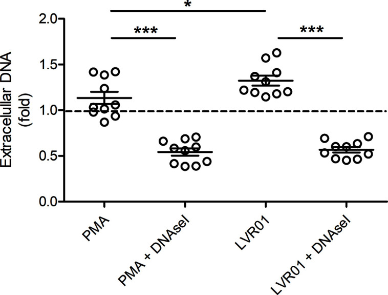
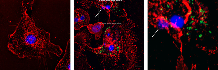
Salmonella comprises two species and more than 2500 serovars with marked differences in host specificity, and is responsible for a wide spectrum of diseases, ranging from localized gastroenteritis to severe life-threatening invasive disease. The initiation of the host inflammatory response, triggered by many Pathogen-Associated Molecular Patterns (PAMPs) that Salmonella possesses, recruits innate immune cells in order to restrain the infection at the local site. Neutrophils are known for killing bacteria through oxidative burst, amid other mechanisms. Amongst those mechanisms for controlling bacteria, the release of Extracellular Traps (ETs) represents a newly described pathway of programmed cell death known as ETosis. Particularly, Neutrophil Extracellular Traps (NETs) were first described in 2004 and since then, a number of reports have demonstrated their role as a novel defense mechanism against different pathogens. This released net-like material is composed of cellular DNA decorated with histones and cellular proteins. These structures have shown ability to trap, neutralize and kill different kinds of microorganisms, ranging from viruses and bacteria to fungi and parasites. Salmonella was one of the first microorganisms that were reported to be killed by NETs and several studies have confirmed the observation and deepened into its variants. Nevertheless, much less is known about their counterparts in other immune cells, e.g. Macrophage Extracellular Traps (METs) and Salmonella-induced MET release has never been reported so far. In this work, we observed the production of METs induced by Salmonella enterica serovar Typhimurium and recorded their effect on bacteria, showing for the first time that macrophages can also release extracellular DNA traps upon encounter with Salmonella Typhimurium. Additionally we show that METs effectively immobilize and reduce Salmonella survival in a few minutes, suggesting METs as a novel immune-mediated defense mechanism against Salmonella infection. Of note, this phenomenon was confirmed in primary macrophages, since MET release was also observed in bone marrow-derived macrophages infected with Salmonella. The evidence of this peculiar mechanism provides new incipient insights into macrophages´ role against Salmonella infection and can help to design new strategies for the clinical control of this transcendental pathogen.

Keywords: MET; Salmonella; defense mechanism; extracellular traps; macrophages.


Conflict of interest statement

The authors declare that the research was conducted in the absence of any commercial or financial relationships that could be construed as a potential conflict of interest.

Figures

Figure 1
Figure 1
MET release in PMA-treated J774A.1 macrophages. Confocal microscopy images of (A) Unstimulated cells, and (B) cells incubated with PMA (200 µM) for 30 minutes. The left panels show maximum intensity z-projection of the image stack; the right panels show 3D models of cells. WGA staining is shown in red and DNA in blue. The arrows point out the presence of METs. The scale bar represents 10 µm.
Figure 2
Figure 2
MET release after Salmonella infection. Confocal microscopy images of J774A.1 macrophage extracellular trap release at t = 10, 30, and 60 min. Upper row: Uninfected cells (Control). Central row: Cells infected with Salmonella enterica serovar Typhimurium LVR01 (40:1). Bottom row: Cells infected with LVR01 pretreated with DNAse I. Maximum intensity z-projections are shown for membrane visualization with WGA staining (red), DNA (blue), and Salmonella (green). The arrows point out MET release. The scale bar represents 10 µm.
Figure 3
Figure 3
Extracellular DNA quantification. Cells were incubated for 30 minutes with PMA or LVR01, with or without DNAse I pretreatment. DNA quantification was assessed with PicoGreen reagent. The dashed line indicates mean value of untreated cells. Results are shown as fold increase of individual replicates (n = 10) and mean ± SD for each evaluated condition. *p ≤ 0.05 and ***p ≤ 0.001, Student T-test. One representative experiment of 6 independent ones performed.
Figure 4
Figure 4
Salmonella trapped in MET of J774A.1 macrophages. Live imaging was performed by confocal microscopy and Salmonella were tracked by image analysis. (A) Fluorescent image at t = 0 s with DNA (blue) and Salmonella (green) channels shows a J774A.1 macrophage (dashed line on the right) and a MET (dashed line on the left). Within the MET, 9 bacteria with high intensity values were identified. (B) Inverse grey scale color table for DNA channel is shown for t = 231 s together with the 15 tracked trajectories for Salmonella. The positions of each bacterium at the beginning and the end of the trajectory are marked by an open and closed circle, respectively. Trajectory lines are colored according to bacteria status: trapped within the MET (grey); free and later trapped (red) and transiently trapped (blue). Trajectories of swimming bacteria are not displayed. The scale bar represents 10 µm.
Figure 5
Figure 5
Percentage of ET releasing macrophages after Salmonella infection. Cells were infected for 10, 30 or 60 minutes with Salmonella, MOI 1, 10 or 40. Number of selected fields was established so as to allow counting a minimum of 150 cells in each condition. Results are shown as mean ± SEM for each evaluated condition. * indicates significant differences between infected and uninfected cells in the same timepoint, *p ≤ 0.05, **p ≤ 0.01, Student T-test. One representative experiment of 2 independent ones performed.
Figure 6
Figure 6
Percentage of Salmonella death by METs. Salmonella was co-cultured for 60 minutes with J774A.1 cells, in presence or absence of DNAse I. Cells were lysed and bacteria counted. Results are shown as mean ± SD for each evaluated condition (n = 8). *p ≤ 0.05, Student T-test. One representative experiment of 4 independent ones performed.
Figure 7
Figure 7
Bone marrow derived macrophages infected with Salmonella enterica serovar Typhimurium LVR01 (40:1). Left panel: Uninfected cells (Control). Center panel: 30 minutes of infection. Right panel: zoomed-in image from central panel (dashed square). The images are maximum intensity z-projections of all the slices in the stack in the three channels. In red: membrane, blue: DNA, green: Salmonella. The arrows point out the presence of METs. The scale bar represents 10 µm.

References

    1. Aulik N. A., Hellenbrand K. M., Czuprynski C. J. (2012). Mannheimia Haemolytica and its Leukotoxin Cause Macrophage Extracellular Trap Formation by Bovine Macrophages. Infect. Immun. 80 (5), 1923–1933. 10.1128/IAI.06120-11 - DOI - PMC - PubMed
    1. Azevedo E. P., Guimaraes-Costa A. B., Torezani G. S., Braga C. A., Palhano F. L., Kelly J. W., et al. . (2012). Amyloid Fibrils Trigger the Release of Neutrophil Extracellular Traps (Nets), Causing Fibril Fragmentation by NET-associated Elastase. J. Biol. Chem. 287 (44), 37206–37218. 10.1074/jbc.M112.369942 - DOI - PMC - PubMed
    1. Barnhart M. M., Chapman M. R. (2006). Curli Biogenesis and Function. Annu. Rev. Microbiol. 60, 131–147. 10.1146/annurev.micro.60.080805.142106 - DOI - PMC - PubMed
    1. Betancor L., Pereira M., Martinez A., Giossa G., Fookes M., Flores K., et al. . (2010). Prevalence of Salmonella Enterica in Poultry and Eggs in Uruguay During an Epidemic Due to Salmonella Enterica Serovar Enteritidis. J. Clin. Microbiol. 48 (7), 2413–2423. 10.1128/JCM.02137-09 - DOI - PMC - PubMed
    1. Bianchi M., Hakkim A., Brinkmann V., Siler U., Seger R. A., Zychlinsky A., et al. . (2009). Restoration of NET Formation by Gene Therapy in CGD Controls Aspergillosis. Blood 114 (13), 2619–2622. 10.1182/blood-2009-05-221606 - DOI - PMC - PubMed

Publication types