

Generation of a squamous cell carcinoma mouse model for lineage tracing of BMI1+ cancer stem cells
- PMID: 33982017
- PMCID: PMC8081985
- DOI: 10.1016/j.xpro.2021.100484
Generation of a squamous cell carcinoma mouse model for lineage tracing of BMI1+ cancer stem cells
Abstract
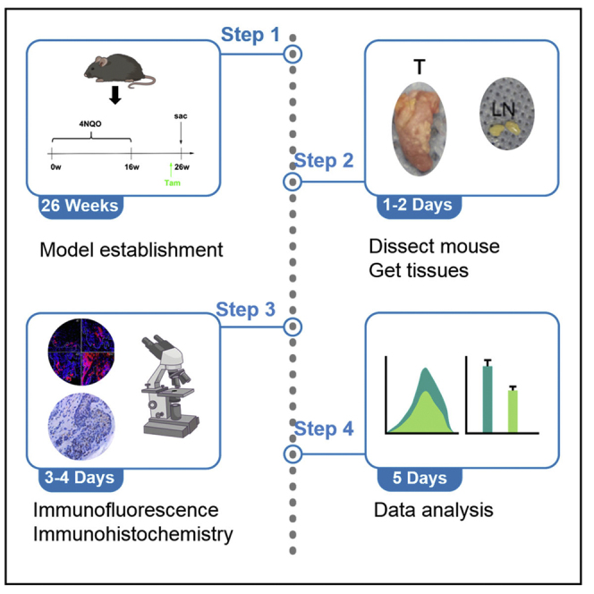
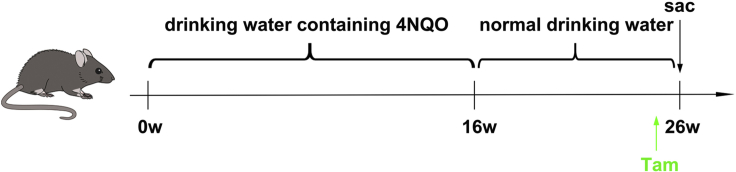
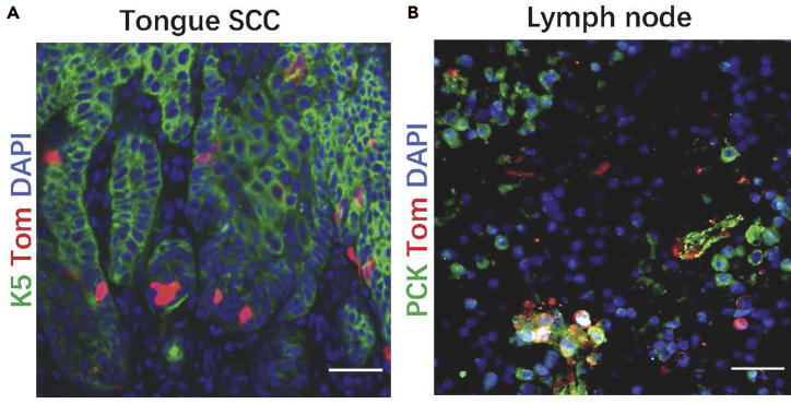
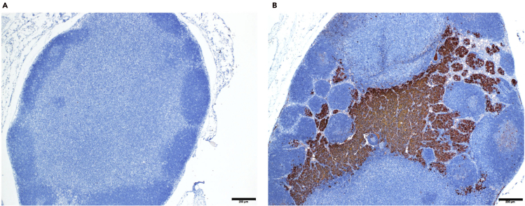
BMI1-expressing cancer stem cells (CSCs) play a key role in the development, progression, therapy resistance, recurrence, and metastasis of head and neck squamous cell carcinoma (HNSCC). Here, we present a chemically-induced HNSCC mouse model, genetically and pathologically similar to human HNSCC. This protocol describes how to use genetic lineage tracing based on the Cre-loxP recombination strategy, which allows us to study the regulation and targeting of BMI1+ CSCs in primary tumors and lymph node metastases. For complete details on the use and execution of this protocol, please refer to Chen et al. (2017) and Jia et al. (2020).
Keywords: Cancer; Microscopy; Model Organisms; Stem Cells.
© 2021 The Author(s).
Conflict of interest statement
The authors declare no competing interests.
Figures
Similar articles
-
Targeting BMI1+ Cancer Stem Cells Overcomes Chemoresistance and Inhibits Metastases in Squamous Cell Carcinoma.Cell Stem Cell. 2017 May 4;20(5):621-634.e6. doi: 10.1016/j.stem.2017.02.003. Epub 2017 Mar 9. Cell Stem Cell. 2017. PMID: 28285905 Free PMC article.
-
Co-targeting BMI1 and MYC to eliminate cancer stem cells in squamous cell carcinoma.Cell Rep Med. 2025 May 20;6(5):102077. doi: 10.1016/j.xcrm.2025.102077. Epub 2025 Apr 15. Cell Rep Med. 2025. PMID: 40239645 Free PMC article.
-
METTL3-mediated ALDH m6A methylation regulates the malignant behavior of BMI1+ HNSCC stem cells.Oral Dis. 2024 Apr;30(3):1061-1071. doi: 10.1111/odi.14609. Epub 2023 May 30. Oral Dis. 2024. PMID: 37249063
-
The role of BMI1 as a biomarker of cancer stem cells in head and neck cancer: a review.Oncology. 2014;86(4):199-205. doi: 10.1159/000358598. Epub 2014 Apr 30. Oncology. 2014. PMID: 24800958 Review.
-
Cancer stem cells in head and neck squamous cell carcinoma: a review of current knowledge and future applications.Head Neck. 2012 Jun;34(6):894-9. doi: 10.1002/hed.21801. Epub 2011 Aug 17. Head Neck. 2012. PMID: 21850700 Review.
Cited by
-
The CTBP2-PCIF1 complex regulates m6Am modification of mRNA in head and neck squamous cell carcinoma.J Clin Invest. 2023 Oct 16;133(20):e170173. doi: 10.1172/JCI170173. J Clin Invest. 2023. PMID: 37643007 Free PMC article.
References
Publication types
MeSH terms
Substances
Grants and funding
LinkOut - more resources
Full Text Sources
Other Literature Sources
Medical
Molecular Biology Databases
