. 2021 Aug;126(8):1017-1029.
doi: 10.1007/s11547-021-01359-3. Epub 2021 May 12.

The role of imaging in acute pancreatitis

Affiliations

The role of imaging in acute pancreatitis

Maria Gabriella Brizi et al. Radiol Med. 2021 Aug.

Abstract

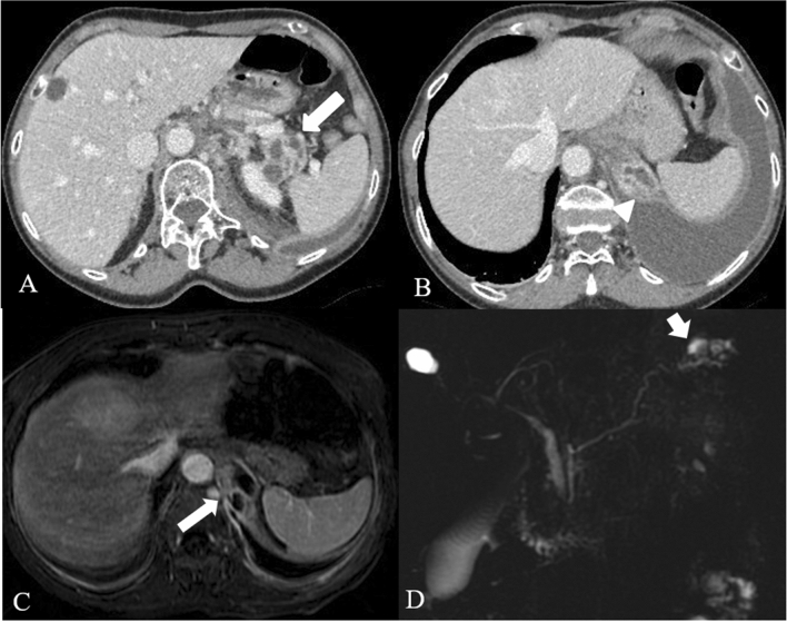
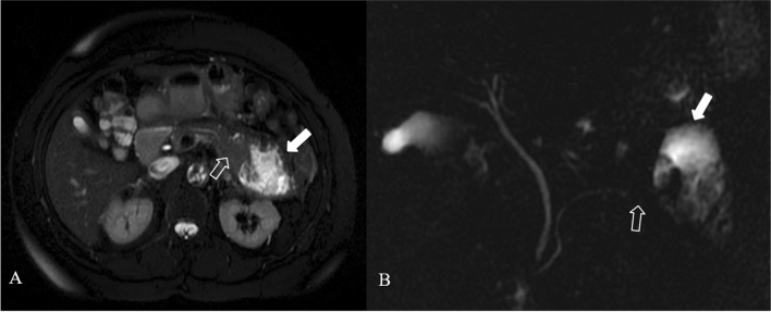
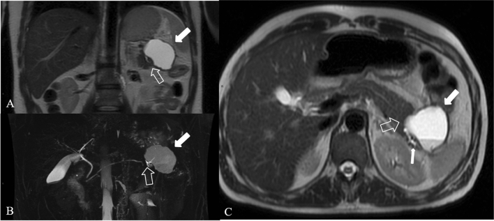
Acute pancreatitis is one of the most commonly encountered etiologies in the emergency setting, with a broad spectrum of findings that varies in severity from mild interstitial pancreas to severe forms with significant local and systemic complications that are associated with a substantial degree of morbidity and mortality. In this article the radiological aspect of the terminology and classification of acute pancreatitis are reviewed. The roles of ultrasound, computed tomography, and magnetic resonance imaging in the diagnosis and evaluation of acute pancreatitis and its complications are discussed. The authors present a practical image-rich guide, applying the revised Atlanta classification system, with the goal of facilitating radiologists to write a correct report, and reinforcing the radiologist's role as a key member of a multidisciplinary team in treating patients with acute pancreatitis. Computed tomography is the most performed imaging test for acute pancreatitis. Nevertheless, MRI is useful in many specific situations, due to its superiority soft tissue contrast resolution and better assessment of biliary and pancreatic duct, for example in the ductal disconnection. The purpose if this article is to review recent advances in imaging acquisition and analytic techniques in the evaluation of AP.

Keywords: Acute pancreatitis; Computed tomography (CT); Interstitial edematous pancreatitis; MRI; Magnetic resonance cholangiopancreatography (MRCP); Necrotizing Pancreatitis.


Conflict of interest statement

The authors declare that they have no competing interests.

Figures

Fig. 1
Fig. 1
Interstistial acute pancreatitis in 17-year-old man, after cannabinoids abuse: axial IV contrast-enhanced CT scan shows mild diffuse enlargement of the whole pancreatic gland with poorly defined contours (arrow); the enhancement of the pancreatic parenchyma is normal and there are no foci of glandular necrosis
Fig. 2
Fig. 2
Acute pancreatitis in a 61 years-old man. The axial contrast-enhanced CT scan after 3 days from the onset (a), in pancreatic arterial phase, shows edematous pancreas (arrows), with disomogeneous and reduced enhancement, with no necrotic area in the contest. It is also evident mild peripancreatic fatty stranding (empty arrow). TC study after nine days of the onset (b) shows heterogeneous contrast enhancement of the pancreatic parenchyma, with focal hypovascular areas due to the necrosis (arrows)
Fig. 3
Fig. 3
Necrotizing Pancreatitis in a 39 years-old man, with acute abdominal pain and sepsis. Axial contrast-enhanced CT scans during the arterial phase (a) and portal phase (b) show enlarged pancreas with poorly defined contours and decreased enhancement of the pancreatic parenchyma (arrows), surrounded by heterogeneous fluid collection (empty arrows in b)
Fig. 4
Fig. 4
Splenic artery pseudoaneurism in a 75 years-old man with RAP. The contrast-enhanced CT scans in the arterial phase axial MIP (a), coronal MIP MPR (b) and sagittal MPR (c) show splenic artery pseudoaneurism (white arrows), in a necrotizing pancreatitis with evident peri-pancreatic collection (empty arrows) extending up to anterior pararenal space of the retroperitoneum, gastrohepatic and gastrosplenic
Fig. 5
Fig. 5
Pseudocyst: 61 years old man, with history of alcohol abuse and hospitalized for necrotizing pancreatitis. Contrast-enhanced CT scan after 4 weeks from the onset, show a hyperdense collection in the retroperitoneum, behind the head of the pancreas and the duodenum (arrow head) suggesting previous bleeding. No signs of active bleeding were evident. Other pseudocysts are localized in the peri-pancreatic fat, between body and tail (empty arrows). Wirsung duct is also dilated (thin arrow)
Fig. 6
Fig. 6
Necrotizing Pancreatitis. The same case as the previous in Fig. 3, after 1 month of the onset of the symptoms, and 1 week after surgical necrosectomy, the contrast-enhanced CT scans in the portal phase show a peri-pancreatic collection with bubbles air and drainage tubes in the collection (empty arrows). Coronal MPR image (a) shows the extention of the collection. Axial scan (b) show diffuse alterations of the CE of the parenchyma, due to necrosis (arrow)
Fig. 7
Fig. 7
ANCs in biliary AP in a 74 years-old man. The contrast-enhanced CT axial scan (a) shows the presence of an acute necrotic fluid collection (ANCs) interesting the head and the body of the pancreatic gland (white arrows), extending in the fat surrounding the gland up to the gastrohepatic ligament. The sagittal MPR (b) and coronal (c) better show the extention of the collection
Fig. 8
Fig. 8
Necrotizing AP in a 74 years-old man with a history of AP, 3 weeks after the onset. The contrast-enhanced CT axial scan (a) and the coronal MPR (b) show the presence of collection in the lesser sac up to the lesser curvature of the stomach (white arrows). The contrast-enhanced CT axial scan (c) and the coronal MPR (d) 5 days after the previous examination show the drainage of the collection by cystogastrostomy (empy arrow), and the presence of gas-collection (thin arrow in c)
Fig. 9
Fig. 9
Walled off necrosis. 73 years old woman undergoing statins and amiodarone treatment. T2 weighted MR image (a) and post-gadolinium T1w scan (b), show the presence of a heterogeneous encapsulated fluid collection suggesting walled-off necrosis (WON); the WON extends in the pancreatic and peripancreatic area, up to the left anterior pararenal space and left paracolic space (arrows). Diffuse hyperintensity on T2w images of the pancreatic parenchyma indicates the presence of parenchymal edema (empty arrow in a)
Fig. 10
Fig. 10
WON evolution. Acute necrotizing pancreatitis in a 61-years-old man. Four weeks after the onset axial CECT scan shows the presence of an encapsulated collection (WON) in the peripancreatic fat near the body and the tail of the gland (arrows)
Fig. 11
Fig. 11
Fistula with pleural cavity in a 66 years-old woman with acute necrotizing pancreatitis. Multiple encapsulated fluid collections in the tail of the pancreatic parenchyma (arrow in a), as for necrotic spots. These collections extend upt to the left hemidiaphragm with pleural fistula and pleural effusion (arrowhead in b). Axial (c) post contrast T1w MR images and MRCP (d) confirm the pleural fistula (arrows)
Fig. 12
Fig. 12
Pseudocyst in a 36 years-old woman, with AP, 12 weeks from the onset. Axial scan (a) and coronal scan (b) show the hyperintense collection in T2 weighted image (empty arrow) with dilated pancreatic duct (white arrow). MRCP (c) shows bile duct dilatation (arrowhead), due to the compression from the collection
Fig. 13
Fig. 13
ACNs in a 72 years-old woman. T2 weighted MR image (a) and MRCP (b) 2 weeks after the onset show a heterogeneous collection in the pancreatic tail (white arrow), where there is a focal interruption of the Wirsung duct (empty arrow)
Fig. 14
Fig. 14
Disconnected pancreatic duct syndrome in a 41 years-old man. Coronal (a), axial (c) T2 weighted MR images and MRCP (b) 3 weeks after the onset of the symptoms show focal interruption of the Wirsung duct (empty arrow)—upstream pancreatic duct dilatation (thin arrow in c)—and a heterogeneous encapsulated fluid collection in the pancreatic tail (white arrow)

References

    1. Petrov MS, Yadav D. Global epidemiology and holistic prevention of pancreatitis. Nat Rev Gastroenterol Hepatol. 2019;16(3):175–184. doi: 10.1038/s41575-018-0087-5. - DOI - PMC - PubMed
    1. Petrov MS. Editorial: abdominal fat: a key player in metabolic acute pancreatitis. Am J Gastroenterol. 2013;108(1):140–142. doi: 10.1038/ajg.2012.384. - DOI - PubMed
    1. Yadav D, Papachristou GI, Whitcomb DC. Alcohol-associated pancreatitis. Gastroenterol Clin North Am. 2007;36(2):219–vii. doi: 10.1016/j.gtc.2007.03.005. - DOI - PubMed
    1. Xiao AY, Tan ML, Wu LM, Asrani VM, Windsor JA, Yadav D, Petrov MS. Global incidence and mortality of pancreatic diseases: a systematic review, meta-analysis, and meta-regression of population-based cohort studies. Lancet Gastroenterol Hepatol. 2016;1(1):45–55. doi: 10.1016/S2468-1253(16)30004-8. - DOI - PubMed
    1. Pendharkar SA, Mathew J, Petrov MS. Age- and sex-specific prevalence of diabetes associated with diseases of the exocrine pancreas: a population-based study. Dig Liver Dis Off J Ital Soc Gastroenterol Ital Assoc Study Liver. 2017;49(5):540–544. doi: 10.1016/j.dld.2016.12.010. - DOI - PubMed

LinkOut - more resources