Pulsatile load and wasted pressure effort are reduced following an acute bout of aerobic exercise
- PMID: 33982596
- PMCID: PMC8325608
- DOI: 10.1152/japplphysiol.00946.2020
Pulsatile load and wasted pressure effort are reduced following an acute bout of aerobic exercise
Abstract
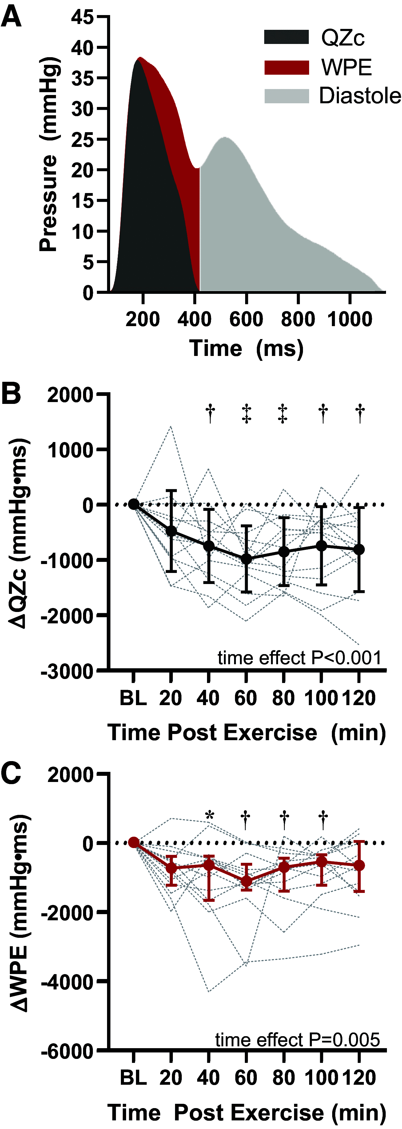
Following aerobic exercise, sustained vasodilation and concomitant reductions in total peripheral resistance (TPR) result in a reduction in blood pressure that is maintained for two or more hours. However, the time course for postexercise changes in reflected wave amplitude and other indices of pulsatile load on the left ventricle have not been thoroughly described. Therefore, we tested the hypothesis that reflected wave amplitude is reduced beyond an hour after cycling at 60% V̇o2peak for 60 min. Aortic pressure waveforms were derived in 14 healthy adults (7 men, 7 women; 26 ± 3 yr) from radial pulse waves acquired via high-fidelity applanation tonometry at baseline and every 20 min for 120 min postexercise. Concurrently, left ventricle outflow velocities were acquired via Doppler echocardiography and pressure-flow analyses were performed. Aortic characteristic impedance (Zc), forward (Pf) and backward (Pb) pulse wave amplitude, reflected wave travel time (RWTT), and wasted pressure effort (WPE) were derived. Reductions in aortic blood pressure, Zc, Pf, and Pb were all sustained postexercise whereas increases in RWTT emerged from 60 to 100 min post exercise (all P < 0.05). WPE was reduced by ∼40% from 40 to 100 min post exercise (all P < 0.02). Stepwise multiple regression analysis revealed that the peak ΔWPE was associated with ΔRWTT (β = -0.57, P = 0.003) and ΔPb (β = 0.52, P = 0.006), but not Δcardiac output, ΔTPR, ΔZc, or ΔPf. These results suggest that changes in pulsatile hemodynamics are sustained for ≥100 min following moderate intensity aerobic exercise. Moreover, decreased and delayed reflected pressure waves are associated with decreased left ventricular wasted effort after exercise.NEW & NOTEWORTHY We demonstrate that pulsatile load on the left ventricle is diminished following 60 min of moderate intensity aerobic exercise. During recovery from exercise, the amplitude of the forward and backward traveling pressure waves are attenuated and the arrival of reflected waves is delayed. Thus, the work imposed upon the left ventricle by reflected pressure waves, wasted pressure effort, is decreased after exercise.
Keywords: afterload; arterial load; left ventricle; postexercise hypotension.
Conflict of interest statement
D.G.E. has research grants from the National Institutes of Health. J.A.C. has recently consulted for Bayer, Sanifit, Fukuda-Denshi, Bristol-Myers Squibb, Johnson & Johnson, Edwards Life Sciences, Merck, and the Galway-Mayo Institute of Technology. He received University of Pennsylvania research grants from National Institutes of Health, Fukuda-Denshi, Bristol-Myers Squibb, and Microsoft. He is named as inventor in a University of Pennsylvania patent for the use of inorganic nitrates/nitrites for the treatment of Heart Failure and Preserved Ejection Fraction. He has received payments for editorial roles from the American Heart Association and the American College of Cardiology. He has received research device loans from Atcor Medical, Fukuda-Denshi, Uscom, NDD Medical Technologies, Microsoft, and MicroVision Medical.
Figures


References
-
- Romero SA, McCord JL, Ely MR, Sieck DC, Buck TM, Luttrell MJ, MacLean DA, Halliwill JR. Mast cell degranulation and de novo histamine formation contribute to sustained postexercise vasodilation in humans. J Appl Physiol (1985) 122: 603–610, 2017. doi: 10.1152/japplphysiol.00633.2016. - DOI - PMC - PubMed
Publication types
MeSH terms
Grants and funding
LinkOut - more resources
Full Text Sources
Other Literature Sources
Medical

